© 2016, IJCSMC All Rights Reserved 442 Available Online atwww.ijcsmc.com
International Journal of Computer Science and Mobile Computing
A Monthly Journal of Computer Science and Information Technology
ISSN 2320–088X
IMPACT FACTOR: 5.258
IJCSMC, Vol. 5, Issue. 6, June 2016, pg.442 – 447
HISTOGRAM BASED COLOR
IMAGE SEGMENTATION
Anchal Malhotra
1, Dr. Nripendra Narayana Das
21. M.Tech Scholar, 2. Professor
Department of Computer Science and Engineering
(Specialization in Image Processing)
Rawal Institute of Engineering and Technology, Faridabad, India
[email protected] 1 , [email protected] 2
Abstract— Due to rapid growth in digital media, image processing become an important area of research. Image segmentation is a part of image processing steps. With the improvement in computer processing and increasing demand of color images segmentation is more concerned field of research. Although many segmentation techniques have been developed for image segmentation. But still there is lack of general purpose segmentation. This project is based on histogram based approach for segmenting the images. In order to segment the images multi threshold histogram are used. The color image will be coarsely represented using several bins. Coarse representation uses the spatial information from a Histogram based windowing process. K-Means is used to cluster the coarse image data.
Keywords— Histogram; image segmentation; Image segmenter; K-Mean.
I. INTRODUCTION
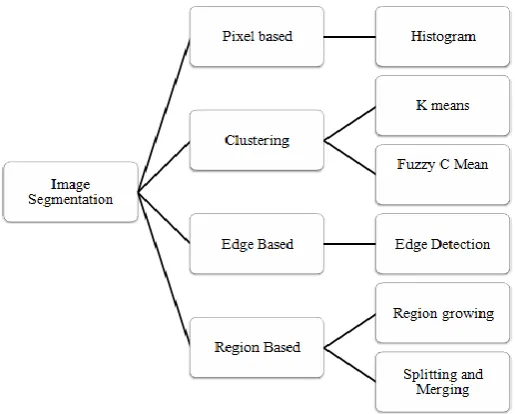
In image segmentation the digital images are partitioned into multiple regions. Each region may having different color, and texture. Image segmentation may be done either in spatial domain or in transform domain. In this system segmentation based on pixel domain approach are used. The result of image segmentation is either a set of regions that together cover up the entir e image or a set of contours extracted from the image [1]. One direct way to segment an image is by applying an edge detection technique, especially if the image consists of clear objects on a different intensity background. However, this method can fail, if the image contains noise. It is usually necessary to conduct validation experiments so as to quantify the performance of a segmentation technique. Although different types of image segmentations techniques are used. But there is no unique segmentation technique exists for all images. Image Segmentation Techniques are broadly classified in following category as shown in Figure 1.
i. Pixel based : In this process a digital image is segmented into multiple segments or groups of pixels, also known as super pixels. Histogram based segmentation works in this category.
© 2016, IJCSMC All Rights Reserved 443
iii. Edge based With this method, the edges which are detected in an image are assumed to represent object boundaries, and used to identify these objects.
iv. Region based Where an edge based technique may attempt to find the object boundaries and then locate the object itself by fulfilling them in, a region based method takes the opposite approach Structural Segmentation Methods. These methods utilize information about the structural features of the image to implement segmentation of the target image. Some of the common structural methods include edge-detection, graph searching, deformable models, is on surface and level set. We highlight some.
Figure 1 Types of Segmentation Techniques
© 2016, IJCSMC All Rights Reserved 444
facilitates the use of three types of user inputs: 1) foreground and background seed input, 2) soft constraint input, and 3) hard constraint input, as well as their combinations. To support the context of their research model WenxianYang et al[13] argued that one common fault in the existing interactive image segmentation algorithms is the lack of more intellectual ways to understan d the intention of user inputs. Ping-Feng Chen et al[14] proposed a novel model to jointly segment and register objects of interest in layered images. Since the Layered images refer to imageries taken from different perspectives and possibly by different sensors, the registration and segmentation are therefore the two main tasks which contribute to the bottom level, data alignment, of the multi sensor data fusion hierarchical structures.
Figueiredo et al[15] proposal expected to introduce a variation image segmentation method for assess the aberrant crypt foci (ACF) in the person colon captured in vivo by endoscopy. The proposed segmentation technique enhanced the active contours without edges model of Chan and Vese to account forthe ACF's particular structure. Level sets to represent the segmentation boundaries and discretize in space by finite elements and in (artificial) time by fixed difference sare employed. Yazdanpanah et al [16] presented a semi-automated segmentation algorithm to detect intra-retinal layers inOCT images acquired from rodent models of retinal degeneration. The proposed segmentation technique was adapted Chan-Vese's energy-minimizing active contours without edges for the OCT images, which inturn suffered from low contrast and were highly tarnished by noise. Further Dana et. al. [17] the adaptive integration of the colour and texture attributes in the development of complex image descriptors is highlighted. The substantial interest shown by the research community in colour–texture-based segmentation is mainly motivated by two factors. Delu Zeng et al[18] considered the task of object segmentation and achieve in a novel manner that backed by the Poincar map method in a defined vector field in view of dynamical systems. n interpolated swirl and attract flow ( F) vector field is first generated for the observed image. Then, the states on the limit cycles. In Nguyen, et. al [19] proposed a robust and accurate interactive method based on the recently developed continuous-domain convex active contour model. The proposed method exhibits many desirable properties of an effective interactive image segmentation algorithm, including robustness to user inputs and different initializations, the ability to produce a smooth and accurate boundary contour, and the ability to handle topology changes. Experimental results on a benchmark data set show that the proposed tool is highly effective and outperforms the state-of-the-art interactive image segmentation algorithms.
Based on these literature survey it is clear that no single algorithm can be considered good for all applications and all images [1-21]. Segmentation methods used for color images can be divided into two main categories: feature-space based techniques (clustering methods and histogram multi-thresholding, and image-domain based techniques. The latter are further divided into pixel-similarity based algorithms and pixel-difference based algorithms. Image-domain based techniques exploit the pixel context interaction which increase computational complexity.
Rest of paper organized as section II represents detailed methodology and experimental results and finally section III concludes the paper.
II. METHODOLOGY USED
in this experiments segmentation based on color thresholder and Image segmenters are used. The detailed description of these techniques are described in [22] .
A. A Image Segmentation Using the Color Thesholder
The Color Thresholder applications lets us threshold color images by manipulating the color components of these images, based on different color spaces. Using this app, we can create a segmentation mask for a color image.
B. Image Segmenter
© 2016, IJCSMC All Rights Reserved 445
Table 1 Results of Histogram based image segmentation
Sl. No. Input Image Segmented Image
1
2
3
The result obtained from color thresholding is shown in
Figure 2- 3
Figure 2 Color segmentation of images.
© 2016, IJCSMC All Rights Reserved 446
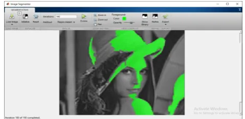
The result obtained from image segmenter is shown in
Figure 4.
Figure 4 Region based segmentation having threshold 150.
III. CONCLUSION
In this paper an unsupervised image segmentation strategy based on histogram, color thresholding, and image segmenter has been performed. The main colclusion drawn from this thesis is highlighted below: An exhaustive analysis of image segmentation techniques has been carried out. Main strategies perform the various combinations have been identified and a classification of these has been proposed. The following segmentation methods of pixel based, edge based and region based methods of image segmentation has been discussed. This systematic comparison study is helpful for individual researchers to do research in the ground of image segmentation. These Image segmentation techniques are extremely efficient particularly, graph based image segmentation methods which comes under the third category i.e., region based methods. Medical image processing is one of the main dynamic research topics in image processing. Most modern research in image segmentation has tinted the potential of graph based techniques for medical applications. In order to implement graph theory in image segmentation professionally in particular in medical image processing we need to set up implementation between mathematical outstanding junior scientists and biological scientists, and describe the sketch to build up the new tools on this domain. The motivation should be on the study of properties of Euler graphs, minimal spanning trees, Fuzzy graphs, shortest paths trees, minimal cuts and Normalized cuts and we re-examine these ideas for image segmentation purposes. From this comparison study we conclude that there is no universal segmentation method that can be executed for all types of images, but a number of techniques do healthier than others for particular types of images representative better performance can be attained by selecting appropriate algorithm or combination of suitable techniques.
REFERENCES
[1] Hall L.O., Bensaid A.M., Bezdek J.C. Clarke L.P., Velthuizen R.P.Silbiger M.S., ― Comparison of Neural Network and Fuzzy Clustering Techniques in egmenting Magnetic Resonance mages of the Brain‖, EEE Transactions on Neural Networks, Volume: 3, Pages: 672–682, 1992.
[2] chalkoff R.J., Pattern Recognition: ― tatistical, tructural and Neural Methods‖, John Wiley and ons, 1992.
[3] Viergever M. and Lobregt ., ― Discrete Dynamic Contour Model‖, Medical maging, an EEE Transactions, Volume: 14, Pages: 12–24, 1995. [4] Pathak S. D., Grimm P. D., Chalana V., and Kim Y.,Pubic Arch Detection in Transrectal Ultrasound Guided Prostate Cancer Therapy, IEEE Transactions on
Medical Imaging, Volume: 17, Pages: 762-771, 1998.
[5] Pham D. L, Prince J. L. and Xu C, ―Current Methods in Medical mage egmentation‖, Biomedical Engineering, nnual Review, Volume: 2, Pages: 315-38, 2000.
[6] Shapiro L. G. and Stockman G. C., Computer Vision, Prentice-Hall Publications, New Jersey, 2001.
[7] Zhang, Y. J, ― n Overview of mage and Video egmentation in the last 40 years‖, 6th nternational ymposium on ignal Processing and Its Applications Proceedings, Pages: 144-151, 2001.
[8] N. Paragios, O. Mellina-Gottardo, and V. Ramesh, ―Gradient vector flow fast geometric active contours,‖ EEE Transactions on Pattern nalysis and Machine Intelligence, no. 3, pp. 402–407, 2004.
[9] Xu R., and Wunsch D., ― urvey of Clustering lgorithms‖, Neural Networks EEE Transaction, Volume: 16, May 2005.
[10] Chunming Li; Chiu-Yen Kao; Gore, J.C.; Zhaohua Ding; , "Minimization of Region-Scalable Fitting Energy for Image Segmentation," Image Processing, IEEE Transactions on , vol.17, no.10, pp.1940-1949, Oct. 2008; doi: 10.1109/TIP.2008.2002304;
© 2016, IJCSMC All Rights Reserved 447
[12] Srinivasa, G.; Fickus, M.C.; Yusong Guo; Linstedt, A.D.; Kovacevic, J.; , "Active Mask Segmentation of Fluorescence Microscop e Images," Image Processing, IEEE Transactions on , vol.18, no.8, pp.1817-1829, Aug. 2009; doi: 10.1109/TIP.2009.2021081
[13] Wenxian Yang; Jianfei Cai; Jianmin Zheng; Jiebo Luo;, "User-Friendly Interactive Image Segmentation Through Unified Combinatorial User Inputs," Image Processing, IEEE Transactions on , vol.19, no.9, pp.2470-2479, Sept. 2010; doi: 10.1109/TIP.2010.2048611
[14] Ping-Feng Chen; Krim, H.; Mendoza, O.L.; , "Multiphase Joint Segmentation-Registration and Object Tracking for Layered Images," Image Processing, IEEE Transactions on , vol.19, no.7, pp.1706-1719, July 2010; doi: 10.1109/TIP.2010.2045164
[15] Figueiredo, I.N.; Figueiredo, P.N.; Stadler, G.; Ghattas, O.; Araujo, A.; , "Variational Image Segmentation for Endoscopic Hu man Colonic Aberrant Crypt Foci," Medical Imaging, IEEE Transactions on , vol.29, no.4, pp.998-1011, April 2010; doi: 10.1109/TMI.2009.2036258
[16] Yazdanpanah, A.; Hamarneh, G.; Smith, B.R.; Sarunic, M.V.; , "Segmentation of Intra -Retinal Layers From Optical Coherence Tomography Images Using an Active Contour Approach," Medical Imaging, IEEE Transactions on , vol.30, no.2, pp.484-496, Feb. 2011; doi: 10.1109/TMI.2010.2087390
[17] Ilea, Dana E., and Paul F. Whelan. "Image segmentation based on the integration of colour–texture descriptors—A review." Pattern Recognition 44.10 (2011): 2479-2501.
[18] Delu Zeng; Zhiheng Zhou; Shengli Xie; , "Image Segmentation Based on the Poincaré Map Method," Image Processing, IEEE Transactions on , vol.21, no.3, pp.946-957, March 2012; doi: 10.1109/TIP.2011.216840
[19] Nguyen, Thi Nhat Anh, et al. "Robust interactive image segmentation using convex active contours." Image Processing, IEEE Transactions on 21.8 (2012): 3734-3743.
[20] Kumar, Rajiv, and Kiran Kumar Ravulakollu. "Handwritten Devnagari Digit Recognition: Benchmarking On New Dataset." Journal of Theoretical & Applied Information Technology 60.3 (2014).
[21] Kumar, Rajiv, Amresh Kumar, and Pervez Ahmed. "A benchmark dataset for devnagari document recognition research." 6th International Conference on Visualization, Imaging and Simulation (VIS’13). 2013.