Western University Western University
Scholarship@Western
Scholarship@Western
Electronic Thesis and Dissertation Repository
2-26-2018 3:00 PM
Ultrasound-Augmented Laparoscopy
Ultrasound-Augmented Laparoscopy
Uditha Lakmal Jayarathne
The University of Western Ontario
Supervisor Terry M. Peters
The University of Western Ontario
Graduate Program in Biomedical Engineering
A thesis submitted in partial fulfillment of the requirements for the degree in Doctor of Philosophy
© Uditha Lakmal Jayarathne 2018
Follow this and additional works at: https://ir.lib.uwo.ca/etd
Part of the Other Biomedical Engineering and Bioengineering Commons
Recommended Citation Recommended Citation
Jayarathne, Uditha Lakmal, "Ultrasound-Augmented Laparoscopy" (2018). Electronic Thesis and Dissertation Repository. 5219.
https://ir.lib.uwo.ca/etd/5219
This Dissertation/Thesis is brought to you for free and open access by Scholarship@Western. It has been accepted for inclusion in Electronic Thesis and Dissertation Repository by an authorized administrator of
Abstract
Laparoscopic surgery is perhaps the most common minimally invasive procedure for many diseases in the abdomen and thorax. Since the laparoscopic camera provides only the surface view of the internal organs, in many procedures, surgeons use laparoscopic ultrasound (LUS) to visualize deep-seated surgical targets. Conventionally, the 2D LUS image is visualized in a display spatially separate from that which displays the laparoscopic video. Therefore, reasoning about the geometry of hidden targets requires mentally solving the spatial alignment and resolving the modality differences, which are cognitively very challenging. Moreover, the mental representation of hidden targets in space acquired through such cognitive mediation may be error prone, and cause incorrect actions to be performed.
To remedy this, advanced visualization strategies are required where the US information is visualized in the context of the laparoscopic video. To realize such visualization schemes, efficient computational methods are required (i) to accurately align the US image coordinate system with that centred in the camera, (ii) to accurately represent 3D information of hidden targets from a series of 2D US images, and (iii) to blend the US information with the surface image provided by the camera such that the surgeons perceive the geometry of hidden targets accurately.
In this thesis, a complete pipeline is described to visualize hidden targets, imaged by a LUS probe, in 3D in the context of the laparoscopic video. A novel method to register US images with a coordinate system centred in the camera is detailed with an experimental in-vestigation into accuracy bounds in representing an imaged target in this coordinate system. This method eliminates the requirement for extrinsic tracking devices in the operating room (OR), significantly reducing both the financial and logistical overhead. An improved method to blend US information with the surface view provided by the camera is also presented with an experimental investigation into the accuracy of perception of the target locations in space.
The work presented here, together with concurrent development in related fields, will en-able image-guidance in laparoscopic soft-tissue surgery. The suggested improvements will
increase both the efficiency and the safety of many minimally invasive abdominal procedures.
Keywords: Laparoscopic Surgery, Laparoscopic Ultrasound, Visualization, Pose Estima-tion, Ultrasound ReconstrucEstima-tion, Direct Volume Rendering, Psychophysical Evaluation
Acknowledgements
During the past six years, from the day I started my graduate work at the Robarts Research Institute to the day I finished compiling this thesis, I received enormous support from a large group of people. Many of them contributed by providing financial support, involving in dis-cussions, or by arranging travel to various destinations throughout the world.
I would like to start by acknowledging my supervisor, Dr. Terry Peters, for giving me the opportunity to pursue my graduate studies in his work-class research laboratory. He encour-ages every student to think outside the box and solve clinically impactful problems. Terry makes sure that we have access to the best research facilities required to undertake world class research. I appreciate Terry’s trust in me to represent his lab in many international conferences, and I will never forget the education I received from him. In no specific order, I would also like to thank Dr. Elvis Chen, John Moore, Dr. Ali Khan, and Dr. Xiongbiao Luo, Terry’s research staff, for their support, either in the development of software tools, setting up experiments, or lengthy hours of theoretical discussions. Special thanks should go to Dr. Roy Eagleson, Dr. Hanif Ladak, and Dr. Stephen Pautler for steering me in the right direction being in my advi-sory committee. I also express my gratitude to Christine Ellwood, and the administrative staff at the Robarts Research Institute for their assistance in all the paperwork.
Since I first came to London, many people helped me with many logistical issues. My sincere gratitude goes to Janette Wallace, Jackie Williams, and many people in the Sri Lankan community living in London for making this city feels like home. Also, special recognition is given to my academic siblings Dr. Kamyar Abhari, Dr. John Baxter, Dr. Jonathan McLeod, (Dr.-to-be) Golafsoun Ameri, and Adam Ranking for their enormous support and many hours of useful discussions.
Finally, I would like to thank my family. Even though I am thousands of miles away from home, endless love and support from my parents, my sister and brother always helped me to stay focussed in my academic goals even under most stressful situations. Most of all, I thank my beautiful wife, Yashmi, for her endless love. Her support in realizing my academic dreams
was tremendous. She was always by my side in many difficult situations, and I will never forget her commitment during the past three years of our married life.
This work was supported financially by the Canadian Institute of Health Research (CIHR), the Natural Science and Engineering Research Council (NSERC), the Canadian Foundation for Innovation (CFI), and the University of Western Ontario.
Dedicated to my parents, Yashmi, and specially to our future ...
“Believe nothing, unless it agrees with your own reason”
- Lord Buddha
Contents
Abstract ii
Acknowledgments iv
List of Figures xi
List of Tables xii
List of Abbreviations xiii
1 Introduction 1
1.1 Image-guided Minimally Invasive Surgery . . . 2
1.2 Visualization in Image-guided Surgery . . . 3
1.3 Assessment of IGS Systems . . . 5
1.4 Image Guidance in Soft-tissue Surgery . . . 6
1.5 Laparoscopic Surgery . . . 7
1.6 Robot-Assisted Laparoscopic Surgery . . . 8
1.7 Intra-operative Ultrasound in Laparoscopic Surgery . . . 10
1.8 Cognitive Processes in Ultrasound-guided Action . . . 13
1.9 Perceiving 3D Form from Cross-sectional Images . . . 15
1.10 In Situ Visualization of US . . . 17
1.11 Motivation: Ultrasound-augmented Laparoscopy . . . 19
1.12 Hypothesis and Research Questions . . . 21
1.13 Thesis Outline . . . 22
1.13.1 Robust, Intrinsic Tracking of a Laparoscopic Ultrasound Probe . . . 23
1.13.2 Accuracy in Freehand 3D US Reconstruction with Robust Visual Track-ing . . . 23
1.13.3 Visualizing Ultrasound In the Context of Laparoscopy . . . 24
Bibliography 24 2 Robust, Intrinsic Tracking of a Laparoscopic Ultrasound Probe 32 2.1 Introduction . . . 32
2.1.1 Related Work . . . 33
2.1.2 Contributions . . . 35
2.2 Methods . . . 36
2.2.2 Simultaneous Pose and Correspondence from a Single View . . . 38
2.2.3 Handling Not-Detected Points . . . 41
2.2.4 Priors . . . 42
2.2.5 Extension to Multiple Views . . . 43
2.3 Experiments and Results . . . 47
2.3.1 Experimental Setup . . . 47
2.3.2 Comparison to the Optical Tracking-based Reference . . . 50
2.3.3 US-Video Overlay . . . 50
2.3.4 Results . . . 51
2.4 Discussion . . . 56
2.5 Conclusions . . . 57
Bibliography 58 3 Accuracy in Freehand 3D-US Reconstruction with Robust Visual Tracking 63 3.1 Introduction . . . 63
3.2 Methods . . . 65
3.2.1 Robust Intrinsic Tracking . . . 66
3.2.2 US Calibration . . . 69
3.2.3 Realtime 3D Ultrasound Reconstruction . . . 71
Implementation . . . 72
3.2.4 Experiments . . . 73
3.3 Results . . . 74
3.4 Discussion . . . 78
3.5 Conclusions . . . 79
Bibliography 79 4 Visualizing Ultrasound In the Context of Laparoscopy 82 4.1 Introduction . . . 82
4.2 Related Work . . . 83
4.2.1 Contributions . . . 84
4.3 Methodology . . . 85
4.3.1 Real-time 3D US Reconstruction . . . 85
4.3.2 Blending the US Volume with the Camera Image . . . 86
Implementation . . . 87
4.3.3 Experimental Setup . . . 90
4.4 Experiments and Results . . . 92
4.4.1 Experiment I: Monoscopic Viewing . . . 95
Experiment . . . 95
Results . . . 95
4.4.2 Experiment II: Stereoscopic Viewing . . . 99
Experiment . . . 99
Results . . . 99
4.6 Conclusion . . . 105
Bibliography 105 5 Conclusion 109 5.1 Concurrent Development . . . 111
5.2 A Look Into the Future . . . 112
5.2.1 Future Image-guided Soft-tissue Surgery . . . 112
5.2.2 Fusing Pre-operative and Intra-operative Imaging . . . 113
Bibliography 113 Appendices 116 A Anisotropically Scaled ICP 117 B Copyright Releases 119 B.1 Releases for Material in Chapter 2 . . . 120
B.2 Releases for Material in Chapter 3 and 4 . . . 126
Vita 130
List of Figures
1.1 Visualization in Image-guided Surgery . . . 4
1.2 Laparoscopic Surgery . . . 7
1.3 daVinciSurgical Robotic System . . . 9
1.4 Stereoscopic camera and the surgeons console in adaVincirobotic system . . . 10
1.5 Laparoscopic Ultrasound Probes . . . 11
1.6 TileProTMdisplay . . . 12
1.7 Cognitive transformations in visually-guided action . . . 14
1.8 The Sonic Flashlight device . . . 18
1.9 In situvisualization of US in laparoscopy: Concept . . . 20
2.1 Search for Pose and Correspondence Simultaneously . . . 37
2.2 Search Space Managed by a Tree Data-Structure . . . 41
2.3 Simultaneous Pose and Correspondence with Multiple Views . . . 45
2.4 Clinical LUS Probe with a Fiducial Pattern Attached, Mock-probe, and US Calibration . . . 49
2.5 Errors in the Computed Estimates . . . 53
2.6 TRE Maps for each Method . . . 54
2.7 Qualitative Demonstration for the Efficacy of the Pose Estimation Framework . 55 3.1 LUS Probe with Fiducial Pattern Attached, PVA-C Phantom, and US Calibra-tion Tool . . . 68
3.2 Data Capture for US Calibration . . . 70
3.3 US Images Approximated by 2D Plans in 3D Ultrasound Reconstruction Method 72 3.4 Histogram of Distances between Centerline of Tubular Structures in CT Vol-ume, and the Registered US Volume . . . 75
3.5 Geometric Accuracy of Freehand 3D US Reconstruction . . . 76
3.6 Centerline Alignment Error Projected to the Nearest Voxel on the Lumen Wall . 77 4.1 Distance Dependent Transparency Function . . . 88
4.2 Steps in Keyhole-blending Method . . . 89
4.3 The Experimental Setup for the Perceptual Study . . . 91
4.4 PVA-C Phantom under Different Visualization Modes . . . 94
4.5 Quantitative Results of the Perceptual Study . . . 98
4.6 Subjective Ranking Based on the NASA TLX . . . 102
List of Tables
3.1 Fiducial distance error and the centerline distance error after the registration . . 75
List of Abbreviations
2D
Two Dimensional (typicaly (x,y) domain)
3D
Three Dimension (typically (x,y,z) domain)
DoF
Degrees of Freedom
MIS
Minimally Invasive Surgery
US
Ultrasound
IGS
Image Guided Surgey
CT
Computed Tomography
MRI
Magnetic Resonance Imaging
OR
Operating Room
DVR
Direct Volume Rendering
HMD
Head Mounted Display
SPECT
Single-photon Emision Tomography
CCD
Charge-coupled Device
HD
High Definition
LUS
Laparoscopic Ultrasound
VATS
Video-assisted Thoracic Surgery
PnP
Perspective-n-Point
ICP
Iterative Closest Point
FLE
Fiducial Localization Error
EKF
Extended Kalman Filter
GMM
Gaussian Mixture Model
UT
Unscented Transform
SRUKF Square-Root Kalman Filter
RMSE
Room Mean Squared Error
TRE
Target Registration Error
VTK
Visualization Took Kit
Chapter 1
Introduction
Today, surgery is the only curative treatment for many life threatening diseases. During a surgical procedure, the surgeon navigates to the proximity of the diseased region in an organ, and executes a curative surgical action (excision, suturing, thermally destroying diseased tissue etc.). To help access the target region manually, visualize the pathology with the surgeons’ direct vision and apply the curative treatment, traditionally, surgeries are performed with large incisions, hence the term open-surgery. These incisions not only cause significant hemorrhage, pain, and increased healing time, they elevate the risk of post-operative complications like infections and hernia, which may require secondary interventions.
To minimize undesired side-effects in open surgery, minimally invasive surgical (MIS) ap-proaches were introduced over time to treat many diseases in the brain, heart, and the abdomen. In contrast to the large incisions in open surgery, the MIS procedures are performed through small incisions that are optimal for the surgical task. Therefore, hemorrhage, risk for infections and post-operative complications can be drastically reduced. Even though MIS approaches bring significant benefits to the patient, they require advanced surgical skills to be performed safely: Small incisions obstruct the surgeons direct vision while the direct access to the pathol-ogy is restricted. Therefore, the surgeons require reliable, indirect means to visualize, and access the organs of interest. Traditionally, real-time imaging modalities such as optical,
2 Chapter1. Introduction
sound (US), and fluoroscopy replace the surgeon’s direct vision while catheters or miniature instruments that are manipulated from outside the patient’s body are used to perform surgical actions indirectly. For instance, in minimally invasive surgery to repair aotic/mitral value in the heart, fluoroscopy and transesophageal echocardiography, a real-time US stream obtained from a probe inserted through the esophagus, provides real-time imaging of the surgical site. Surgi-cal actions are performed at the distal end of a catheter [1], or miniaturized special instrument [2]. Conventionally, the real-time images are viewed in a display placed at a distance from the patient. Therefore, additional cognitive efforts are necessary to fuse information from the images, and coordinate the required action at the distal ends of the tools. In addition, images provided by some modalities are difficult to interpret. For example, US images often contain speckle noise, and are very difficult to interpret without a-prioriknowledge about the imaged anatomy. Fluoroscopy lacks soft-tissue contrast, hence,a-prioriknowledge may be necessary to prevent accidentally hitting a critical structure. Thus, fusing anatomical knowledge with the images to interpret them, relating the images to the patient’s anatomy, and coordinating the actions at the distal tips of the instruments is an essential skill the surgeons need to master in order to safely perform surgeries using MIS approaches.
1.1
Image-guided Minimally Invasive Surgery
1.2. Visualization inImage-guidedSurgery 3
anatomical landmarks [3]. In this manner, pre-operative plans may be brought into the surgical suite enabling easy translation of the plans into actual surgery. With pre-operative images, sur-gical tools, and other imaging sources registered with the patient, surgeons now have wealth of information to compensate for the lost direct vision in MIS.
IGS systems have improved the safety of many minimally invasive procedures. They have become an essential part of the operating room (OR), particularly in neuro and orthopaedic surgeries. The relatively static, rigid nature of the anatomical regions of interest makes the registration processes simple and easy, which is one of the reasons behind wide adaptation of these systems by neuro and orthopaedic surgical communities. When the environment is deformable, the registrations can be updated by registering the intra-operative images with the pre-operative images using deformable registration methods [4]. However, such techniques are typically computationally expensive, and hence may not be suitable for realtime applications. Nevertheless, several attempts to extend the use of IGS systems to highly deformable, dynamic tissue environments can be found in the literature [5, 6].
1.2
Visualization in Image-guided Surgery
Visualizing multiple images registered to a common coordinate system is a challenging task. To avoid unnecessary visual clutter in the visual field that may degrade the surgeon’s performance, only the most relevant information should be presented to the surgeon at the appropriate time during the procedure. To mitigate this situation, advanced visualization techniques have been developed as outlined by Seilhorst et al. [7], making IGS more effective and safe.
4 Chapter1. Introduction
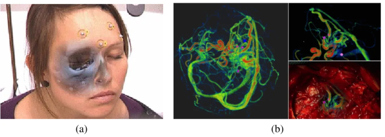
(a) (b)
Figure 1.1: (a) A CT image registered to the patient is rendered in a video see-through HMD [8] cIEEE 2007, (b) Volume rendering of blood vessels visualized in the context of an image captured by a camera affixed to the surgical microscope in brain surgery [9] cSpringer 2015 whole volume, some systems employ manual, interactive or fully-automatic [14] segmentation methods to extract a regions of interest, and render. Several attempts to render information in stereoscopic displays can be found in the literature [15], with the intention of improving depth perception, and thereby improving the accuracy of the guided surgical task. A major drawback in these systems is that significant cognitive efforts are necessary to relate the visualization to the action site. The cognitive processes involved in this may result in erroneous surgical actions to be performed that may end up in undesired consequences.
1.3. Assessment ofIGS Systems 5
consequences.
1.3
Assessment of IGS Systems
The performance of an IGS system depends not only on the technical aspects of the system, but also on how the surgeon interacts with the system and the patient. Therefore, assessing an IGS system performance is a complex procedure. Jannin and Korb [22] define six levels of IGS system assessment depending on the assessed property of the system. At the lowest level, technical parameters of the system such as the accuracy, precision, latency etc. are assessed under laboratory conditions. At level two, the therapeutic/diagnostic reliability of the system is evaluated under simulated laboratory environments such as phantom studies. At level three, surgical performance is assessed to determine the efficacy of the system in the clinic. Levels 4-6 assess patient outcomes, economic aspects, and social, legal, and ethical aspects based on data gathered routinely in the clinic at multiple centers. Thus, thorough evaluation of IGS systems at all these levels requires studies over a lengthy period involving patients at multiple clinical facilities.
Before an IGS technology is tested for performance in surgery involving patients, its tech-nical parameters and reliability should be evaluated under laboratory conditions. Techtech-nical parameters of the system can be assessed objectively fairly easily using highly accurate mea-suring systems. However, assessment of the reliability of a system is a complex process since the experiments should have control over fairly large parameter space. To maintain high de-gree of control over the experiment, the surgical scenario may be simulated at a cost of losing realism. On the other hand, experiments can be conducted in more realistic surgical setting, but at a cost of loosing the control over the experiment. It is important to assess the system under different testing environments to gain a thorough understanding about the system.
6 Chapter1. Introduction
study human factors, but the results may vary among different subject groups. For instance, experienced surgeons may be better/worse at certain surgical tasks compared to resident sur-geons, and produce completely different results. Therefore, subject demographics should be taken into consideration during experimental design. There are certain human factors that can-not be quantitatively measured. Subjective assessments using a ranking system such as the NASA Task Load Index (NASA TLX) [23] may help in these cases.
1.4
Image Guidance in Soft-tissue Surgery
1.5. LaparoscopicSurgery 7
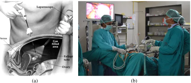
(a) (b)
Figure 1.2: (a) a cross section of the abdomen during a laparoscopic intervention. The sur-geon holds the laparoscopic camera while he/she operates on the patient using a laparoscopic instrument. Typically the abdominal cavity is filled withCO2 to increase the working space,
(b) conventionally, the laparoscopic video, monocular in this case, is displayed in a monitor that is placed away from the patient. The surgeon has to operate on the patient while his eyes are focused on the display, thus decoupling his/her action from the perception.
1.5
Laparoscopic Surgery
8 Chapter1. Introduction
the advancement of video technology, most ORs today are equipped with digital laparoscopic cameras and high definition (HD) video displays. This not only improves the visual fidelity of the captured anatomy, but also allows manipulation, enhancement, and storage of the captured videos for improved surgical guidance, teaching and training purposes.
Compared to the open surgical approach for many procedures [26–29], laparoscopy of-fers significantly lower pain, haemorrhage, and fewer post-operative complications, but with comparable clinical outcomes to conventional surgical procedures. Despite these advantages however, it presents several significant challenges to the surgeon. The laparoscopic video, displayed in a monitor remote from the patient, dissociates surgeon’s perception from his ac-tions. In addition, the monoscopic display used even in modern ORs today, offers limited depth perception. As a result, surgeons must master skills to infer depth from monocular depth cues, and execute actions at a location that is spatially disassociated from their perception. Moreover, surgical actions performed with long, slender instruments not only limit the range of motion, but also requires the mastery of difficult motor skills to compensate for thefulcrum effect[30]. These instruments significantly reduce tactile sensation, making it very difficult to determine the forces exerted at the tip of the instrument accurately. Nevertheless following years of train-ing, surgeons learn to operate under these conditions and perform complex procedures on a daily basis.
1.6
Robot-Assisted Laparoscopic Surgery
1.6. Robot-AssistedLaparoscopicSurgery 9
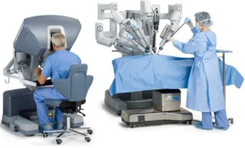
Figure 1.3: daVincisurgical robotic system. The surgeon sits at the console where his gestures are captured and converted to commands that control the slave-robot at the patient site. At the console the surgeon gets stereoscopic visual feedback. Typically these system have four robotic arms: one to hold the stereoscopic laparoscopic camera, two arms controlling surgical instruments, and one auxiliary arm. Image courtesy of Intuitive Surgical Inc.
without fatigue impairing his/her performance. The surgeon’s hand gestures are captured by the mechanical gesture tracking system (Fig. 1.4(b)) at the surgeon’s console, allowing the manipulators at the distal ends of the robotic arms to be driven interactively. Thus, these systems effectively replicate the surgeon’s gestures at the wristed laparoscopic instruments with the ability to scale the magnitude of their motion as desired.
10 Chapter1. Introduction
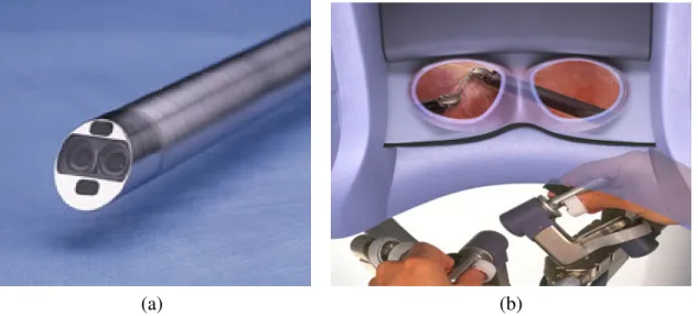
(a) (b)
Figure 1.4: (a) stereoscopic laparoscopic camera used in thedaVincisurgical system. It has a baseline of 5-6mm. A fiber-optic light source illuminates the surgical site, (b) stereoscopic dis-play and the gesture capturing hardware at the surgeon’s console of adaVincisurgical system. surgical tasks to be performed with significantly fewer errors compared to the performance with monoscopic viewing[34]. Thus, these systems assist both experienced and novice surgeons to perform otherwise very difficult surgeries safely and efficiently.
1.7
Intra-operative Ultrasound in Laparoscopic Surgery
1.7. Intra-operativeUltrasound inLaparoscopicSurgery 11
(a) (b)
Figure 1.5: (a) laparoscopic ultrasound probe with an articulated tip. The linear transducer array provides a 2D US image while the manual controller at the handle enables articulation of the tip to reach otherwise difficult space, (b) drop-in (or pick-up) US probe with a curvi-linear transducer array, designed specifically for robot-assisted surgical procedures. The probe has a grooved ridge that fits a laparoscopic/robotic grasper, enabling it to be picked up and manipulated. Image courtesy of BK Medical Systems Inc.
ultrasound probes that can be picked up and manipulated by a grasper tool were introduced with application to robot-assisted surgery (Fig. 1.5(b)). Since these probes provide improved maneuverability and surgeon autonomy, they are preferred over conventional versions in many robot-assisted surgical procedures[36].
12 Chapter1. Introduction
Figure 1.6: The TileProTMdisplay (bottom) indaVincisurgeon’s console, with the primary
dis-play providing the camera view (top). This secondary disdis-play allows the surgeons to visualize pre-operative/intra-operative images, or physiological measurements during an intervention. Image courtesy of Intuitive Surgical Inc.
Planning and execution of many critical surgical tasks benefit from the use of ultrasound during laparoscopic interventions. In laparoscopic and robot-assisted partial nephrectomy, tu-mor margins and resection plans for bothendophytictumours (that have grown into the tissue) andexophytictumours (that have grown outward beyond the organ surface) can be
1.8. CognitiveProcesses inUltrasound-guidedAction 13
reduced[43]. Even though the simultaneous use of these two modalities enable numerous ad-vantages, the conventional method of mental fusion of information may result in surgical errors, due to the error-prone cognitive processes involved. In oncologic surgery, these errors could occur in the form of a positive resection margin, or hitting a major surgical structure, in laparo-scopic cholecystectomy, bile-duct damage, or severe bleeding. Typically, these intraoperative complications are life threatening.
1.8
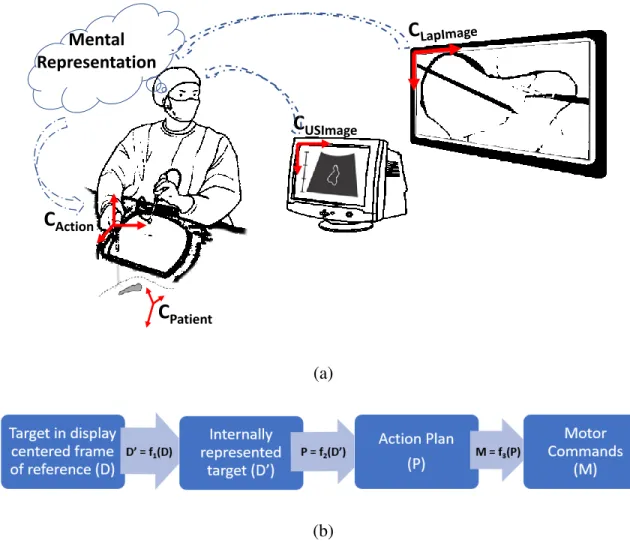
Cognitive Processes in Ultrasound-guided Action
A human reaching action guided by the visual system can be described by a sequence of trans-formations [44] (Fig. 1.7(b)): (1) the transformation that construct a mental representation of the target in space with visual inputs, (2) the transformation that produces an action plan with specific movement parameters based on the mental representation, and (3) the transformation that converts the action plan to motor commands to execute the action. Under transformation (1), image-centered target representationDis transformed to an internal spatial representation
D0 with respect to the perceptual frame of reference. For actions guided by the direct vision, this transformation causes no alternation to the input information because the target is already in the perceptual frame of reference. This internal representation is then mapped to the action centered frame of reference, and an action plan consisting of specific parameters is determined under transformation (2). The mapping (3) uses these parameters, and transforms them to spe-cific motor commands that execute the specified action. Any of these mappings is subject to error while accumulation of error across various stages is also a possibility.
14 Chapter1. Introduction
C
USImageC
ActionC
PatientMental Representation
(a)
(b)
Figure 1.7: (a) Schematic of laparoscopic surgery where laparoscopic ultrasound is used to visualize subcutaneous structures: An internal representation is constructed from the image in-formation read from the two images, based on which surgical actions are planned and executed, (b) Cognitive transformations in visually-guided action: Target represented in image reference frame is transformed to an internal representation. Based on this representation, an action plan is derived based on which appropriate motor commands are derived
1.9. Perceiving3D Form fromCross-sectionalImages 15
experience [47].
When a surgeon uses an US image visualized on a monitor that is spatially dissociated from that displays the laparoscopic video to guide a surgical action (Fig. 1.7(a)), additional mental transformations are required to construct an accurate mental representation of the target in the perceptual reference frame. This mental transformation involves at least mental translation, and mental scaling to account for the scaling difference between two displays. In practice, however, mental rotations are always involved, requiring significant taxing cognitive resources, and can easily cause excessive cognitive load [48, 49]. Given limited cognitive resources, as well as timing constrains associated with certain surgical actions [50], these cognitive processes may be vulnerable to error. Such errors directly affect the accuracy of the mental representations, and may propagate to the action plan, and eventually to the surgical action. Erroneous surgical actions could have life threatening consequences, hence avoiding them is of utmost importance. In addition to the errors in the mental representation, errors in the other parts of the trans-formation chain depicted in (Fig. 1.7(a)) could lead to surgical errors as well. A major source of error could be the transformation from the hand-centered frame of reference to the end-effector centered frame of reference. Similar to the cognitively mediated spatial representation of the targets, the learning of this mapping requires access to limited cognitive resources. Their scarcity, and time constraints may result in learning an erroneous mapping that may lead to un-desirable consequences. However, with extensive training and with the ergonomically designed surgical robotic systems, the occurrence of such error can be reduced drastically.
1.9
Perceiving
3
D Form from Cross-sectional Images
16 Chapter1. Introduction
images. These images are either visualized by conventional methods such as scrolling through a stack of tomographic images, or by using volume visualization method such as ray tracing [51]. However, due to the highly deformable nature of the organs (e.g. kidney, liver, lung etc.) and the surrounding soft-tissue environment in the thoraco-abdominal cavity, this pre-operatively constructed mental representation may not accurately represent the reality during a laparoscopic surgery.
1.10. InSituVisualization ofUS 17
localized, they are combined to construct a representation of the entire image.
When US, visualized in a separate display, is used to understand the 3D form of hidden targets during a laparoscopic procedure, the common frame of reference for localization is that centered on the laparoscopic display. Therefore, the image structures localized in the US dis-play frame of reference must be mentally transformed to the frame of reference of the laparo-scopic display. Such mental transformation, as described above, requires additional cognitive processes, and access to scarce cognitive resources. Surgeons may obtain very limited kines-thetic cues in a laparoscopic environment due to indirect manipulation of the probe. However, it is often the case that a surgical assistant manipulates the LUS probe while the surgeon con-trols other instruments. When this happens, no kinesthetic cues contribute in the localization of the US images, and the process depends entirely on the visual cues. Once the images are localized, the piecemeal representations require storage in the working memory before they are aggregated to construct a complete mental representation. However, the limited cognitive resources, and time constraints associated with certain laparoscopic surgical tasks may render these cognitive processes error-prone, resulting in erroneous mental representations. The ac-curacy of such cognitively mediated representations may also depend on the observers’ spatial ability [58, 59]. To minimize these errors, and reduce the risks of erroneous surgical actions that have life threatening consequences, improvements in visualization methods for this type of image fusion are necessary.
1.10
In Situ Visualization of US
18 Chapter1. Introduction
(a) (b)
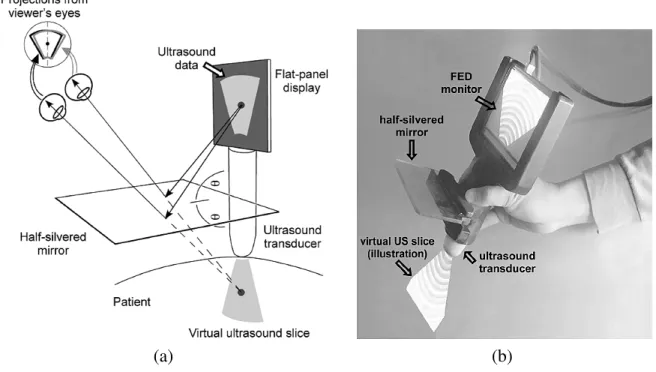
Figure 1.8: (a) schematic diagram of the SonicFlashlight device. The half-silvered mirror places a virtual US image at the correct spatial location allowing (in situ) visualization, (b) a realization of the schematic illustrated in (a). c2005 IEEE
processes that transform information across different frames of reference are eliminated since the perception and action frames of reference are coupled. Perception-action coupling is very important for performing visually-guided action intuitively with a high degree of accuracy.
In laparoscopic surgery, perception and action are already disconnected. However, with extensive learning, the surgeon can maintain a good coupling between the visual perception and the surgical action at the end-effector of the laparoscopic tool. In robot-assisted surgery this coupling may be achieved relatively easily as a consequence of the ergonomic design of these systems. Assuming that a such coupling can be achieved, if US information can also be visualized in the frame of reference of the laparoscopic video display, US guided actions could be more accurate, since the mental representations in such a visualization strategy do not depend on cognitive processes that are vulnerable to error. Such a visualization could be termedhybrid in situvisualization.
1.11. Motivation: Ultrasound-augmentedLaparoscopy 19
visualization. Wu et al. [60, 61] demonstrated that in situ visualization of US helps the ob-servers localize completely hidden targets significantly more accurately compared to theex situ
visualization technique in their study using a hand-held apparatus called the Sonic Flashlight that allows the US image to be visualized at the imaged location (Fig. 1.8). Further experiments showed that thein situvisualization technique significantly improves the understanding of the 3D form of hidden objects compared to theex situvisualization strategy[56, 62]. They attribute these improvements to the accurate internal representations the observers acquire through per-ception, in contrast to those constructed through cognitive mediation. These studies focused on non-laparoscopic use of US where the subjects held the probe manually while direct vi-sion to the imaging/action site was allowed. In laparoscopic surgery however, since surgeons visualize the surgical site through a monocular/stereoscopic laparoscopic camera, their visual perception is drastically different from direct vision. Unlike in these studies, surgeons’ per-ception is dissociated from their actions, introducing a new set of variables to consider during experimental design. In addition, in laparoscopic surgery, the surgeon may not be holding the probe himself, hence, unlike in these experiments, kinesthetic cues may not be available to help localize US images. Given these conditions in laparoscopy, further experiments may be required to investigate the the efficacy ofhybrid in situvisualization of US during laparoscopic interventions.
1.11
Motivation: Ultrasound-augmented Laparoscopy
The hypothesis that the mental representations acquired through perception is more accurate than the cognitively mediated ones may apply to LUS as well. However, the efficiency of such representations in laparoscopy depends on two major factors: (1) the accuracy with which US information can be spatially registered with the laparoscopic video frame of reference, and (2) the method of presentation of the US information in the context of the laparoscopic video.
20 Chapter1. Introduction
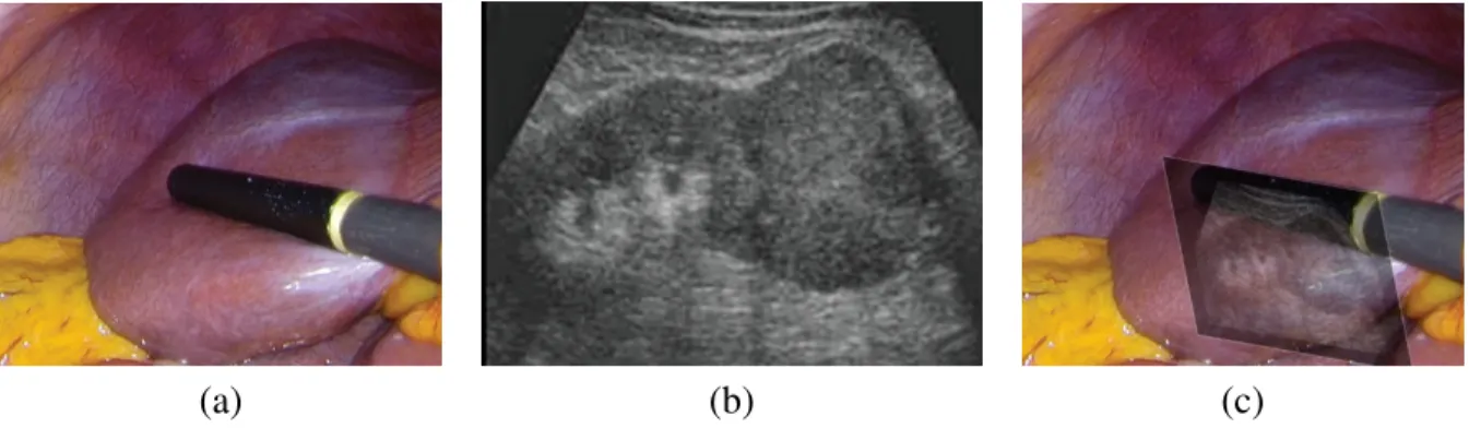
(a) (b) (c)
Figure 1.9: (a) laparoscopic camera image (monocular) showing the imaging tip of a LUS probe lying on an abdominal organ, (b) 2D US image for the probe depicted in (a), and (c) LUS image presented in the context of the laparoscopic camera image. Note that even the image is rendered at the correct scale and pose, it is perceived to float above the rest of the scene
perception, the transformation that maps each US pixel to the frame of reference centered in the laparoscopic camera should be first determined by some external means. In the conventional,
1.12. Hypothesis andResearchQuestions 21
Once registered to the laparoscopic video frame of reference, US image information should be presented to the observer in a manner that allows him/her to perceive the spatial location and the 3D form of a hidden target without ambiguity. A popular method used to achieve this is to texture map the US image to a 2D plane that is placed at the correct spatial position, orientation, and scale[63, 64, 66]. The laparoscopic camera image is set as the background texture to provide the context (Fig. 1.9(c)). This method of presentation conveys ambiguous depth cues resulting in perception that the US image is in front of the rest of the scene. Moreover, the interpretation of the content in the US image in this presentation becomes difficult at certain probe poses. In many laparoscopic applications, the camera is situated directly above the LUS probe due to the standard placement of laparoscopic ports. In such situations, the US image plane is nearly perpendicular to the camera imaging plane, making it difficult to interpret its content if this mode of presentation is used. Moreover, the overlay of a single image still requires cognitively involved mental integration of images for 3D form perception, even though
hybrid in situvisualization helps in localizing the 2D US images in the frame of reference of the laparoscopic video. Reconstructing a 3D image by compounding 2D US images as the surgeon moves the probe over an organ, and presenting 3D US image information instead of 2D images, may be less cognitively taxing since it avoids mental integration. With an appropriate method of blending US information with the camera image, such a method may help surgeons perceive the spatial location as well as the 3D form of a hidden surgical target without cognitive mediation. Surgical actions that rely on mental representations acquired through such a visualization could be significantly less susceptible to error.
1.12
Hypothesis and Research Questions
22 Chapter1. Introduction
compared to the conventionalex situvisualization,hybrid in situvisualization of US improves the surgeon’s ability to perceive the spatial location and the 3D form of hidden surgical targets in laparoscopic interventions. However, as mentioned in the previous section, the improve-ments may depend on the accuracy of spatial registration of laparoscopic video and the LUS video, and the method of presentation of the registered LUS information. Given these fac-tors, the following research questions arise, which should be adequately answered to guide the development of an effectivehybrid in situvisualization strategy.
1. How do we spatially register LUS image the laparoscopic camera image with minimal overhead to the existing OR work-flow? What are the error-bounds in such a registration? 2. How do we reveal 3D information of hidden surgical targets from 2D US without cogni-tively involved visualization approach? What errors should we expect in such a compu-tational approach?
3. How do we present spatially registered US information in the context of the laparoscopic video such that the surgeons perceive the spatial location and the 3D form without ambi-guity?
The answers to these questions pave the path to an effective visualization pipeline with which we can test the above mentioned hypothesis that is specific to laparoscopic interventions. The testing of this secondary hypothesis may strengthen (or weaken) the primary hypothesis which it is based on, and will help us understand visually-guided human action better.
1.13
Thesis Outline
1.13. ThesisOutline 23
with the laparoscopic video stream, and effectively visualize LUS information in the context of the laparoscopic video, that are superior to the state-of-the-art. These algorithms are described in detail in Chapter 2 through 4. Moreover, in Chapter 4, a set of experiments is described that involve experienced US users, and attempts to test the hypothesis mentioned in Section 1.12. The results of these experiments supports the hypothesis, but further experiments may be required to prove their validity in the clinic.
1.13.1
Robust, Intrinsic Tracking of a Laparoscopic Ultrasound Probe
Chapter 2 describes a novel method to spatially register the LUS information with real-time laparoscopic video, that involves a special fiducial pattern attached to the curved back surface of the LUS probe. Compared to other methods based on extrinsic tracking methods, such as magnetic tracking, this method adds minimal overhead to the existing OR work-flow. The algorithm uses monocular laparoscopic images to estimate the 6DoF pose of the probe with respect to the camera, and by using priors on the pose, and on the probe motion, it runs in video frame-rates. It is robust to partial occlusion of the fiducial pattern, and demonstrates sub-milimeter target registration errors against an optical tracking-based reference. The chapter also describes how the proposed method can be extended to stereo/multi-view camera images with application to robot-assisted laparoscopic interventions.
1.13.2
Accuracy in Freehand
3
D US Reconstruction with Robust Visual
Tracking
24 Chapter1. Introduction
accurate. Both quantitative and qualitative results are presented.
1.13.3
Visualizing Ultrasound In the Context of Laparoscopy
Bibliography
[1] J. J. Gallagher, R. H. Svenson, J. H. Kasell, L. D. German, G. H. Bardy, A. Broughton, and G. Critelli, “Catheter technique for closed-chest ablation of the atrioventricular con-duction system,”New England Journal of Medicine, vol. 306, no. 4, pp. 194–200, 1982. PMID: 7054682.
[2] J. T. Moore, M. W. A. Chu, B. Kiaii, D. Bainbridge, G. Guiraudon, C. Wedlake, M. Cur-rie, M. Rajchl, R. V. Patel, and T. M. Peters, “A navigation platform for guidance of beat-ing heart transapical mitral valve repair,”IEEE Transactions on Biomedical Engineering, vol. 60, pp. 1034–1040, April 2013.
[3] R. R. Shamir, L. Joskowicz, S. Spektor, and Y. Shoshan, “Localization and registration accuracy in image guided neurosurgery: a clinical study,”International Journal of Com-puter Assisted Radiology and Surgery, vol. 4, p. 45, Oct 2008.
[4] N. Hata, T. Dohi, S. Warfield, W. Wells, R. Kikinis, and F. A. Jolesz, “Multimodality de-formable registration of pre- and intraoperative images for mri-guided brain surgery,” in
Medical Image Computing and Computer-Assisted Intervention — MICCAI’98: First In-ternational Conference Cambridge, MA, USA, October 11–13, 1998 Proceedings(W. M. Wells, A. Colchester, and S. Delp, eds.), pp. 1067–1074, Berlin, Heidelberg: Springer Berlin Heidelberg, 1998.
[5] A. N. Sridhar, A. Hughes-Hallett, E. K. Mayer, P. J. Pratt, P. J. Edwards, G.-Z. Yang, A. W. Darzi, and J. A. Vale, “Image-guided robotic interventions for prostate cancer,”
Nature Reviews Urology, vol. 10, pp. 452–462, June 2013.
[6] C. Schneider, S. Thompson, J. Totz, Y. Song, K. Gurusamy, S. Ourselin, D. Stoyanov, M. Clarkson, D. Hawkes, and B. Davidson, “18. a pilot study evaluating the overlay display method for image guidance in laparoscopic liver surgery,” European Journal of Surgical Oncology (EJSO), vol. 42, no. 9, p. S72, 2016.
[7] T. Sielhorst, M. Feuerstein, and N. Navab, “Advanced medical displays: A literature review of augmented reality,” Journal of Display Technology, vol. 4, pp. 451–467, De-cember 2008.
[8] C. Bichlmeier, F. Wimmer, S. M. Heining, and N. Navab, “Contextual anatomic mimesis hybrid in-situ visualization method for improving multi-sensory depth perception in med-ical augmented reality,” in2007 6th IEEE and ACM International Symposium on Mixed and Augmented Reality, pp. 129–138, November 2007.
26 BIBLIOGRAPHY [9] M. Kersten-Oertel, I. Gerard, S. Drouin, K. Mok, D. Sirhan, D. S. Sinclair, and D. L.
Collins, “Augmented reality in neurovascular surgery: feasibility and first uses in the operating room,” International Journal of Computer Assisted Radiology and Surgery, vol. 10, pp. 1823–1836, November 2015.
[10] J. Moore, C. Clarke, D. Bainbridge, C. Wedlake, A. Wiles, D. Pace, and T. Peters, “Image guidance for spinal facet injections using tracked ultrasound,” in Medical Image Com-puting and Computer-Assisted Intervention – MICCAI 2009: 12th International Confer-ence, London, UK, September 20-24, 2009, Proceedings, Part I(G.-Z. Yang, D. Hawkes, D. Rueckert, A. Noble, and C. Taylor, eds.), pp. 516–523, Berlin, Heidelberg: Springer Berlin Heidelberg, 2009.
[11] G. Ameri, J. S. H. Baxter, D. Bainbridge, T. M. Peters, and E. C. S. Chen, “Mixed reality ultrasound guidance system: a case study in system development and a cautionary tale,”
International Journal of Computer Assisted Radiology and Surgery, August 2017. [12] J. Beyer, M. Hadwiger, S. Wolfsberger, and K. Bhler, “High-quality multimodal volume
rendering for preoperative planning of neurosurgical interventions,” IEEE Transactions on Visualization and Computer Graphics, vol. 13, pp. 1696–1703, November 2007. [13] I. Viola, A. Kanitsar, and M. E. Groller, “Importance-driven volume rendering,” in
Pro-ceedings of the Conference on Visualization ’04, VIS ’04, (Washington, DC, USA), pp. 139–146, IEEE Computer Society, 2004.
[14] D. L. Pham, C. Xu, and J. L. Prince, “Current methods in medical image segmentation,”
Annual Review of Biomedical Engineering, vol. 2, no. 1, pp. 315–337, 2000. PMID: 11701515.
[15] T. A. N. Hernes, S. Ommedal, T. Lie, F. Lindseth, T. Langø, and G. Unsgaard, “Stereo-scopic Navigation-Controlled Display of Preoperative MRI and Intraoperative 3D Ul-trasound in Planning and Guidance of Neurosurgery: New Technology for Minimally Invasive Image-Guided Surgery Approaches,” min - Minimally Invasive Neurosurgery, vol. 46, pp. 129–137, June 2003.
[16] H. Fuchs, M. A. Livingston, R. Raskar, D. Colucci, K. Keller, A. State, J. R. Crawford, P. Rademacher, S. H. Drake, and A. A. Meyer, “Augmented reality visualization for la-paroscopic surgery,” in Medical Image Computing and Computer-Assisted Intervention — MICCAI’98: First International Conference Cambridge, MA, USA, October 11–13, 1998 Proceedings(W. M. Wells, A. Colchester, and S. Delp, eds.), pp. 934–943, Berlin, Heidelberg: Springer Berlin Heidelberg, 1998.
BIBLIOGRAPHY 27 [18] I. Kuhlemann, M. Kleemann, P. Jauer, A. Aschweikard, and F. Ernst, “Towards x-ray free endovascular interventions using hololens for on-line holographic visualisation,”
Healthcare Technology Letters, vol. 4, pp. 184–187(3), October 2017.
[19] P. J. Edwards, A. P. King, C. R. Maurer, D. A. D. Cunha, D. J. Hawkes, D. L. G. Hill, R. P. Gaston, M. R. Fenlon, A. Jusczyzck, A. J. Strong, C. L. Chandler, and M. J. Gleeson, “Design and evaluation of a system for microscope-assisted guided interventions (magi),”
IEEE Transactions on Medical Imaging, vol. 19, pp. 1082–1093, November 2000. [20] M. Kersten-Oertel, I. Gerard, S. Drouin, K. Mok, D. Sirhan, D. Sinclair, and D. L. Collins,
“Augmented reality in neurovascular surgery: First experiences,” inAugmented Environ-ments for Computer-Assisted Interventions: 9th International Workshop, AE-CAI 2014, Held in Conjunction with MICCAI 2014, Boston, MA, USA, September 14, 2014. Pro-ceedings(C. A. Linte, Z. Yaniv, P. Fallavollita, P. Abolmaesumi, and D. R. Holmes, eds.), pp. 80–89, Cham: Springer International Publishing, 2014.
[21] M. Kersten-Oertel, I. J. Gerard, S. Drouin, K. Mok, D. Sirhan, D. S. Sinclair, and D. L. Collins, “Augmented reality for specific neurovascular surgical tasks,” inAugmented En-vironments for Computer-Assisted Interventions: 10th International Workshop, AE-CAI 2015, Held in Conjunction with MICCAI 2015, Munich, Germany, October 9, 2015. Pro-ceedings(C. A. Linte, Z. Yaniv, and P. Fallavollita, eds.), pp. 92–103, Cham: Springer International Publishing, 2015.
[22] P. Jannin and W. Korb, “Assessment of image-guided interventions,” in Image-Guided Interventions: Technology and Applications(T. Peters and K. Cleary, eds.), pp. 531–549, Boston, MA: Springer US, 2008.
[23] S. G. Hart and L. E. Staveland, “Development of NASA-TLX (Task Load Index): Results of Empirical and Theoretical Research,”Advances in Psy., pp. 139–183, 1988.
[24] B. Fuerst, J. Sprung, F. Pinto, B. Frisch, T. Wendler, H. Simon, L. Mengus, N. S. van den Berg, H. G. van der Poel, F. W. B. van Leeuwen, and N. Navab, “First Robotic SPECT for Minimally Invasive Sentinel Lymph Node Mapping,”IEEE Transactions on Medical Imaging, vol. 35, pp. 830–838, March 2016.
[25] M. Hatzinger, S. T. Kwon, S. Langbein, S. Kamp, A. H¨acker, and P. Alken, “Hans Chris-tian Jacobaeus: Inventor of human laparoscopy and thoracoscopy.,”Journal of endourol-ogy, vol. 20, pp. 848–50, November 2006.
[26] H. J. Bonjer, C. L. Deijen, G. A. Abis, M. A. Cuesta, M. H. G. M. Van Der Pas, E. S. M. De Lange-De Klerk, A. M. Lacy, W. A. Bemelman, J. Andersson, E. Angenete, J. Rosenberg, A. Fuerst, and E. Haglind, “A Randomized Trial of Laparoscopic versus Open Surgery for Rectal Cancer,”New England Journal of Medicine, vol. 14372, no. 2, pp. 1324–32, 2015.
[27] I. S. Gill, L. R. Kavoussi, B. R. Lane, M. L. Blute, D. Babineau, J. R. Colombo, I. Frank, S. Permpongkosol, C. J. Weight, J. H. Kaouk, M. W. Kattan, and A. C. Novick, “Compar-ison of 1,800 Laparoscopic and Open Partial Nephrectomies for Single Renal Tumors,”
28 BIBLIOGRAPHY [28] M. Lesurtel, D. Cherqui, A. Laurent, C. Tayar, and P. L. Fagniez, “Laparoscopic
ver-sus open left lateral hepatic lobectomy: a case-control study,” Journal of the American College of Surgeons, vol. 196, no. 2, pp. 236–242, 2003.
[29] W. J. Scott, M. S. Allen, G. Darling, B. Meyers, P. A. Decker, J. B. Putnam, R. W. Mckenna, R. J. Landrenau, D. R. Jones, R. I. Inculet, and R. A. Malthaner, “Video-assisted thoracic surgery versus open lobectomy for lung cancer: A secondary analysis of data from the American College of Surgeons Oncology Group Z0030 randomized clinical trial,”The Journal of Thoracic and Cardiovascular Surgery, vol. 139, no. 4, pp. 976–983, 2010.
[30] A. G. Gallagher, N. McClure, J. McGuigan, K. Ritchie, and N. P. Sheehy, “An Ergonomic Analysis of the Fulcrum Effect in the Acquisition of Endoscopic Skills,” Endoscopy, vol. 30, pp. 617–620, September 1998.
[31] J. H. Kaouk, G.-P. Haber, R. Autorino, S. Crouzet, A. Ouzzane, V. Flamand, and A. Villers, “A Novel Robotic System for Single-port Urologic Surgery: First Clinical Investigation,”European Urology, vol. 66, pp. 1033–1043, December 2014.
[32] F. C. Holsinger, “A flexible, single-arm robotic surgical system for transoral resection of the tonsil and lateral pharyngeal wall: Next-generation robotic head and neck surgery,”
The Laryngoscope, vol. 126, pp. 864–869, April 2016.
[33] J. S. Lam, J. Bergman, A. Breda, and P. G. Schulam, “Importance of surgical margins in the management of renal cell carcinoma.,” Nature clinical practice. Urology, vol. 5, pp. 308–17, June 2008.
[34] Y. Munz, K. Moorthy, A. Dosis, J. D. Hernandez, S. Bann, F. Bello, S. Martin, A. Darzi, and T. Rockall, “The benfits of stereoscopic vision in robotic-assisted performance on bench models,”Surgical Endoscopy, vol. 18, pp. 611–616, April 2004.
[35] E. D. Light, S. F. Idriss, K. F. Sullivan, P. D. Wolf, and S. W. Smith, “Real-Time 3D Laparoscopic Ultrasonography,”Ultrasonic Imaging, vol. 27, pp. 129–144, July 2005. [36] B. F. Kaczmarek, S. Sukumar, R. K. Kumar, N. Desa, K. Jost, M. Diaz, M. Menon, and
C. G. Rogers, “Comparison of Robotic and Laparoscopic Ultrasound Probes for Robotic Partial Nephrectomy,”Journal of Endourology, vol. 27, pp. 1137–1140, September 2013. [37] J. Shepard, R. N. and Metzler, “Mental Rotation of Three-Dimensional Objects,”Science,
vol. 171, pp. 701–703, 1971.
[38] A. Bundesen, C. and Larsen, “Visual Transformation of Size,”Journal of Experimental Psychology: Human Perception and Performance, vol. 1, pp. 214–220, 1975.
BIBLIOGRAPHY 29 [40] H. Wada, T. Anayama, K. Hirohashi, T. Nakajima, T. Kato, T. K. Waddell, S. Keshavjee, I. Yoshino, and K. Yasufuku, “Thoracoscopic ultrasonography for localization of sub-centimetre lung nodules.,”European journal of cardio-thoracic surgery : official journal of the European Association for Cardio-thoracic Surgery, vol. 49, pp. 690–7, February 2016.
[41] M. Khereba, P. Ferraro, A. Duranceau, J. Martin, E. Goudie, V. Thiffault, and M. Liber-man, “Thoracoscopic localization of intraparenchymal pulmonary nodules using direct intracavitary thoracoscopic ultrasonography prevents conversion of VATS procedures to thoracotomy in selected patients.,”The Journal of thoracic and cardiovascular surgery, vol. 144, pp. 1160–5, November 2012.
[42] K. A. Perry, J. A. Myers, and D. J. Deziel, “Laparoscopic ultrasound as the primary method for bile duct imaging during cholecystectomy,” Surgical Endoscopy, vol. 22, pp. 208–213, January 2008.
[43] J. Machi, J. O. Johnson, D. J. Deziel, N. J. Soper, E. Berber, A. Siperstein, M. Hata, A. Patel, K. Singh, and M. E. Arregui, “The routine use of laparoscopic ultrasound de-creases bile duct injury: a multicenter study,”Surgical Endoscopy, vol. 23, pp. 384–388, February 2009.
[44] B. Wu, R. L. Klatzky, and G. Stetten, “Learning to Reach to Locations Encoded from Imaging Displays.,” Spatial cognition and computation, vol. 8, pp. 333–356, October 2008.
[45] P. M. Fitts, “Perceptual-Motor Skill Learning,” in Catagories of Human Learning, pp. 381–391, Academic Press Inc., 1964.
[46] R. L. Klatzky, B. Wu, D. Shelton, and G. Stetten, “Effectiveness of augmented-reality visualization versus cognitive mediation for learning actions in near space,”ACM Trans-actions on Applied Perception, vol. 5, pp. 1–23, January 2008.
[47] M. Wilson, J. McGrath, S. Vine, J. Brewer, D. Defriend, and R. Masters, “Psychomotor control in a virtual laparoscopic surgery training environment: gaze control parameters differentiate novices from experts.,” Surgical endoscopy, vol. 24, pp. 2458–64, October 2010.
[48] J.-S. Hyun and S. J. Luck, “Visual working memory as the substrate for mental rotation.,”
Psychonomic bulletin&review, vol. 14, pp. 154–8, February 2007.
[49] D. J. Prime and P. Jolicoeur, “Mental Rotation Requires Visual Short-term Memory: Evidence from Human Electric Cortical Activity,” Journal of Cognitive Neuroscience, vol. 22, pp. 2437–2446, November 2010.
30 BIBLIOGRAPHY [51] P. S. Calhoun, B. S. Kuszyk, D. G. Heath, J. C. Carley, and E. K. Fishman,
“Three-dimensional Volume Rendering of Spiral CT Data: Theory and Method,”RadioGraphics, vol. 19, pp. 745–764, May 1999.
[52] F. Z¨ollner, “Ueber eine neue Art anorthoskopischer Zerrbilder,”Annalen der Physik und Chemie, vol. 193, pp. 477–484, January 1862.
[53] E. M. Palmer, P. J. Kellman, and T. F. Shipley, “A theory of dynamic occluded and illusory object perception.,” Journal of Experimental Psychology: General, vol. 135, pp. 513– 541, November 2006.
[54] R. Fendrich, J. W. Rieger, and H.-J. Heinze, “The effect of retinal stabilization on anortho-scopic percepts under free-viewing conditions,”Vision Research, vol. 45, pp. 567–582, March 2005.
[55] J. S. Girgus, L. H. Gellman, and J. Hochberg, “The effect of spatial order on piece-meal shape recognition: A developmental study,” Perception&Psychophysics, vol. 28, pp. 133–138, March 1980.
[56] B. Wu, R. L. Klatzky, and G. D. Stetten, “Mental visualization of objects from cross-sectional images.,”Cognition, vol. 123, pp. 33–49, April 2012.
[57] J. M. Loomis, R. L. Klatzky, and S. J. Lederman, “Similarity of Tactual and Visual Picture Recognition with Limited Field of View,”Perception, vol. 20, pp. 167–177, April 1991. [58] M. Hegarty, M. Keehner, C. Cohen, D. R. Montello, and Y. Lippa, “The Role of Spatial
Cognition in Medicine: Applications for Selecting and Training Professionals.,” in Ap-plied spatial cognition: From research to cognitive technology., pp. 285–315, Mahwah, NJ, US: Lawrence Erlbaum Associates Publishers, 2007.
[59] M. Lanca, “Three-Dimensional Representations of Contour Maps,” Contemporary Edu-cational Psychology, vol. 23, pp. 22–41, Janurary 1998.
[60] B. Wu, R. L. Klatzky, D. Shelton, and G. D. Stetten, “Psychophysical evaluation of in-situ ultrasound visualization.,” IEEE transactions on visualization and computer graphics, vol. 11, no. 6, pp. 684–93, 2005.
[61] R. L. Klatzky, B. Wu, and G. Stetten, “Spatial Representations From Perception and Cog-nitive Mediation: The Case of Ultrasound.,”Current directions in psychological science, vol. 17, pp. 359–364, December 2008.
[62] B. Wu, R. L. Klatzky, and G. Stetten, “Visualizing 3D objects from 2D cross sectional images displayed in-situ versus ex-situ.,” Journal of experimental psychology. Applied, vol. 16, pp. 45–59, March 2010.
[63] C. L. Cheung, C. Wedlake, J. Moore, S. E. Pautler, and T. M. Peters, “Fused video and ultrasound images for minimally invasive partial nephrectomy: A phantom study,” in
BIBLIOGRAPHY 31 (T. Jiang, N. Navab, J. P. W. Pluim, and M. A. Viergever, eds.), pp. 408–415, Berlin, Heidelberg: Springer Berlin Heidelberg, 2010.
[64] X. Kang, M. Azizian, E. Wilson, K. Wu, A. D. Martin, T. D. Kane, C. A. Peters, K. Cleary, and R. Shekhar, “Stereoscopic augmented reality for laparoscopic surgery.,” Surg. En-dosc., pp. 1–9, 2014.
[65] M. Feuerstein, T. Reichl, J. Vogel, A. Schneider, H. Feussner, and N. Navab, “Magneto-optic tracking of a flexible laparoscopic ultrasound transducer for laparoscope augmenta-tion,” inMedical Image Computing and Computer-Assisted Intervention – MICCAI 2007: 10th International Conference, Brisbane, Australia, October 29 - November 2, 2007, Pro-ceedings, Part I (N. Ayache, S. Ourselin, and A. Maeder, eds.), pp. 458–466, Berlin, Heidelberg: Springer Berlin Heidelberg, 2007.
Chapter 2
Robust, Intrinsic Tracking of a
Laparoscopic Ultrasound Probe
This chapter is adapted from the papers,
• Jayarathne U.L., McLeod A.J., Peters T.M., Chen E.C.S. (2013) Robust Intraoperative US Probe Tracking Using a Monocular Endoscopic Camera. In: Mori K., Sakuma I., Sato Y., Barillot C., Navab N. (eds) Medical Image Computing and Computer-Assisted Intervention MICCAI 2013. MICCAI 2013. Lecture Notes in Computer Science, vol 8151. Springer, Berlin, Heidelberg
• Jayarathne U.L., Luo X., Chen E.C.S., Peters T.M. (2015) Simultaneous Estimation of Feature Correspondence and Stereo Object Pose with Application to Ultrasound Aug-mented Robotic Laparoscopy. In: Linte C., Yaniv Z., Fallavollita P. (eds) AugAug-mented Environments for Computer-Assisted Interventions. MICCAI 2015. Lecture Notes in Computer Science, vol 9365. Springer, Cham
• Uditha L. Jayarathne, Elvis C.S Chen, John Moore, Terry M. Peters, ”Robust, Intrinsic Tracking of a Laparoscopic Ultrasound Probe for Ultrasound-augmented Laparoscopy”, IEEE Transactions on Medical Imaging, (submitted)
2.1
Introduction
In many laparoscopic procedures, surgeons use laparoscopic ultrasound (LUS) to visualize sur-gical targets hidden deep inside organs. For example, in laparoscopic resection tasks of many
2.1. Introduction 33 endophytictumors, LUS is used to determine the resection margins, and to gain better under-standing about the 3D form of the tumor and its surrounding [1–3]. During these procedures, the 2D ultrasound (US) image is conventionally displayed separately from the laparoscopic video. During conventional laparoscopic surgery, this second display is usually on the ultra-sound machine itself, while in robot-assisted surgery with thedaVincisystem the US image is presented in a separate display panel known as the TileProTM. In either case, the ultrasound
image and the laparoscopic video are spatially dissociated. In addition, these two modalities provide two distinct types of information: laparoscopy, being a projective imaging modality, provides a perspective projection of the scene, while the ultrasound image provides a tomo-graphic view into the tissue. Therefore, reasoning about the geometry of the hidden surgical targets requires mentally solving the spatial alignment problem and the resolving the modality differences, which is cognitively very challenging [4, 5]. Mental representations of the hidden surgical targets in space acquired through such cognitive mediation are error-prone [6], and may cause incorrect surgical actions to be performed. Hybrid in situvisualization where US information is displayed in the frame of reference of the laparoscopic video enables mental representations of the targets through perception [7] which are more accurate. Such visualiza-tions require the US image to be mapped to the camera coordinate system which involves a six degrees of freedom (6DoF) rigid transformation to be solved at camera frame rate.
2.1.1
Related Work
Many attempts to solve the rigid transformation that maps US image to a camera centered co-ordinate system can be found in the literature. A popular approach is to employ an extrinsic
34 Chapter2. Robust, IntrinsicTracking of aLaparoscopicUltrasoundProbe
the probe handle. With application to robot-assisted surgery, one could compute this transform based on kinematic tracking of the camera and the robotic manipulator that holds the US probe [12]. However, such approaches require the US probe to be rigidly attached to the robotic arm, so that the transformation of the imaging tip with respect to the kinematic coordinate system can be computed by aggregating the pose of the end-effector with the constant transforma-tion from the end-effector to the imaging tip. This constant transformation can be determined
apriori through a calibration process. Despite its popularity, extrinsic-tracking-based solu-tions adds financial and logistical overhead to the existing operating room (OR) work-flow. In addition, the accuracy of such solutions may be affected by error accumulation in long trans-formation chains involved, or by the factors in the OR that affect the robustness of tracking. In particular, the accuracy of magnetic-tracking-based solutions may be affected by the presence of ferromagnetic materials in most ORs.
2.1. Introduction 35
be solved accurately and efficiently [18–20]. In stereo-laparoscopy, where two views of the fiducial pattern are available simultaneously, the corresponding points localized in each image can be triangulated to obtain a 3D point-cloud in the camera coordinate system. The pose is then determined by registering this point-cloud with the fiducial point cloud by using an algo-rithm such asiterative closest point(ICP) [21] that solves for the 3D-3D correspondence, and the rigid transformation simultaneously.
Methods that rely on highly localized point-fiducials effectively solve two computational problems; (a) the 3D-to-2D point correspondence problem, and (b) the pose estimation prob-lem. The use of a planar and structured fiducial geometry enables easy computation of a solu-tion to the correspondence problem. For this reason the best performing methods use fiducial patterns arranged in a planar-grid [13–15, 17]. Since the fiducial geometry is known and sim-ple, the 2D coordinate of an undetected fiducial can be interpolated to improve the robustness of tracking. However, most of the clinically used LUS probes are cylindrical in shape, mak-ing the planar fiducial pattern difficult to use. In addition, a planar pattern has a very limited tracking range. In a recent paper, Zhang et al. [16] described a method in which the fiducial pattern could cover the entire curved, back surface of a clinically used LUS probe. However, the method makes several assumptions that may not be true in practical surgery. In particular, it is not clear how the proposed method handles fiducial occlusions (either due to physical occlu-sion, or the feature detector being insensitive), as well as outliers, all of which are unavoidable in practice.
2.1.2
Contributions
36 Chapter2. Robust, IntrinsicTracking of aLaparoscopicUltrasoundProbe
framework [22], that allows easy integration ofpriorson the pose based on the topology of the pose space, and the probe motion. Incorporation of strong priors based on probe motion allows the algorithm to converge to a solution quickly, enabling real-time performance. Moreover, the uncertainty of the solution is also reported, which is very useful in acquiring a comprehensive understanding about the tracking performance. The chapter includes a detailed description of a monocular image-based method as well as its extension to stereo/multi-view imaging models, an empirical investigation into the efficacy of the proposed methods, and a detailed discussion including limitations and insight into the future.
2.2
Methods
Since most clinical LUS probes do not provide adequate visual cues to be tracked in 6DoF, a special fiducial pattern is attached to the curved back surface of the probe. One could attach any textured pattern to the probe, and employ state-of-the-art detectors such as SIFT [23]/SURF [24] to localize interest points in image space. This will allow the correspondence problem to be solved explicitly using the descriptors computed by SIFT/SURF for each of the detected interest points. However, as demonstrated by Zeisl et al. [25], such features may introduce significant Fiducial Localization Error (FLE) that may propagate to the pose estimates. There-fore, to minimize the FLE, the fiducials in the pattern used in this work are localized at the intersection of two high contrast edges.
In the following subsections I describe the mathematical details of the pose estimation algo-rithm. This discussion assumes that the the laparoscopic camera image is free from geometric distortions while the camera is approximated by thepinholemodel with its intrinsic matrixA
known through an appropriate calibration method. When multiple views are involved, the pose
Pcof one camera relative to the other is assumed to be known, in addition to the intrinsic matrix
38 Chapter2. Robust, IntrinsicTracking of aLaparoscopicUltrasoundProbe
2.2.1
Preliminaries
Let us assume that each fiducial marker (model poitns) in the pattern in 3D space, is determined with respect to a local coordinate system by some means, and that the coordinate of theithpoint is given by Mi. Let the cardinality of themodel points setbe N. When the fiducial pattern is
visible in thenthframe of a camera, an image processing routine (feature detector) is applied
to the image to localize the fiducial points in the camera image. Letujbe the 2D coordinate of
the jthfiducial localized in the image space. Occlusions of some fiducials, and outliers in the image space due to imperfections of the feature detector are allowed, hence, the cardinality of the two point-sets may not be equal. The rigid posePnof the fiducial pattern with respect to the
camera in thenthframe (Fig. 2.1(a)), is parameterized as a 6D vectorPn = [r1,r2,r3,tx,ty,tz]T
with three parameters representing rotations1, and three parameters representing translation in
3D space. Given the model pointsM, feature detector responseu, and the camera intrinsicsA, the objective is to estimatePn.
2.2.2
Simultaneous Pose and Correspondence from a Single View
Let us begin by assuming aGaussian prioron the pose represented by its mean posePn, and its
6x6 covariance matrixΣnp for thenthcamera frame. Section 2.2.4 details how these priors are
computed in this tracking framework. Given this prior, the image space locationmi of model
pointMi is given by the Eq. (2.1) while the corresponding image space covarianceΣiis given
by Eq. (2.2),
mi =Pro j(Pn,Mi) (2.1)
Σi = J(Mi)ΣnpJ(Mi)T +R (2.2)
where Pro j(Pn,Mi) is the operator that projects the ith model point with pose Pn, J(.) is its
Jacobian, andRis a 2x2 diagonal matrix representing isotropic measurement uncertainty. Σi
2.2. Methods 39
defines a search region for a putative match, and I only consider 2D points such that
(mi−uj)TΣi(mi−uj)≤Ψ (2.3)
with Ψ = 3 giving 99% confidence in matching. Thus, the search space for a match for Mi
reduces to an elliptical region (Fig. 2.1(b)). Inside the search region for putative matches, several feature points may be found, each of which is equally probable to be a correct match. Hypothesizing that one such point uj is the correct match, the pose and its covariance are
updated using Extended Kalman Filter (EKF) equations, Eq. (2.4) and Eq. (2.5).
P0n =Pn+K(uj−mi); (2.4)
Σ0p
n =(I−KJ(Pn))Σn, (2.5)
where,Kis the optimal Kalman gain andIis an identity matrix.