Copyright 0 1982 by the Genetics Society of America
POLYCOMBLIKE: A GENE THAT APPEARS T O BE REQUIRED
FOR THE NORMAL EXPRESSION OF THE BITHORAX AND
ANTENNAPEDIA GENE COMPLEXES OF DROSOPHILA
MELANOGASTER
IAN McKENZIE DUNCAN'
Division of Biology, California Institute of Technology, Pasadena, California 91125 Manuscript received December 30, 1981
Revised copy accepted May 1,1982
ABSTRACT
A newly identified gene is described that is required for the maintenance of normal identities in many of the body segments of the fly. The effects of mutants in this gene, which is called Polycomblike (Pcl), suggest that its wild- type allele functions in the regulation of the bithorax gene complex (BX-C) and the Antennapedia gene complex (ANT-C). Evidence in favor of this idea derives from (1) the close correspondence between segmental transformations caused by Pcl mutants and those caused by dominant gain-of-function mutants in the BX-C and ANT-C, (2) the interactions observed between Pcl mutants and mutants in these complexes, and (3) the dependence upon BX-C and ANT-C dosage of the severity of at least one of the transformations caused by Pcl mutants. Arguments are presented that the control of the BX-C and ANT-C by Pcl+ is negative in nature. The results of clonal analysis experiments indicate that, at least for the BX-C, Pcl+ exerts this control until late in development. Since the wild-type allele of another gene, called Polycomb (Pc), has previously been shown to have many of the same properties as Pcl+, it appears that the BX-C and perhaps also the ANT-C are continuously regulated during develop- ment by at least two and probably several other genes.
HE morphological specialization of most body segments of Drosophila
T
appears to be under the direct control of a number of genes that are organized into two clusters or gene complexes. The differentiation of posterior segments, including at least those from the third thoracic through the most posterior abdominal segment, requires the activities of genes located within the bithorax complex (BX-C). This gene complex has been studied extensively by E. B. LEWIS (for reviews see LEWIS 1978, 1981a, 1981b) who has shown that the activities of the BX-C genes promote the development of posterior characteris- tics; all known loss-of-function mutants within the BX-C cause particular segments, or structures within them, to develop as if they were located more anteriorly in the animal. LEWIS has mapped many inactivation-type mutants by recombination and has found a striking correlation: the order of mutants within the BX-C is the same, with one exception, as the order on the animal of the body segments that these mutants affect. The study of embryos deficient for portions of the BX-C has led LEWIS to propose that early in normal development the genes of the BX-C become activated in sequence along the chromosome as one proceeds posteriorly in the animal. Evidence has been presented (LEWIS1978) that once activated, at least two, and perhaps all, of the BX-C genes ' Present address: Department of Biology, Washington University, St. Louis, Missouri 63130.
remain active in all more posterior segments. To explain this ordered regulation,
LEWIS has proposed that each structural gene in the BX-C is controlled by adjacent cis-regulatory regions that are in some way able to sense location along the anteroposterior axis. These cis-regulatory sites are thought to be identified by dominant gain-of-function mutants in the BX-C, all of which cause poste- riorly directed segmental transformations. LEWIS (1978) has presented genetic evidence that at least two of these gain-of-function mutants cause such trans- formations by altering the regulation of adjacent BX-C genes so that they are expressed more anteriorly than they normally are.
Recently, T. KAUFMAN and his colleagues (KAUFMAN, LEWIS and WAKIMOTO
1980) have presented evidence that the anterior portion of the fly, including the head region and the first and second thoracic segments, is under the control of another gene cluster that they have named the Antennapedia complex (ANT-
C). Although the regulatory rules for this gene complex have not yet been well characterized, certain similarities to the BX-C exist. For example, cis-regulatory mutants appear to occur in the ANT-C (DENELL et al. 1981; STRUHL 1981a), and at least one of the genes in this complex appears to be regulated along the body axis in a fashion similar to that seen for some of the BX-C genes (DUNCAN,
1982).
A central problem is how the genes of the ANT-C and BX-C come to be differentially expressed along the body axis. LEWIS (1978) has proposed how such spatial control of the BX-C may occur. He has found that mutants at the Polycomb (Pc) locus, which is not closely linked to either the ANT-C or the BX-
C, have properties suggesting that this gene is required in normal development to inactivate BX-C functions in anterior body segments. Thus, many or all of the BX-C genes appear to be expressed in many or all of the body segments of embryos deficient for Pc+, resulting in transformations of these segments toward the most posterior, or eighth, abdominal segment. Posteriorly directed transfor- mations are also seen in adult animals carrying only one dose of Pc+. These include partial conversions of wing to haltere and patchy transformations of the first, fourth, fifth and sixth abdominal segments to the next most posterior segments. Since the severity of many of these transformations has been shown to depend on BX-C dosage (LEWIS 1978; DUNCAN and LEWIS 1981), it seems likely that these transformations result from the defective regulation of the BX-C and are not a direct consequence of the inactivation of Pc+.
To explain the effects of Pc mutants, LEWIS (1978) has proposed that Pc' is a negative regulator of BX-C genes and that early in development a gradient of Pc+ activity is set up along the body axis with its high point in the anterior of the animal. In this model, LEWIS considers the genes of the BX-C to be differentially sensitive to repression by Pc+, with the consequence that BX-C genes with increasing sensitivities to repression by Pc+ become sequentially activated as one proceeds posteriorly in the animal.
CONTROL OF THE BX-C AND ANT-C 51
In order to better understand how the ANT-C and BX-C are controlled, a systematic search has been made for other genes that appear to act as regulators of these gene complexes. In this report, the properties of one of the genes identified in this search are described. This locus appears to be very similar in function to Pc+ and has, therefore, been called Polycomblike (Pcl). Mutants in this gene cause a number of segmental transformations that correspond closely to those caused by the known dominant gain-of-function mutants in the ANT- C and BX-C. This and the observation that the severity of particular segmental transformations in Pcl-mutant animals depends upon ANT-C and BX-C dosage suggest that Pel+, like Pc+, is required for the normal control of these gene complexes. Clonal analysis experiments indicate that, at least for the BX-C, Pcl+
exerts this control until late in development.
MATERIALS AND METHODS
Culture conditions: Routine cultures were maintained in half-pint milk bottles or in vials on the medium of LEWIS (1960) at 25°C and approximately 80% humidity. Cultures used in the mutagenesis and clonal analysis experiments were supplemented with live baker’s yeast. Unless otherwise indicated, all mutants are described in LINDSLEY and GRELL (1968).
Jsolation of mutants: Canton-S (C-S) males of various ages were treated with ethyl methanesulfo- nate (EMS) as described by LEWIS and BACHER (1968) or were X-rayed with 4200 r using a Westinghouse 250-kv industrial unit (dose rate 400 r/min, 220 kv, 14 mA, unfiltered). These males were mated to In(3R)85C-E;88E-F,Pc3/Df(3R)Ubx’Og (both chromosomes described in LEWIS 1980) females and were transferred to fresh media every second day until the sixth, when they were discarded. The In(3R)85C-E;88E-F chromosome was used here to effectively balance Pc3 with respect to Df(3R)Ubx”. The number of offspring was estimated by counting all animals emerging from every tenth bottle. Although progeny from these crosses were examined to detect mutations that interact with either Pc3 or Df(3R)Ubx’Og, only those interacting with Pc3 are described in this report. Likely mutant-bearing individuals were mated to In(3R)85C-E;88E-F,Pc3/TM1 animals and those breeding true were kept in stock balanced by C y 0 or TMI.
Mapping: All mutant chromosomes were examined in salivary gland preparations by standard methods. In order to detect translocations with heterochromatic breakpoints each mutant was tested for meiotic segregation in males from the markers net (chromosome Z), sbd’ (chromosome 3) and spap’ (chromosome 4). Second-chromosome mutants were mapped by recovering crossovers between mutant-bearing chromosomes and an a1 dp b pr c px sp chromosome. Both crossover types were recovered in each interval and were tested for their interactions with Pc3.
Clonal analysis: Cultures destined for irradiation of larvae were maintained in half-pint milk bottles that had been cut transversely just above the level of the food. The two halves of the bottle were kept sealed together with “Time” tape except during irradiation. All cultures were treated with approximately lo00 r using a Westinghouse 250 kv unit (dose rate 400 r/min, 220 kv, 14 mA, unfiltered) and only those animals treated 24-28 hr before pupation were examined. Abdomens and wings were mounted and inspected for clones as described below.
Adult morphological examinations: Flies were preserved in a solution of one part glycerol in three parts 95% alcohol by volume. Heads and legs were dissected away from the bodies of preserved flies, cooked for approximately 5 min in 10% KOH solution at the temperature of boiling water, boiled in distilled water, dehydrated in n-propanol, and mounted in euparal “Vert” on a slide under a coverslip. Wings were plucked from flies, washed in distilled water, dehydrated in n- propanol, and mounted in euparal ‘Vert”. Spermathecae were dissected from the abdomens of freshly killed females, boiled briefly in 10% KOH, placed under a coverslip, and photographed while still in 10% KOH solution. Abdomens were pulled from the thoraces of preserved flies with forceps, split mid-dorsally with a razor blade, and placed in a drop of 10% KOH on a microscope slide. These were arranged with forceps so that the hemitergites were spread out as in a fan. They were then squashed under coverslips and placed on a slide warmer at 51’C for 3 hr. After this, abdomens were washed off the slide in a stream of distilled water, dehydrated in n-propanol, and mounted in euparal “Vert”. All preparations were examined with a compound microscope.
Plexiglas cages and embryos were collected on pallets containing media prepared as follows (recipe from Dr. DANIELLE THIERRY-MIEG): 1000 ml distilled water and 30 g agar were autoclaved in one flask, while 500 g fresh baker’s yeast and 500 ml 3.75% acetic acid (vinegar) were autoclaved in another. These were then combined and neutral red was added to color. Just before use, methyl p- hydroxybenzoate was dissolved in 95% ethanol and added (2 g dissolved in 20 ml ethanol/liter). Eggs were collected for 12 hr and counted. Unhatched eggs were scored at 48 hr and the number showing no sign of development (presumably unfertilized eggs) was noted. The chorion and vitelline membranes of embryos to be examined were removed by manipulating the animal on the sticky surface of ”Time” tape. Embryos were mounted in a drop of nine parts lactic acid to one part 95% ethanol (LEWIS 1978), left on a slide warmer at 51OC for approximately 24 hr, and examined by phase contrast microscopy.
RESULTS
Isolation of mutants and their genetic characterization: To search for genes involved in regulating the BX-C and ANT-C, mutants were selected that enhance the frequency or extent of the segmental transformations found in Pc3/+ adults. Pc3 heterozygotes often show partial transformations of antenna to leg, second and third leg to first leg, and wing to haltere. Posteriorly directed conversions of the first, fourth, fifth and sixth abdominal segments are also often seen. Because of these incomplete transformations, it was thought that Pc3 hetero- zygotes might be very sensitive to the presence of any mutants that further reduce the extent of BX-C repression or ANT-C regulation. Accordingly, after X-ray or EMS mutagenesis of parental males, Pc3/+ progeny were examined for the strong expression of any Pc-associated segmental transformation. Of the mutants found, only those that act as general enhancers of Pc3 and that are located on the second chromosome are described in this report.
From approximately 7800 animals examined after X-ray mutagenesis, one second-chromosome mutant that enhances all Pc-associated transformations was recovered. Examination of about 6800 individuals after EMS mutagenesis led to the isolation of three such mutants. All mutant-bearing chromosomes are normal in salivary gland preparations and the results of segregation tests indicate that none carry translocations with heterochromatic breakpoints.
The four mutants appear to be allelic. They fail to complement one another (heterozygous combinations are either lethal or surviving adults show marked segmental transformations) and each mutant maps to approximately the same locus at map position 84 (based on a combined total of 135 recombinants between c and px). For reasons presented below, the gene identified by these mutants has been called Polycomblike (Pcl).
Two deletions for PcJ have been recovered by E. B. LEWIS in the course of experiments in which X-ray-induced mutants were selected that enhance the normally weak wing-to-haltere transformation present in flies of the genotype
CONTROL OF THE BX-C AND ANT-C 53
the development of sex comb teeth on the posterior legs of some males) and partial transformations of their fourth abdominal segments into fifth segments (as shown by the development of pigmented patches on the fourth tergites of some males). Pcl is, therefore, a haplo-abnormal gene.
Tests of allelism of Pcl mutants with known homeotic mutants located in 2R indicate that Pcl is a previously undescribed gene. None of the Pcl mutants interacts with Hqxaptera (located distal to en by LEWIS, unpublished) in heter- ozygotes. Df(2R)lZB does not interact with Opthalmoptera-B (OptB) (LEWIS and
BACHER 1969) or OptG (GARCIA-BELLIDO 1969) in the presence or absence of eyD (which enhances the expression of OptB). The cytological location of Pcl rules out allelism with engrailed (KORNBERG 1981) or bicaudal (NUSSLEIN-VOLHARD 1977).
Properties of Polycomblike mutant alleles: The Polycomblike point mutants have been named Pcl’, Pcl’, PcP, and Pc14. Pcl’ (the only X-ray-induced mutant) and Pcl’ behave identically. They interact strongly with Pc3 and are lethal when homozygous or when heterozygous with one another. It is likely that these alleles are amorphic or extremely hypomorphic because they are indistinguish- able from Df(2R)ZZB and Df(ZR)7B in complementation tests. Pc13 and Pc14 are weak alleles, each surviving when homozygous or when heterozygous with Pcl’
or Pcl’. Viable genotypes show in the adult partial transformations of the second and third legs to first legs and posteriorly directed transformations of the abdominal tergites. In order of decreasing severity of segmental transformations these genotypes are:
Pcl’/Pcl3>Pcl’/Pcl4>Pcl3/Pcp>Pcl3/Pcl4>Pcl4/Pcl4.
This sequence covers a broad range; whereas Pc14 homozygotes are almost wild type, the Pcl’/Pc13 combination is semi-lethal with survivors showing extreme trans- formations of their abdominal tergites and posterior legs. Although no striking differences were seen when heterozygous combinations of Pcl alleles were raised at 18O, 26’ and 29O, combinations including Pc14 show slightly more extreme leg transformations when raised at 29O, indicating that this allele is weakly temperature sensitive. Pcl’/Pc14 adults show roughly the same frequency of transformation (at least for the conversion of fourth abdominal tergite to fifth) whether they are produced by Pcl’/+ or Pcl’/Pc14 mothers.Description of adult segmental transformations caused by Pcl mutants: Pcl’/ Pc13 adults were chosen for detailed morphological examination. Such animals are severely affected and most die as pharate adults. Rarely, outgrowths of leg tissue (as evidenced by the presence of bracted bristles) occur on the antennae of these animals. Otherwise, all head structures appear normal. The prothorax, including the first leg and dorsal prothorax as defined by FERRIS (1950), appears normal as do the humerus and mesonotum. The fifth wing vein often fails to reach the posterior wing margin, which possibly indicates a weak transforma- tion of wing to haltere (see clonal analysis results). The dorsal metathorax appears normal.
compartment of the second leg femur and a row of large bristles that develops at the ventral edge of this region. The presence of trichomes but not bristles over most of the posterior compartment of the second leg femur indicates that this region is at least partly composed of normal second leg cuticle (the homologous region in the first leg femur has many more bristles and in the third leg has no trichomes). Third leg characteristics that occur in the posterior second legs of Pcl’/Pc13 animals include: the frequent absence of the posterior apical coxal bristle (normally present only on first and second legs), the presence of very fine bristles in the proximal central portion of the femur posterior compartment (normally found only on third legs), and the presence of transverse rows of bristles in the posterior of the basitarsus (also normally found only on the third leg).
As was found for the second leg, the transformation of third leg to first leg in
Pcl’/Pc13 animals occurs primarily in the anterior compartment. This transfor- mation is complete distally, with sex combs of 9-13 teeth forming in the basitarsus, but is incomplete proximally, with the coxa being only slightly affected. The posterior compartment of the third leg appears almost normal. There is however, some indication of a transformation of this region to first or
perhaps second leg. Occasionally, a bristle forms in the central region of the trochanter posterior compartment, a feature characteristic of first and second legs. Also, small groups of trichomes often occur in the central portion of the femur posterior compartment (which is normally devoid of trichomes) and the bristles at the ventral edge of this region are slightly enlarged (a first leg characteristic). The major transformations seen in legs of Pcl’/Pc13 animals are shown in Figure 1, where midventral views of basitarsae from the three legs of mutant and wild-type males are compared.
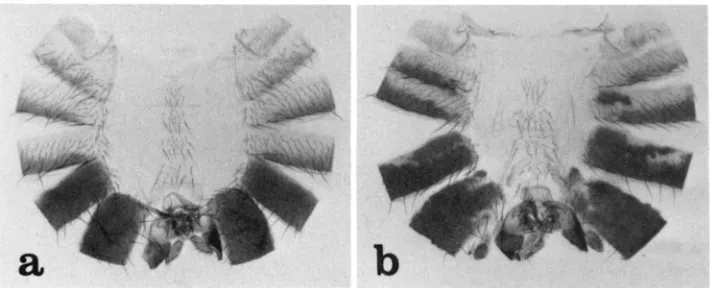
All of the abdominal tergites of Pcl’/Pc13 animals show variegated transfor- mations toward the posterior. The first abdominal tergite, which is unique in normally having only small bristles, often contains patches of cuticle that are transformed to second, third, or perhaps fourth tergite (these are indistinguish- able in small patches). No fifth tergite tissue (recognizable in males by black pigmentation and presence of trichomes) has ever been seen in the first tergite. The second, third and fourth tergites often show patches of cuticle transformed to fifth or sixth (recognizable by absence of trichomes) tergites. The fifth tergite is usually largely transformed to sixth and the sixth to seventh. In males, which normally have no seventh tergite (SANTAMARIA and GARCIA-BELLIDO 1972), the latter transformation results in the loss of the sixth tergite. The transformations described for the tergites of Pcl’/Pc13 animals also occur ventrally and can be seen by changes in sternite morphology. The abdominal cuticle from a typical mutant male is compared with that from a wild-type male in Figure 2.
CONTROL OF THE BX-C AND ANT-C 55
._
56
FIGURE 2.-Abdominal cuticles from a wild-type male ( a ) and a PcI’/PcP male (b). Note in (b) the patch of second, third or fourth abdominal segment cuticle in, the first tergite and the patch of fifth or sixth segment cuticle in the third tergite. The fourth and fifth tergites are almost entirely transformed to fifth and sixth tergites, as evidenced by changes in pigmentation and trichome pattern. The sixth tergite is very reduced, presumably because this segment is largely transformed to the seventh, which in males has no tergite. The genitalia are normal. The sternites in (b) show posteriorly directed transformations similar to those described for the tergites.
Spermathecae from mutant and wild-type females are shown in Figure 3. The internal genitalia of males are normal.
Effects of Pcl mutants on embryogenesis: The data presented in Table 1 show that Pcl’ hemizygotes or homozygotes die as embryos. These animals appear to complete embryogenesis and their cuticular structures are indistinguishable from wild type (for a description of the cuticle of wild-type embryos see
LOHS-
SCHARDIN et al. 1979). Pcl’-homozygous embryos are morphologically wild type whether they come from mothers that are Pcl’/+ or Pcl’/Pc14. Animals hetero- zygous for Df(2R)HB and Df(2R)7B die as late embryos, but are grossly abnormal in morphology, presumably because of the absence of genes other than Pcl.Pcl’ hemizygotes that are heterozygous or homozygous for Pc3 have also been constructed. Regardless of the parental origin of Pc3, genotypically Pcl’/ Df(2R)11B;Pc3/+ embryos are wild type in morphology. Pcl’/Df(2R)11B;Pc3/Pc3 embryos, however, show very extreme posteriorly directed segmental transfor- mations. Such animals are considerably more affected than are Pc3 homozygotes that are PcP. Posterior spiracle-like structures (characteristic of the eighth abdominal segment in wild type) appear in most segments. The mouth hook apparatus is almost completely undeveloped and a region of the head, probably from the labial segment, usually develops an abdominal-type ventral setal belt. Behavior of epidermal clones homozygous for Pcl’: Animals of the genotype al dp pwn Pcl’/+;mwh/+ were irradiated with approximately 1O00 r 24-48 hr before pupation and clones marked by pwn (2-58, GARCIA-BELLIDO and DAPENA 1974) or mwh were scored in the adult abdomen and wing. The results of this and a control experiment are presented in Table 2. In the following discussion all clones marked by pwn are assumed to be homozygous for the more distally located Pcl’.
CONTROL OF THE BX-C A N D ANT-C 57
FIGURE 3.-Internal genital structures of ( 0 ) a wild-type (C-S) and (b) a Pcl'/PcP female. Wild- type females possess two spermathecae (S) and two parovaria (P), whereas Pcl'/PcP females lack parovaria and have four spermathecae. In general, one pair of spermathecae is significantly smaller than the other in Pcl'/Pcl' females. The smaller spermathecae are often incompletely formed, presumably because they represent an intermediate stage in the transformation of parovaria into spermathecae.
TABLE 1
Embryonic lethality of Pcl' hemizygotes and homozygotes
Cross
Embryos un-
Unfertil- hatched % dead Total ems ized ems' at 48 hr embryos
Df(2R)IIB/C-S Po X PcI'/CYO 66 288 15 77 28
Df(2R)llB/C-S 99 X C-S 66 369 17 12 3
a1 dp b pr c Pcl '/C-S Po x Pcl' px sp/CyO 66 288 7 67 24 a1 dp b pr c Pcl'/Pcl' 99 x a1 dp b pr c Pcl'/CyO 66 416 56 75 21
TABLE 2
Behavior of Pcl'/Pcl' clones in the abdominal tergites and wing
Experimental genotype: Control genotype:
01 dp pwn Pcl' px sp/+;mwh/+ 01 dp pwn/+;mwh/+
trans- total formed
pwn pwn mwh pwn mwh
Tergite clones clones clones N" clones clones N"
Ab1 0 d
Ab2 0 d
Ab3 0 d
Ab4 P d
Ab5 P d
Ab6 P d Wing'
15 0
6 0
12 3 11 7 12 3 17 13 11 7
9 7
14 14
9 8
9 6
0 0
83 41
23 193 16 184 19 199 16 200 24 200 21 200 22 198 19 200 29 194 18 200 10 200 2 199 215 196
9 13 26 12 14 16 12 18 8 17 11 6 72
33 194 18 183 29 200
19 198 32 200 27 199 28 200 30 200 25 200
23 200 18 200 8 200 168 196
N = number of hemitergites or wings examined.
'Wing clones were from ad dp pwn Pcl'/+;mwh/+ and a1 dp pwn/+;mwh/+ animals.
Pcl', segmental transformations are almost entirely restricted to cells marked by pwn, indicating autonomy of Pcl+ action. As illustrated in Figure 4, more than one segmental transformation can often be recognized within each clone. Included in Table 2 are the proportions of Pcl' clones in each tergite that are detectably transformed to the posterior. Because the second, third and fourth tergites appear identical (at least over most of their surfaces), clones in these segments must be transformed to the fifth or sixth segments in males (recogniz- able by black pigmentation) or the sixth or seventh segments in females (recognizable by absence of trichomes) to be detectably transformed. This difference between the sexes probably explains why Pcl' clones in the second and third abdominal segments of females show a lower frequency of transfor- mation than do clones in these segments of males. (Because the fifth tergite has a lower trichome density than more anterior tergites, one might expect that transformations to this segment could be seen in females. In practice, however, it is difficult to score trichome density changes in small patches.) In both sexes, clones occurring in the second and third abdominal tergites show a lower frequency of transformation than do clones in the fourth and fifth tergites, probably because short range transformations can be seen only in more poste- rior segments. Transformations of the sixth segment to seventh were detected in females by the loss of trichomes in Pcl' clones occurring in the anterior portion of the sixth tergite. This transformation probably also occurs in males (which have no seventh tergite), resulting in the absence of Pcl' clones in the male sixth tergite. Pcl' clones in the abdominal sternites show the same types of posteriorly directed transformations as do clones in the tergites.
None of the 21 Pcl' clones seen in the first abdominal tergite showed segmental transformations. This result is remarkable because any posteriorly directed transformation would be easily scored in the first abdominal segment.
CONTROL OF THE BX-C AND ANT-C 59
FIGURE 4.-a, A patch of haltere-like cuticle in the wing of an 01 dp pwn Pcl'/+ female irradiated
24-48 hr before pupation. Note the presence of a bristle and small trichomes similar to those that normally occur in the haltere. A vesicle of haltere-type cuticle is also present between the dorsal and ventral surfaces of the wing blade. This sector is probably a Pcl'-homozygous clone which arose by mitotic recombination between the loci of pwn and Pcl. In contrast, most patches of haltere-type cuticle in such wings are clearly marked by pwn. having arisen by mitotic recombina- tion proximal to pwn. b, A third abdominal hemitergite from an al dp pwn Pcl' px sp/+ male irradiated 24-48 hr before pupation. Shown is a clone homozygous for Pcl' and for the cell marker pwn, which causes the truncation of bristles and trichomes. The clone is darkly pigmented because it is transformed to a mixture of fifth and sixth tergite. Around most of the border of the clone, pigmentation extends for a short distance into areas that have normal length trichomes, suggesting that pwn+, Pcl', or the process of pigmentation itself is nonautonomous over short distances. As is often seen in tergites, cells within the clone are not contiguous. In particular, note the migration of pwn/pwn bristles out of the clone and the migration of wild-type bristles into the clone.
transformed to haltere, as indicated by the development of small, densely packed trichomes similar to those that occur on the haltere and, occasionally, of small bristles like those found on the haltere. An example of such a clone is shown in Figure 4. The morphology of these clones is the same in the anterior and posterior compartments of the wing.
Evidence for the interaction of Pcl' with the ANT-C and BX-C: That the segmental transformations present in Pcl'/Pc13 animals resemble closely those caused by certain dominant gain-of-function mutants in the ANT-C and BX-C (see DISCUSSION) suggests that Pcl+ interacts with both of these gene complexes.
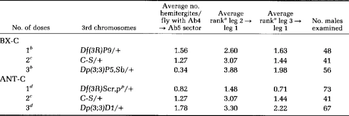
Further evidence supporting this notion has come from experiments which indicate that the severity of segmental transformations in Pcl mutant animals depends upon the dosage of the ANT-C and BX-C. Pcl'/Pc14 males were chosen for these tests because these animals have good viability and show partial transformations of their fourth abdominal tergites to fifth tergites and of their second and third legs to first legs. Rearrangements used in the BX-C dosage tests were Df(3R)Ps(=Df(3R)89E1;89E4-5), which is deficient for the entire BX-C
(LEWIS 1978), and Dp(3;3)P5(=Dp(3;3)89E1,2;90A), which is a direct tandem duplication for the BX-C and surrounding material (LEWIS unpublished). ANT- C dosage was varied using Df(3R)Scr(=Df(3R)84A1;84B1), a deficiency for most
or all of the ANT-C (R. A. LEWIS et al. 1980) and Dp(3;3)DZ(=op(3;3)84A;85A),
a direct tandem duplication that includes at least the Scr and Antp loci of the ANT-C (DUNCAN unpublished). To reduce the effects of differences in genetic background, male parents used in the dosage tests were from stocks maintained as
Pc14pxsp/SMZ;Dp(3;3)P5,Sb/Df(3R)P9
or as Pc14pxsp/SM1;Dp(3;3)D1/ Df(3R)Scr, pp.The results of the dosage tests are presented in Table 3. Within the range of doses tested, the data indicate that the frequency of transformation of fourth abdominal tergite to fifth in Pcl'/Pc14 animals is enhanced by increasing ANT- C dosage but is markedly suppressed by increasing BX-C dosage. DUNCAN and
TABLE 3
Dependence of segmental transformations in Pcl'/Pc14 males on BX-C and ANT-C dosage
Average no.
hemitergites/ Average Average
fly with Ab4 rank" leg 2
-
rank" leg 3 + No. malesNo. of doses 3rd chromosomes -+ Ab5 sector leg 1 lea 1 examined BX-C
lh Df(3R)P9/+ 1.56 2.60 1.63 48
2'
c-s/+
1.27 3.07 1.44 413h Dp(3;3)P5,Sb/+ 0.34 3.88 1.98 56
ANT-C
Id Df(3R)Scr,pP/+ 0.82 1.48 0.71 73
2'
c-s/+
1.27 3.07 1.44 413d Dp(3;3)Dl/+ 1.78 3.30 2.22 67
" T h e number of sex comb teeth occurring on the second and third legs were counted and each male ranked according to the following scale: 0 = 0 teeth, 1 = 1-4 teeth, 2 = 5-8 teeth, 3 = 9-13 teeth, 4 = more than 13 teeth or the presence of more teeth on one leg than could be counted reliably under the dissecting microscope (>7).
From cross of a1 dp b pr c Pcl'/CyO PP X Pc14 pxsp/SMl;Dp(3;3)P5,Sb/Df(3R)P9 $8.
From crass of ol dp b pr c Pcl'/CyO 99 X Pc14 pxsp/SMl;TMl/C-S 86.
CONTROL OF THE BX-C A N D ANT-C 61
LEWIS (1981) have reported a similar effect of varying ANT-C and BX-C dosage in Pc3/+ animals. These authors have also found that increasing ANT-C dosage enhances the frequency of transformation of the first abdominal segment to a more posterior (second, third or fourth) abdominal segment in Pc3 heterozygotes. Indeed, otherwise wild-type animals carrying three or four doses of the ANT-C occasionally show posteriorly directed transformations of the first abdominal segment. The latter two effects of ANT-C duplications have been shown to be suppressed by Df@R)Scr, indicating that the dosage sensitive gene(s) responsible is in or near the ANT-C. Since only specific transformations are promoted by extra doses of the ANT-C, it seems unlikely that these transformations result from the competition of the ANT-C and BX-C for a negative regulator common to all genes in both complexes. Rather, DUNCAN and LEWIS (1981) have suggested that these transformations result from the action of a gene within the ANT-C that is a positive regulator of certain BX-C genes. This idea has received support from the discovery of mutants in the ANT-C (which will be described elsewhere) that cause certain BX-C related segmental transformations.
The dependence upon BX-C and ANT-C dosage of the transformation of the posterior legs to first leg (as measured by sex comb teeth counts) in Pcl’/Pc14 males is similar to that described for Pc3 heterozygotes by DUNCAN and LEWIS
(1981). Increasing BX-C dosage appears to weakly enhance this transformation (at least in the second leg) while increasing ANT-C dosage has little effect. Transformations to first leg are weak in Pcl’/Pc14 males carrying a single dose of the ANT-C, but this effect is to be expected because males with only one dose of the ANT-C also show a marked reduction in the number of sex comb teeth on their first legs (KAUFMAN, LEWIS and WAKIMOTO 1980).
Further evidence for the interaction of Pcl+ with the ANT-C and BX-C is supplied by the observation that Pcl mutants enhance certain dominant gain- of-function mutants in both complexes. Females heterozygous for Pcl’ were mated to males carrying the BX-C mutants Cbx, Uab’, Uab’, Uab4, Hab and Mcp (all described in LEWIS 1978), and the ANT-C mutants (GREEN 1975), SCX, and Msc. Progeny from these crosses showed that PcJ’ causes unequivocal enhancement of Cbx, Mcp, SCX, and Msc. A particularly striking interaction is the enhancement of Cbx: Pel'/+; Cbx/+ animals show an almost complete wing-to-haltere transformation, whereas Cbx/+ flies usually show this trans- formation in the posterior of the wing only. Pcl’ has also been shown to partially suppress the haltere-to-wing transformation present in animals homozygous for the recessive loss-of-function BX-C mutant bxd.
The relation of Pcl+ to Pc+: In Table 4 representative data are shown illus- trating that Pcl’ and Pc3 markedly enhance one another. This interaction does not depend upon the Pcl or Pc alleles used nor does it depend upon their parental origin (data not shown). For each of the segmental transformations scored, the interaction appears to be synergistic rather than additive, suggesting that Pcl’ and Pc+ are related in function. That Pc+ and Pcl’ have qualitatively different functions, however, is indicated by the results presented in Table 5. In these experiments, extra doses of Pc+ were introduced using Dp(3;3)C126, a direct tandem duplication for the material from approximately 78D to 79B
TABLE 4
The interaction between Pcl' ond Pc3
Avg. no hemiter- Avg. rank" Avg. rank' Avg. rank" Avg. rank" gites/fly Genotvpe N" ant + leg wing
-
halt. leg 2 + leg 1 leg 3 -+ leg 1 Ab4-
Ab5Pcl ]/+e
s
28 0 0 0.18 0.04 0.07P 28 0 0
Pc:j/+f
s
40 0 0.50 0.95 0.55 0.63O 41 0.5 0.80
Pcl '/+;Pc3/+g
s
32 0.34 1.72 3.75 2.41 1.97P 39 1.08 2.05
N = number of animals scored.
Each animal was assigned a rank based on its more extremely affected antenna according to the following scale: 0 = wild type, 1 = tiny outgrowth, 2 = moderate outgrowth, and 3 = large outgrowth.
Each animal assigned rank based on more extremely affected wing according to the following scale: 0 = wild type, 1 = posterior wing slightly serrated, 2 = posterior wing serrated along most of posterior edge, 3 = posterior wing crumpled with wing abnormally curved, and 4 = haltere cuticle obvious at posterior of wing.
As in Table 3.
e From cross of C-S PO X Pcl'/CyO 88. From cross of Pc3/TMI 99 X C-S 68.
From cross of Pc3/TMI 99 X Pcl'/CyO 88.
TABLE 5
Dependence of segmental transformations in Pcl'/Pc14 moles on Pc+ dosage
~
~ ~~
Avg. no. hemi- tergites/fly
No. of with Ab4
-
Avg. rank" Avg. rank" No. males ex-Pc+ doses 3rd chromosomes Ab5 sector lea 2 + leg 1 lee 3 -+ lee 1 amined
2"
c-s/+
1.27 3.07 1.44 4136 Dp(3;3)C126, in ri pp/+ 0.84 2.59 1.11 56
4b Dp(3;3)CZ26, in ri pp/ 0.74 2.36 0.83 47
Dp(3;3)CZ26, in ri pp
~
As in Table 3.
From cross of 01 dp b pr c Pclf/+;Dp(3;3)C226, in ri pp/+ PP x Pc14 px sp/Pc14 px sp;Dp(3;3)C126, in ri pp/TM3, Sb Ser 88.
four doses of Pc+ there appears to be a trend towards suppression of segmental transformations in these animals. Dp(3;3)CZ26 is a moderately large duplication, however, and this suppression may result from genes other than Pc+ that are contained within it. In any case, Pc+ activity does not appear to be able to substitute for PcJ+ activity, at least within the range of doses tested.
DISCUSSION
CONTROL OF THE BX-C AND ANT-C 63
type allele of this locus is involved in the control of the ANT-C and BX-C, two gene clusters thought to be directly responsible for the specialization of most body segments.
The main line of evidence for the involvement of PcJ+ in the control of the ANT-C and BX-C is that the segmental transformations caused by Pcl mutants closely correspond to those caused by dominant gain-of-function mutants in these complexes. For example, the transformation of antenna into leg, which is occasionally seen in Pcl’/Pc13 animals, is also caused by a number of dominant mutants in the ANT-C which, for the most part, have been considered Anten- napedia (Antp) alleles. Transformations of second and third legs into first legs are caused by two other dominant ANT-C mutants called Extra sex comb (SCX) and Multiple sex comb (Msc). Although each of these mutants is lethal when homozygous, Scx/Msc heterozygotes survive to adulthood and show much stronger leg transformations than those seen in animals carrying either mutant alone. Microscopic examination of the posterior legs from Scx/Msc animals has revealed a pattern of conversion to first leg that is almost identical to that found in Pcl’/Pc13 animals [with the exception that, because of a n Scr mutation associated with Msc (R. A. LEWIS et al. 1980), the number of sex comb teeth and bristles of the anterior tarsal transverse rows is reduced in all legs of Scx/Msc as compared to Pcl’/PcJ3 animals]. In the second legs of Scx/Msc animals the anterior compartment is almost completely transformed to first leg, whereas the posterior compartment is only partially so, as evidenced by the development of an incomplete set of first leg bristles in the femur posterior compartment. The anterior compartment of the third legs of Scx/Msc animals is completely transformed to first leg distally, but is almost normal proximally. The third leg posterior compartment appears to be unaffected. The preferential transforma- tion of the anterior compartments of the second and third legs to first leg is also seen in males heterozygous for Pc mutants and, at least for the third leg, apparently also in males homozygous for l(3)2902°x736*“ after exposure to the restrictive temperature in the third larval instar (SHEARN, HERSPERGER and
HERSPERGER 1978). Unlike PcJ’/PcJ3 animals, Scx/Msc adults do not show any transformation of second leg to third leg.
The remaining transformations described for PcJ mutant animals correspond to those caused by dominant gain-of-function mutants located in the BX-C. Transformation of the second thoracic segment to third thoracic segment, as seen in the wing (in PcJ’/PcJ’ clones) and in the posterior compartment of the leg (in PcJ’/PcP adults), is caused by the BX-C mutant Cbx’ (LEWIS 1981b). (Cbx’, although causing a partial transformation of anterior and posterior wing to haltere, does not affect the posterior compartment of the second leg.) Posteriorly directed transformations of the abdominal segments are caused by mutants in the Uab group of the BX-C (which transform the first abdominal segment to a more posterior segment) and by Mcp (which converts the fourth abdominal segment into the fifth). Finally, the transformation of parovaria into spermathecae, as described for PcJ’/Pc13 females, is often seen in Pc3/Uab’ females (approximately 50% of such females show at least one parovarium transfmmed to a spermatheca). This transformation has not been seen in either Pc3/+ or Uab’/+ females (over 30 of each type examined). Although cytologi- cally normal, Uab’ suppresses recombination over most of the BX-C (LEWIS
least two breakpoints within the BX-C. Because heterozygotes for Pc3 and other mutants of the Uab type do not show transformation of parovaria to sperma- thecae, it seems likely that this transformation is promoted by a different breakpoint of Uab' than is the transformation of the first abdominal segment to the second. Moreover, because Pc3/Df(3R)P9 (Df(3R)Pg is deficient for the entire BX-C) females do not show transformation of parovaria to spermathecae (over 30 females examined), it is likely that Uab' promotes this transformation by a gain rather than a loss of function in the BX-C. This conclusion is supported by the finding of the reverse transformation, the conversion of spermathecae into parovaria, in females heterozygous for In(3LR)Uab4 and T(2;3)P20 (these rear- rangements, which both have breakpoints within the BX-C, are described in
LEWIS 1978). In(3LR)Uab4/T(2;3)P20 animals have been described by LEWIS
(1978) as showing transformations of the third, fourth and fifth abdominal segments to second abdominal segments. Based on these transformations, LEWIS has argued that In(3LR)Uab4 and T(2;3)P20 inactivate at least partially a BX-C gene or genes required for the normal development of posterior abdominal segments. It seems likely that the inactivation of these genes is also responsible for the observed transformation of spermathecae into parovaria.
The conversion of parovaria to spermathecae is unique among transforma- tions caused by BX-C gain-of-function mutants in that it is anteriorly directed; evidence from fate mapping in Drosophila (SCHUPBACH, WIESCHAUS and No-
THIGER 1978) and from embryological examination of other dipterans (DUBEN- DORFER 1970, 1971; EMMERT 1972) indicates that spermathecae lie anterior to
parovaria at the time of their formation. Based on extensive studies of the internal reproductive structures of species in Drosophila and related genera, Dr.
L. THROCKMORTON (personal communication; see also THROCKMORTON 1962) has formed the opinion that spermathecae have evolved from parovaria-like struc- tures. The transformations of parovaria to spermathecae described above may be consistent, therefore, with the generalization that the activities of BX-C genes promote the development of evolutionarily more advanced characteristics.
CONTROL OF THE BX-C AND ANT-C 65
The nature of the control of the ANT-C by Pc+ and Pcl+ is not as clear as that of the BX-C. Although cases for Loth positive and negative control of the ANT- C by these genes can be made, in at least two instances the available evidence argues in favor of negative control. DENELL et al. (1981) and STRUHL (1981a) have presented evidence that the ANT-C mutant Ns (and perhaps all dominant mutants of the Antp type) causes the transformation of antenna into leg by turning on an ANT-C gene in the antenna, where in normal development it would be inactive. That animals mutant for Pc or Pcl also show this transfor- mation suggests that Pcl+ and Pc+ are required to repress the activity of this gene in the antenna.
The transformations of second and third leg to first leg seen in Pcl- and Pc- mutant animals can also be interpreted as resulting from hyperactivation of an ANT-C gene. Examination of Figure 1 reveals that the first tarsal segments (basitarsae) of the three legs of wild type show a striking symmetry. Several transverse rows of bristles (one of which is modified in males to form the sex comb) are present in the anterior of the first leg basitarsus whereas a similar set of transverse rows is located in the posterior of the third leg basitarsus. No transverse rows are present on the first tarsal segment of the second leg. R. A.
LEWIS et al. (1980) have found that animals heterozygous for mutations that inactivate the Scr gene of the ANT-C show a marked reduction in the number of bristles per anterior transverse row in the first leg basitarsus. The author has quantitated this effect by counting the number of bristles present in the penultimate distal transverse row in the first leg basitarsae of males carrying one, two and three doses of the ANT-C. In this bristle row, Df(3R)Scr/+ males show an average of 4.6 bristles, wild-type (C-S) males show 7.5 bristles, and Dp(3;3)DI/+ males show 9.4 bristles (20 basitarsae of each genotype were examined). Similarly, it has been determined that the number of bristles per posterior transverse row in the third leg basitarsus is dependent upon BX-C dosage. The average number of bristles in the penultimate distal transverse row in the third leg basitarsae of Df(3R)P9/+ males is 4.5; for wild-type (C-S) males it is 6.1, and for Dp(3;3)P5/+ males it is 7.4 (based on counts of 20, 22 and 12
basitarsae, respectively). It appears, therefore, that the activity of an ANT-C gene (Scr+) promotes the formation of anterior tarsal transverse rows whereas the activity of a BX-C gene or genes promotes the formation of posterior tarsal transverse rows. In normal development, these genes appear to be inactive in the second leg, which has no transverse bristle rows. In Pcl'/Pc13 animals, however, transverse bristle rows develop in both the anterior and posterior of the second leg basitarsus, suggesting that Pcl' functions in this leg to repress the activities of Scr+ and of one or more BX-C genes. In Pc-mutant animals, transverse bristle rows have been found in the anterior, but not the posterior, of the second leg basitarsus. Pc+, therefore, can also be thought of as a repressor of Scr+ activity in the second leg. Although the model that Pcl' and Pc+ are negative regulators of Scr+ is attractive, the observations of WAKIMOTO and
BX-C genes in the anterior and posterior portions of the thoracic segments. Evidence for such differential expression of Antp+ (DUNCAN 1982) and of certain BX-C genes (MORATA and KERRIDCE 1981; LEWIS 1978) has already been de- scribed.
Although the similar phenotypes and strong mutual enhancement of Pcl and
Pc mutants suggest that these genes have very similar functions, it appears that these functions are not identical. One line of evidence suggesting this is that Pc
and Pcl mutants exert their major effects on different body structures. For example, animals heterozygous for Pc mutants have more strongly affected wings and antennae and much more weakly transformed legs and abdomens than do Pcl'/Pc13 animals. The results of clonal analysis experiments also indicate that these genes have different functions. When induced late in devel- opment, abdominal clones of cells homozygous for Pcl' show variable short- range transformations toward the posterior. Similar clones lacking Pc+ activity uniformly transform to what appears to be anal plate (STRUHL 19810; DUNCAN
and LEWIS 198l), thought to be the most posteriorly derived structure in the external cuticle of the adult fly (SCHUPBACH, WIESCHAUS and NOTHIGER 1978). Since Pcl' appears to be a null allele, these observations indicate that Pcl+
activity is not absolutely required for the maintenance of BX-C gene repression, whereas Pc+ activity probably is so required. Because the clones examined in these experiments were large, it seems unlikely that the differences in behavior of clones lacking Pcl+ and those lacking Pc+ are due to persistence of Pcl+
product. That extra doses of Pc+ have no major effect on the phenotype of Pcl'/ Pc14 animals supports the idea that Pc+ and Pcl+ perform different functions.
The discovery of Pcl raises the question of whether other genes exist that are similar in function to Pc+. In fact, three other such genes have been identified by selecting mutants that act as dominant enhancers of Pc3 and by selecting mutants that modify the expression of certain BX-C mutants. These genes will be described in future reports. In addition, two genes have been described in the literature that appear similar in function to Pc+. One of these is a gene called extra sex combs (esc). STRUHL (1981b) has recently reported that this gene exerts a maternal effect such that homozygous mutant progeny from homozygous mutant mothers die as mature embryos that show transformations of most of their body segments toward the most posterior abdominal segment. Animals homozygous for Pc mutants show similar transformations (LEWIS 1978; DUNCAN
CONTROL OF THE BX-C AND ANT-C 67
suppress the development of the dorsal prothorax (as defined by FERRIS 1950), effects that have not been seen in animals mutant for Pc or Pcl. Although it is not clear why the fly should require several genes that are similar to Pc+ in function, the existence of these genes should not be surprising in view of the number of genes known to be involved in other developmental processes (for example sex determination; see BAKER and RIDGE 1980).
In addition to negative control by a number of genes, the functions of the BX-
C appear to be under the positive control of at least one gene. This is Regulator of bithorax (Rg-bx) (see GARCIA-BELLIDO and CAPDEVILA 1978), a gene discovered and localized to 88B on the polytene chromosome map by E. B. LEWIS (1968). Animals mutant for this gene show a number of segmental transformations that correspond to those caused by loss-of-function mutants in the BX-C. Patchy transformations of the haltere to wing and of the posterior abdominal segments to more anterior ones are often seen in animals heterozygous for amorphic alleles of Rg-bx. Homozygotes for such alleles die as late embryos or first instar larvae that show anteriorly directed transformations of their posterior abdom- inal segments (DUNCAN and LEWIS 1981). INGHAM and WHITTLE (1980) have described a hypomorphic Rg-bx allele called trithorax (trx) that when homo- zygous or hemizygous causes segmental transformations that are strikingly similar, but opposite in direction, to those seen in Pcl’/Pc13 adults. For example, trx homozygotes show anteriorly directed conversions of the abdominal seg- ments and partial transformations of the first and third legs to second legs.
Females homozygous for trx also show extra spermathecae (INGHAM and WHIT-
TLE 1980), although it is not clear if these arise by transformation of parovaria,
as they do in Pc11/Pc13 females. The opposite orientations of most of the transformations caused by Rg-bx and Pcl mutants suggest that these genes control in opposite ways many of the same genes in the ANT-C and BX-C.
The picture that is emerging from recent work on genes controlling the specialization of body segments in Drosophila is that segmental identities are determined directly by genes that are clustered within the ANT-C and BX-C. Many genes that affect segmental character are located outside of these clusters, but several, if not all, of these genes can be thought of as regulators of genes within the two complexes. These regulators appear to be of at least two types: those that are active only early in development and those that act continuously. At present, our working hypothesis is that early in embryogenesis one or more of the continuously acting regulators comes to be expressed differentially along the body axis, a process which may be controlled by the early acting regulators. The activities of the continuously acting gene(s) could be regulated, for example,
so that they grade from anterior to posterior in the animal, as suggested for Pc+
to act as intermediaries between a few controlling genes like Pc+ and Pcl+ and the large batteries of genes that presumably must be regulated to elaborate structures characteristic of each body segment.
I thank DANIELA BONAFEDE, MADELINE CROSBY and MARGIT LOHS-SCHARDIN for critical reading of the manuscript and L. CRAYMER, E. B. LEWIS and L. THROCKMORTON for helpful discussions. This work was supported by a grant from the Helen Hay Whitney Foundation and Grant HD 06331 from the United States Public Health Service.
LITERATURE CITED
BAKER, B. S. and K. A. RIDGE, 1980 Sex and the single cell. I. On the action of major loci affecting sex determination in Drosophila melanogoster. Genetics 94: 383-423.
DENELL, R. E., K. R. HUMMELS, B. T. WAKIMOTO and T. C. KAUFMAN, 1981 Developmental studies of lethality associated with the Antennapedia gene complex in Drosophila melanogaster. Dev. Biol. 81: 43-50.
DUBENDORFER, A., 1970 Entwicklungsleistungen transplantierter Genital- und Analanlagen von
Untersuchungen zum Anlageplan und Determinationszustand der wei- blichen Genital- und Analprimordien von Musca domestico L. Wilhelm Roux' Arch. 168: 142- 168.
Musca domestico und Phormia regina. Experientia 2 6 1158-1160.
DUBENDORFER, A., 1971
DUNCAN, I., 1982
DUNCAN, I. and E. B. LEWIS, 1981
Localization of bithorax complex (BX-C) and Antennapedia complex (ANT-C) gene activities along the body axis of Drosophila. Genetics 100: S20.
Genetic control of body segment differentiation in Drosophila. In: Developmental Order: Its Origin and Regulation, the 40th Symposium of the Society for Developmental Biology. Edited by S. SUBTELNY. Alan Liss, Inc., New York. In press.
Entwicklungsleistungen abdominaler Imaginalscheiben von Calliphora ery- throcephala (Insecta, Diptera). Experimentelle Untersuchungen zur Morphologie des Abdo- mens. Wilhelm Roux' Arch. 169: 87-133.
FERRIS, G. F., 1950 External morphology of the adult. pp. 368-419. In: Biology of Drosophila.
D. melanogaster-linkage data: report of A. Garcia-Bellido. Opt': EMMERT, W., 1972
Edited by M. DEMEREC. John Wiley and Sons, Inc., New York.
Ophthalmoptera of Goldschmidt. Drosophila Inform. Serv. 4 4 52. GARCIA-BELLIDO, A., 1969
GARCIA-BELLIDO, A. and M. P. CAPDEVILA, 1978 The initiation and maintenance of gene activity in a developmental pathway of Drosophila. pp. 3-21. In: The Clonal Basis of Development, the 36th Symposium of the Society for Developmental Biology. Edited by S. SIJBTELNY and I. M. SUSSEX. Academic Press, New York.
GARCIA-BELLIDO, A. and J. DAPENA, 1974 Induction, detection and characterization of cell differ- entiation mutants in Drosophila. Mol. Gen. Genet. 128 117-130.
GEHRING, W., 1970a A recessive lethal (1(4)29) with a homeotic effect in D. melonogaster.
Problems of cell determination and differentiation in Drosophila. pp. 231- 241. In: Problems in Biology: RNA in Development. Edited by E. W. HANLY. University of Utah Press, Salt Lake City.
Conversion a s a possible mechanism of high coincidence values in the centromere region of Drosophila. Mol. Gen. Genet. 139 57-66.
Rev. Suisse Zool. 51: 418-423.
INGHAM, P. and R. WHITTLE, 1980 Drosophila Inform. Serv. 45: 103. GEHRING, W., 1970b
GREEN, M. M., 1975
HADORN, E. and H. GRABER, 1944 Uber einen Drosophila-Stamm mit veranderten Spermatheken.
CONTROL OF THE BX-C AND ANT-C 69
Cytogenetic analysis of chromosome 3 in Drosophila melanogaster: The homoeotic gene complex in polytene chromosome interval 84A- B. Genetics 94: 115-133.
engrailed: A gene controlling compartment and segment formation in Dro-
Bristle patterns and compartment boundaries KAUFMAN, T. C., R. LEWIS and B. WAKIMOTO, 1980
KORNBERG, T., 1981
LAWRENCE, P. A., G. STRUHL and G. MORATA, 1979 sophila. Proc. Natl. Acad. Sci. USA 7 8 1095-1099.
in the tarsi of Drosophila. J. Embryol. Exp. Morphol. 5 1 195-208.
LEWIS, E. B., 1960 A new standard food medium. Drosophila Inform. Serv. 3 4 117-118.
LEWIS, E. B., 1968
LEWIS, E. B., 1978 LEWIS, E. B., 1980
LEWIS, E. B., 1981a
Genetic control of developmental pathways in Drosophila melanogaster. Proceedings XI1 International Congress of Genetics 2 96-97. Science Council of Japan, Tokyo.
A gene complex controlling segmentation in Drosophila. Nature 2 7 6 565-570. New mutants-D. melonogaster. Report of E. B. Lewis. Drosophila Inform. Serv.
Developmental genetics of the bithorax complex in Drosophila. In: Develop- mental Biology Using Purified Genes. ICN-UCLA Symposium on Molecular and Cellular Biology Vol. XXIII. Edited by D. D. BROWN and C. F. Fox. Academic Press, New York.
Control of body segment differentiation in Drosophila by the bithorax gene complex. In: Embryonic Development: Genes and Cells. Proceedings of the IX Congress of the International Society of Developmental Biologists. Edited by M. M. Burger. Alan Liss, Inc., New
York. In press.
Method of feeding ethyl methane sulfonate (EMS) to Drosophila males. Drosophila Inform. Serv. 43: 193.
New mutants (me1anogaster)-Report of E. B. Lewis and F. Bacher: OptB: Opthalmoptera-Bacher. Drosophila Inform. Serv. 4 4 48.
Genetic analysis of the antennapedia gene complex (ANT-C) and adjacent chromosomal segments 84A-84B1,2. Ge- netics 95: 383-397.
Genetic variations of Drosophila melanogaster. Publication No. 627, Carnegie Institute, Washington.
LOHS-SCHARDIN, M., C. CREMER and C. NUSSLEIN-VOLHARD, 1979 A fate map for the larval epidermis of Drosophila melanogaster: Localized defects following irradiation of the blastoderm with a n ultraviolet laser microbeam. Dev. Biol. 73: 239-255.
Sequential functions of the bithorax complex. Nature 2 9 0
Genetic analysis of pattern-formation in the embryo of Drosophila melanogaster. Characterization of the maternal-effect mutant Bicaudal. Wilhelm Roux’ Arch.
55: 207-208.
LEWIS, E. B., 1981b
LEWIS, E. B. and F. BACHER, 1968
LEWIS, E. B. and F. BACHER, 1969
LEWIS, R. A., B. T. WAKIMOTO, R. E. DENELL and T. C. KAUFMAN, 1980
LINDSLEY, D. L. and E. H. GRELL, 1968
MORATA, G. and S. KERRIDGE, 1981 778-781.
NUSSLEIN-VOLHARD, C., 1977
183: 249-268.
SANTAMARIA, P. and A. GARCIA-BELLIDO, 1972 Localization and growth pattern of the tergite anlage of Drosophila. J. Embryol. Exp. Morphol. 28: 397-417.
The embryonic organization of the genital disc studied in genetic mosaics of Drosophila melanogoster. Wilhelm Roux’ Arch. 185: 249- 270.
Genetic analysis of two allelic temperature- sensitive mutants of Drosophila melanogaster both of which are zygotic and maternal-effect lethals. Genetics 8 9 341-353.
STEINER, E., 1976 Establishment of compartments in the developing leg imaginal discs of Drosoph-
STRUHL, G., 1981a A homoeotic mutation transforming leg to antenna in Drosophila. Nature 2 9 2
STRUHL, G., 1981b A gene product required for correct initiation of segmental determination in SCHUPBACH, T., E. WIESCHAUS and R. NOTHIGER, 1978
SHEARN, A., G. HERSPERGER and E. HERSPERGER, 1978
ila melanogaster. Wilhelm Roux’ Arch. 1 8 0 9-30.
635-638.
THROCKMORTON, L. H., 1962 The problem of phylogeny in the genus Drosophila. pp. 207-343. In: Studies in Genetics 11. Research Reports on Drosophila Genetics, Taxonomy and Evolution. Edited by M. R. WHEELER. Publication number 6205 of the University of Texas, Austin.
TOKUNAGA, C. and C. STERN, 1965 The developmental autonomy of extra sex combs in Drosophila melanogaster. Dev. Biol. 11 50-81.
WAKIMOTO, B. T. and T. C. KAUFMAN, 1981 Analysis of larval segmentation in lethal genotypes associated with the antennapedia gene complex in Drosophila melanogaster. Dev. Biol. 81: 51-
64.