Western University Western University
Scholarship@Western
Scholarship@Western
Electronic Thesis and Dissertation Repository
7-2-2015 12:00 AM
Functional Characterization of P3N-PIPO Protein in the Potyviral
Functional Characterization of P3N-PIPO Protein in the Potyviral
Life Cycle
Life Cycle
Hoda Yaghmaiean
The University of Western Ontario Supervisor
Dr. Aiming Wang
The University of Western Ontario Graduate Program in Biology
A thesis submitted in partial fulfillment of the requirements for the degree in Master of Science © Hoda Yaghmaiean 2015
Follow this and additional works at: https://ir.lib.uwo.ca/etd
Part of the Plant Pathology Commons, and the Virology Commons
Recommended Citation Recommended Citation
Yaghmaiean, Hoda, "Functional Characterization of P3N-PIPO Protein in the Potyviral Life Cycle" (2015). Electronic Thesis and Dissertation Repository. 2930.
https://ir.lib.uwo.ca/etd/2930
FUNCTIONAL CHARACTERIZATION OF P3N-PIPO PROTEIN IN THE POTYVIRAL LIFE CYCLE
(Thesis format: Monograph)
by
Hoda Yaghmaiean
Graduate Program in Biology
A thesis submitted in partial fulfillment of the requirements for the degree of
Master of Science
The School of Graduate and Postdoctoral Studies The University of Western Ontario
Abstract
Potyviruses represent the largest genus of plant-infecting viruses and include many
agriculturally important viruses such as Turnip mosaic virus (TuMV), Soybean mosaic
virus (SMV) and Plum pox virus (PPV). The potyviral genome consists of a large open
reading frame (ORF) and a small ORF owing to a translational or transcriptional slippage
in the P3 cistron. The polyproteins encoded by these two ORFs are proteolytically
processed into 11 mature proteins. Recent studies have shown that P3N-PIPO, the
frameshift resulting protein, is a plasmodesmata (PD)-located protein and involved in
potyviral cell-to-cell movement by mediating the targeting of the potyviral CI protein to
PD to form canonical structures for potyviral cell-to-cell movement. In this study, I
introduced in a full-length cDNA infectious clone of TuMV a stop codon or point
mutations into P3N-PIPO without affecting the amino acid sequence of other viral
proteins including P3 and evaluated effects of these mutations on TuMV infection. It was
found that elimination of PIPO or substitution of the positive charged amino acid lysine
with the negatively charged amino acid glutamic acid within PIPO compromises TuMV
cell-to-cell spreading in Nicotiana benthamiana plants. PEG-mediated transfection assay
revealed that virus replication of these mutants is not affected in Nicotiana benthamiana
protoplasts. Moreover, transient co-expression of CI and P3N-PIPO mutants showed that
the PIPO mutants lose the ability to target the TuMV CI protein to plasmodesmata.
Subcellular localization of these PIPO mutants indicated that the substitution mutants
retain their PD-targeting. These data strongly support that the potyviral P3N-PIPO
protein is likely a dedicated protein for potyviral intercellular movement via PD.
Acknowledgments
I would like to thank my supervisor, Dr. Aiming Wang, for his continuous patience and
encouragement throughout my journey of obtaining my degree. I am especially grateful
to him for giving me the opportunity to join his team to work on this project.
I am extremely grateful to my co-supervisor, Dr. Greg Thorn, for his valuable advice,
assistance, and constructive suggestions on my project. I wish to thank my advisory
committee Drs, Sashko Damjanovski and Krzysztof Szczyglowski for their helpful
feedback.
I warmly thank the current and former members of the Wang lab. I wish to extend my
warm appreciation to, Jamie McNeil, Alex Molnar, Dorothy Drew and Shelly Snook for
their exceptional technical assistance at Southern Crop Protection and Food Research
Centre, London, Ontario. I would like to express my gratitude to the staff of the
University of Western Ontario especially Carol Curtis, Arzie Chant for all their help.
My deepest gratitude goes to my parents. I am indebted to their many sacrifices over the
years in providing me the best possible studying opportunity and environment. Their high
expectations have always motivated me to aim higher and work harder in accomplishing
my goals. I also thank my two amazing brothers, for their good humor, understanding,
and insightful counsels during my frustrating moments. Without doubt their support has
Table of Contents
Abstract ... i
Acknowledgments ... ii
List of Tables ... vi
List of Figures ... vii
List of Appendices ... ix
List of Abbreviations ... x
Chapter 1 ... 1
1 Introduction ... 1
1.1 General background about plant viruses ... 1
1.2 The family Potyviridae... 2
1.2.1 General description of Potyviridae ... 2
1.2.2 Genomic organization of potyviruses ... 3
1.2.3 Overview of potyviral functions ... 4
1.2.4 Overlapping essential PIPO gene in the Potyviridae ... 5
1.2.5 General life cycle of Potyvirus ... 6
1.2.6 Potyvirus movement ... 9
1.3 TuMV ... 10
1.3.1 Taxonomy and genome organization ... 10
1.3.2 Host range and transmission ... 12
1.3.3 Disease symptoms ... 12
1.4 Proposed research ... 14
Chapter 2 ... 17
2.2 General molecular biology techniques ... 17
2.2.1 Bacterial strain and cultures ... 17
2.2.2 Antibiotic stock preparation ... 18
2.2.3 Preparation of E.coli competent cells ... 18
2.2.4 Preparation of Agrobacterium tumefaciens competent cells ... 19
2.2.5 Gel electroprosis and DNA isolation ... 19
2.2.6 Restriction enzyme digestion ... 20
2.2.7 Bacterial transformation ... 20
2.2.8 Transformation of Agrobacterium tumefaciens by electroporation ... 20
2.2.9 DNA sequencing ... 21
2.3 TuMV PIPO protein-coding region ... 21
2.4 Plasmid construction for PIPO mutants ... 23
2.5 Transient expression in Nicotiana benthamiana ... 26
2.6 Cell-to-cell movement assay ... 26
2.7 Confocal microscopy ... 27
2.8 Protoplast isolation and transfection assay ... 27
2.9 RNA extraction and Quantitative reverse transcription –PCR ... 28
2.10 P3N-PIPO artificial frame shift for subcellular localization ... 28
2.11 Gateway cloning ... 30
2.12 Yeast related methods ... 32
2.12.1 Yeast strain and culture ... 32
2.12.2 Yeast two hybrid screening ... 32
3.1 Knock-out of the P3N-PIPO expression ... 34
3.2 Synonymous mutations in P3N-PIPO ... 40
3.3 Protoplast transfection and quantitative RT-PCR assay ... 42
3.4 P3N-PIPO mutants lost their ability to target the TuMV CI protein ... 45
3.5 The P3N-PIPO mutants lost the ability for interaction with TuMV CI in PD ... 50
3.6 P3N-PIPO and CI interactions in plant cells co-localized with CI protein and formed a thread like structure ... 54
3.7 The N-terminal portion of TuMV CI protein is responsible for P3N-PIPO interaction ... 56
3.8 The N-terminal portion of TuMV CI protein is necessary for self-assembly . 63 4 Discussion... 66
4.1 The potyviral P3N-PIPO protein is essential for intercellular movement ... 66
4.2Targeting the CI protein in PD is necessary for intercellular movement in potyviruses ... 67
4.3 P3N-PIPO anchors CI at PD likely via an indirect interaction with CI ... 68
4.4 The N-terminal region of TuMV CI protein is responsible for the interaction with P3NPIPO and self-interaction ... 69
4.5 Conclusions and Prospective for Future Research ... 71
References……….81
Appendices………88
List of Tables
Table 2.1 Mutagenic Primers for PIPO mutants and plasmid construction. ... 24
List of Figures
Figure 1.1 Schematic infection cycle of positive-sense RNA viruses ... 9
Figure 1.2 Schematic representation of TuMV genome. ... .12
Figure 1.3 Turnip mosaic virus symptoms on a cabbage leaf of the variety Superdane . ... 14
Figure 2.1 Schematic representation of the TuMV genome.. ... 23
Figure 2.2 Schematic representation of overlap PCR process. ... 25
Figure 2.3 Schematic representation of TuMV P3N-PIPO reading frame ... 30
Figure 3.1 Cell-to-cell movement of TuMV requires P3N-PIPO ... 40
Figure 3.2 Detection of TuMV accumulation in upper newly emerging leaves of Nicotinana benthamiana plants inoculated with PIPO mutants ... 42
Figure 3.3 TuMV P3N-PIPO substitution mutants impaired intercellular movement ... 45
Figure 3.4 Replicaiton assay of TuMV P3NPIPO mutants in Nicotiana benthamiana mesophyll protoplasts ... 48
Figure 3.5 RT-PCR quantification of PIPO mutants RNA in protoplast . ... 49
Figure 3.6 Subcellular localization of TuMV CI and P3N-PIPO proteins in Nicotiana benthamiana leaf cells. ... 51
Figure 3.7 Subcellular localization of TuMV CI in Nicotianabenthamiana leaf cells. ... 52
Figure 3.9 P3N-PIPO substitution mutants keep their ability for PD localization in
Nicotiana benthamiana leaf cells . ... 55
Figure 3.10 TuMV PIPO mutants lose their ability for interaction with CI protein in
planta ... 58
Figure 3.11 P3N-PIPO or P3N-PIPO mutants do not interact with TuMV CI protein
in the yeast two-hybrid assay .. ... 59
Figure 3.12 Colocalization of BIFC complex (P3N-PIPO-YC :: CI-YN) and TuMV
CI-CFP protein in planta. ... 61
Figure 3.13 Multi-sequence alignments of the CI proteins of potyviruses TuMV,
TEV, PPV, PVA and SMV ... 64
Figure 3.14 Schematic representation of full length of TuMV CI protein ... 65
Figure 3.15 The N-terminal region of TuMV CI interacts with P3NPIPO in planta..
... 66
Figure 3.16 The N-terminal region of TuMV CI interacts with P3N-PIPO in the
yeast two-hybrid assay ... 67
Figure 3.17 The N-terminal region of TuMV CI showed interactions with
P3N-PIPO . ... 69
Figure 3.18 The F3CI fragment of TuMV CI interacts with P3N-PIPO in the yeast
two-hybrid assay. ... 70
Figure 3.19 The N-terminal region of TuMV CI is responsible for the
self-interaction in planta ... 72
List of Appendices
Appendix A: List of all the interactions tested by BIFC ... 88
List of Abbreviations
BLAST Basic Local Alignment Search Tool
Bp base pair(s)
C Centigrade
Cat # Catalog Number
cDNA Complimentary DNA
CDS Coding DNA sequences
CFP Cyan fluorescent protein
ddH2O Double-distilled Water
DNA Deoxyribonucleic acid
dNTP Deoxyribonucleic triphosphates
E. coli Escherichia coli
EtOH Ethanol
G Gravitational Force
GFP Green Fluorescent Protein
h Hour(s)
L Liter(s)
LB Luria Broth
M Molar
mg Milligram(s)
mM Millimolar(s)
mL Milliliter(s)
NaCl Sodium Chloride
OD 600 Optical density at 600 nm
PCR Polymerase Chain Reaction
PEG Polyethylene glycol
SDS Sodium Dodecyl Sulphate
TBE Tris/Borate/EDTA Buffer
TAE Tris-acetate-EDTA
Tris Tris-(hydroxylmethyl)-methylamine
U Unit
UV Ultra-violet
Vol Volume
w/v Weight/volume
μL Microliter(s)
μg Microgram(s)
Chapter 1
1 Introduction
1.1 General background about plant viruses
Plant viruses are grouped into 73 genera and 49 families (Hull, 2013). Like all other
viruses, plant viruses are intracellular parasites that can replicated only inside living cells
(Matthews and Hull, 2002; Hipper et al., 2013). All viruses have a very small genome
that encodes a limited number of proteins, and thus depend on host factors for viral
multiplication (Ahlquist et al., 2003; Laliberté and Sanfaçon, 2010). Viruses maintain
very basic fundamental structures and limited properties but can display a great
adaptability in different habitats by genetic variability over time (Thresh, 2006). Viruses
are extremely small and can only be observed under an electron microscope. The simplest
viruses are composed of a small piece of nucleic acid (DNA/RNA) surrounded by a coat
protein (Ahlquist et al., 2003). Only a minority of plant viruses possess double-stranded
DNA (dsDNA) genomes, and some have genomes composed of single-stranded (ss)
DNA. However, the genomes of most plant viruses are composed of RNA. Most of these
genomes are composed of ssRNA that is the same (positive-sense) polarity as the
messenger RNAs of the cell (Laliberté and Sanfaçon, 2010; Hull, 2013). All types of
living organisms, including plants, animals, fungi, and bacteria are hosts for viruses, but
most viruses infect only one type of host. In the case of plant viruses, differences in host
Plants do not move, and so plant-to-plant transmission usually involves vectors such as
insects (e.g., aphids) or mechanical inoculation (Ivanov et al., 2014). Plant viruses cause
many important plant diseases and are responsible for losses in crop yield and quality
around the world (Gergerich and Dolja, 2006). In addition, infection by plant viruses can
cause a variety of symptoms in hosts, such as stunted growth, yellowing, ring spot
(chlorotic or necrotic rings) or necrosis on different parts of the plants tissue (Matthews
and Hull, 2002). Plant virology as a subject of study started in the late nineteenth century,
when Dutch microbiologist Martinus Beijerinck and Russian researcher Dmitrii
Iwanowski were studying the reason behind a mysterious disease of tobacco. Tobacco
mosaic virus (TMV) was the first virus to be discovered over a century ago and was the
first virus ever purified. Since then, over 1000 species of plant viruses have been
identified (Scholthof, 2008).
1.2 The family Potyviridae
1.2.1 General description of Potyviridae
Potyviridae are very important as pathogens globally due to the great economic losses in crops caused by this family. Viruses in this family have also drawn the attention of the research community for years, and much effort has been made to study taxonomy, evolution, structure, functional characterization of viral proteins, diagnosis, control and interaction with hosts (López‐Moya et al., 2009). These types of research have resulted in positive steps for controlling virus propagation and designing antiviral strategies. The
Potyviridae comprise more than 30% of known plant virus species (Fauquet et al., 2005).
important viruses such as Plum pox virus (PPV), Potato virus Y (PVY), Potato virus A
(PVA), Papaya ringspot virus (PRSV), Zucchini yellow mosaic virus (ZYMV), Bean
common mosaic virus (BCMV), Maize dwarf mosaic virus (MDMV), Turnip mosaic
virus (TuMV), Tobacco etch virus (TEV), Lettuce mosaic virus (LMV) and Soybean
mosaic virus (SMV). The genus is named after the type virus Potato virus Y, and, based
on the genome organization, belongs to the picorna-like viruses superfamily
(Urcuqui-Inchima et al., 2001; Wei et al., 2010; King et al., 2012). Potyviruses are flexuous,
rod-shaped particles 680-900 nm long and 11-15 nm wide. They are composed of a single
positive stranded RNA around 10 kb long which is surrounded with a protein shell known
as a capsid, but lack a lipid bilayer (envelope). The capsid is made of approximately 2000
units of a single virally encoded protein, the capsid protein (CP). Potyviruses are
transmitted predominantly by aphids in a non-persistent manner (Urcuqui-Inchima et al.,
2001; Walsh and Jenner, 2002).
1.2.2 Genomic organization of potyviruses
The genomic RNA of potyviruses has a poly-adenine (A) tail of variable length at its 3'
end and a VPg (viral protein genome linked) covalently bound to its 5' end. The genome
codes for a long open reading frame (ORF) flanked by two untranslated regions (UTRs)
(Majer et al., 2014) and also a small ORF owing to a translational or transcriptional
slippage in the P3 cistron (Chung et al., 2008; Rodamilans et al., 2015). The two
polyproteins encoded by the virus are processed co- and post-translationally by three viral
protein), P3N-PIPO (resulting from the frame-shift in the P3 cistron), 6K1 (the first 6
kDa peptide), CI (the cylindrical inclusion protein), 6K2 (the second 6 kDa peptide),
NIa-VPg (nuclear inclusion ‘a’ – viral genome-linked protein; also NIa-VPg), NIa-Pro (nuclear
inclusion ‘a’ protein – the protease), NIb (the nuclear inclusion ‘b’ protein), and CP (coat
protein) (Riechmann et al., 1992; Urcuqui-Inchima et al., 2001; Wei et al., 2010).
1.2.3 Overview of potyviral functions
The first protein, P1, is a proteinase. P1 is a type of serine proteinase that self-cleaves the
P1/HC-PRO junction at a specific conserved motif (Adams et al., 2005; Tena Fernández
et al., 2013). The P1 protein has shown the ability to bind nonspecifically to RNA
(Urcuqui-Inchima et al., 2001; Tena Fernández et al., 2013). P1 is also involved in host
defense suppression. It was shown that the fusion of P1 and HC-Pro enhances viral
pathogenicity through suppression of post-transcriptional gene silencing (PTGS) in the
plant cells (Syller, 2006; Tena Fernández et al., 2013). HC-Pro is a multifunctional
protein and is involved in various steps of the viral life cycle such as interaction with
aphids and virus particles (Roudet-Tavert et al., 2002), self-interaction, amplification and
systemic movement (Syller, 2006; Torres-Barceló et al., 2010; Hipper et al., 2013),
synergism, symptom development and suppression of gene silencing (Mallory et al.,
2001). The P3 protein and P1 together are the most variable proteins in this family
(Urcuqui-Inchima et al., 2001). The P3 protein plays a vital role in pathogenicity through
interaction with other potyviral proteins (Saenz et al., 2000). CI is an important protein
for viral replication. The N-terminal segment of this protein contains several conserved
binding, and RNA helicase activity (Urcuqui-Inchima et al., 2001; Sorel et al., 2014). It
was proposed that the 6k1 protein carries a determinant for pathogenicity. 6k2 is required
for inducing the formation of viral replication vesicles (Grangeon et al., 2013). NIa is
divided into two domains based on functions, the N-terminal VPg domain, and the
C-terminal proteinase domain. NIa is the major proteinase of Potyviruses: it processes the
polyprotein in cis and in trans to produce functional products(Jebasingh et al., 2013).
NIa-VPg (N-terminal part of NIa) is referred to as VPg protein, which is involved in viral
genome replication and movement (Urcuqui-Inchima et al., 2001). RNA polymerase ΙΙ
(RdRp) that can produce complementary viral RNA during the replication process. The
Glycine–Asparagine–Asparagine (GDD) motif is a highly conserved motif that is located
in this protein, and -deletion or mutation of GDD is lethal to the virus (Riechmann et al.,
1992; Urcuqui-Inchima et al., 2001). CP is involved in the protection of viral genomes,
systemic movement and aphid transmission (Blanc et al., 1997; Fernandez-Fernandez et
al., 2002). The recently discovered protein PIPO (pretty interesting potyviridae ORF) is
produced by +2 frameshifting in the P3 cistron. The possible roles of this protein include
cell-to-cell movement and replication or combinations of those functions (Chung et al.,
2008).
1.2.4 Overlapping essential PIPO gene in the Potyviridae
Based on bioinformatics results by MLOGD gene-finding software, Chung et al. (2008)
(Chung et al., 2008). The length of PIPO is quite variable among the different potyvirus
species, ranging from 60 to 115 codons and this variability in length was associated with
the host species, geographic province or other strain features (Hillung et al., 2013).
Previously Chung et al. (2008) and Vijayapalani et al. (2012), presented evidence
indicating that detection of 7 kDa PIPO protein was not successful in TuMV infected
tissues and instead a 25 kDa protein product, consistent with the predicted size for the
fusion protein of the N-terminal region (upstream of the frameshift site) and PIPO, was
detected. More recently, study by Rodamilans et al. (2015) suggested that P3N-PIPO is
produced at least partially through polymerase slippage. They also mentioned that this
possibility, previously considered by Chung et al. could not be demonstrated at that time
likely due to the low rate of nucleotide insertion in this site. Therefore, P3N-PIPO was
chosen for this study.
1.2.5 General life cycle of Potyvirus
The main steps of the Potyvirus life cycle are entry, decoating, translation, replication,
assembly, cell-to-cell and systemic movement and plant to plant transmission (Ivanov et
al., 2014). The Potyvirus genome is expressed as a polyprotein which is processed into
eleven mature proteins. The majority of these proteins are multifunctional proteins
although most of the viral proteins are involved in some way in viral replication
(Matthews and Hull, 2002). Initially, the virus penetrates a host cell by insect vector or
mechanical transmission then uncoats and viral genomic RNAs are released from the
virions into the host cytoplasm and act as templates for translation to produce viral
proteins (which is processed by viral proteases) using the host’s translation machinery
with cellular membranes where host factors are recruited to form the viral replication
complex (VRC). Viral multiplication is carried out by the membrane bound VRC. The
VRC synthesizes a complementary negative-strand RNA [(-)RNA] using the original
genomic RNA as a template. The (-)RNA is then used as a template to synthesize many
new (+)RNAs that undergo more rounds of translation and replication, or move to
adjacent cells, or are encapsidated into virions. In the next step, viral cell-to-cell
movement via plasmodesmata (PD) and formation of PD-associated pinwheel structures
by viral proteins are followed (Wei et al., 2010; Nagy and Pogany, 2012) (Fig. 1.1). For
long-distance, virus must move from the mesophyll via bundle sheath cells, phloem
parenchyma, and companion cells into phloem sieve elements (SEs), and are then
passively transported following the source-to-sink flow of photo assimilates and unloaded
from SEs to sink tissues (distant sites) from which further infection will occur (Hipper et
Figure 1.1 Schematic infection cycle of positive-sense RNA viruses. Virion enters the
plant cells by insect vector. Following virion uncoating, potyviral RNA is translated by
host cell ribosomes into polyprotein. The resulting viral replication proteins then recruit
the (+)RNA to membranous vesicles, where functional viral replication complexes
(VRCs) are assembled. A small amount of negative-sense RNA ((−)RNA) is synthesized
and serves as a template for the synthesis of a large number of (+)RNA progeny. The new
(+)RNAs are released from the VRCs, whereas the (−)RNA is retained. The released
(+)RNAs start a new cycle of translation and replication, become encapsidated, and then
exit the cells move to neighboring cells through plasmodesmata. Entry uncoating virion & disassembly (+)RNA Translation Recruitment & VRC formation
1.2.6 Potyvirus movement
The movement of viruses in plants is divided into three steps: intracellular, intercellular
and long distance movement (Nelson and Citovsky, 2005; Benitez-Alfonso et al., 2010).
Initially, the virus moves intracellularly from the sites of replication to plasmodesmata
(PD). PD are a unique intercellular membranous channel which can control the
intercellular trafficking of micro- and macro- molecules, including plant viruses. This
complicated structure also establishes cytoplasmic and endomembrane continuity
between neighboring cells (Oparka, 2004). The virus further transverses the PD to spread
intercellularly (cell-to-cell movement). Finally, long distance movement of the virus
within the plant happens through phloem sieve tubes and xylem. The plant to plant
transmission happened by aphids (Boevink and Oparka, 2005; Lucas, 2006).
Viral cell-to-cell movement through PD is mediated by virus-encoded factors termed
movement protein (MP) (Heinlein, 2015). The first evidence demonstrating that the
process of plant virus movement is controlled by certain viral proteins came from early
investigations of temperature sensitive mutants of TMV encoding a nonstructural 30 kDa
MP (Taliansky et al., 2008; Harries and Ding, 2011).
Plant viruses can be classified into several groups based on the characteristics of their
intercellular transport. One group, which includes TMV and related viruses, encodes a
single dedicated MP that associates with, and increases the size exclusion limits of, PD
to allow transport of viral genomes through the modified channel (van Regenmortel et al.,
particles, such as Potexviruses, that contain a set of three movement genes called the
triple gene block. These genes encode proteins that are assumed to function coordinately,
but without forming the tubular structure, to transport viral particles or genomes through
PD (Heinlein, 2015). In the case of Potyvirus, no dedicated MP, like those for other
viruses, has been identified. However, some potyviral proteins, including HC-Pro, CI,
VPg, CP, and P3N-PIPO, have been demonstrated to be involved in viral cell-to-cell
movement (Dunoyer et al., 2004; Hofius et al., 2007; Wei et al., 2010; Vijayapalani et al.,
2012; Ivanov et al., 2014)..
1.3 TuMV
1.3.1 Taxonomy and genome organization
TuMV was ranked the second most important virus infecting field-grown vegetables.
Like other viruses in this genus, TuMV is transmitted by aphids (van Regenmortel et al.,
2000). This family, belonging to the picorna-like virus super group, includes plant and
animal viruses (Culley et al., 2003). Seventy-six isolates of TuMV were collected from
around the world, mostly from Brassica and Raphanus crops, but also from several
non-brassica species (Ohshima et al., 2002). TuMV virions are non-enveloped, flexuous
rod-shaped particles of ~720 nm long and 15-20 nm wide (Walsh and Jenner, 2002) (Fig.
1.2). The TuMV genomic RNA is 9830 nucleotides in length. The molecular mass of
viral proteins is P1 (40 kDa), HC-Pro (52 kDa), P3 (40 kDa), P3N-PIPO (25 kDa), 6K1
(6 kDa), CI protein (72 kDa), 6K2 (6 kDa), VPg protein (22 kDa), NIa (27 kDa), NIb (60
kDa) and the CP (33 kDa) as shown in (Fig. 1.2) (Walsh and Jenner, 2002; Chung et al.,
tool of enormous potential to study the molecular genetics of TuMV (Sánchez et al.,
1998).
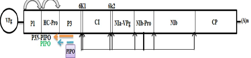
Figure 1.2 Schematic representation of TuMV genome. The viral RNA is symbolized
by the horizontal line, linked to a single VPg at the 5′ end and to a poly (A) tail at the 3′
end. The poly protein is processed by three different proteinases to 11 mature proteins.
The black arrows indicate the cleavage site. P3N-PIPO results from the translation of the
5′ sequence of TuMV P3 cistron followed by a small open reading frame protein
1.3.2 Host range and transmission
The wide host range of TuMV is one of its remarkable features. At least 318 species of
plants including both dicots and monocots, in more than 43 families, are known to be
infected by TuMV (Walsh and Jenner, 2002). TuMV is known to be particularly
damaging in brassicas (members of the mustard family or Brassicaceae) in parts of Asia,
North America and Europe. It has caused serious losses in arable crops, including oilseed
rape (Brassica napus) and vegetable crops, including swede/rutabaga (B. napus), turnip,
Chinese cabbage (B. rapa), cabbage, Brussel sprouts, cauliflower, kohlrabi and collards
(B. oleracea) (Nguyen et al., 2013). It also infects and causes damage in many
non-brassica crops and ornamentals, including radish, lettuce, endive, escarole, horseradish,
peas, courgette, rhubarb, statice (Limonium) and stock (Matthiola) (Walsh and Jenner,
2002). TuMV occurs in many parts of the world, including the temperate and tropical
regions of Africa, Asia, Oceania and North and South America (Nguyen et al., 2013).
This virus is transmitted to plants through 40-50 species of aphids in a non-persistent
manner.
1.3.3 Disease symptoms
TuMV infection results in different symptoms depending on the plant, such as mosaic,
mottling, chlorotic rings or color break on stems, foliage, flowers and fruits. The
infection can also cause severe stunting of young plants and represents an important
reduction of yield. Malformations of leaf, stem and fruit can also be observed as well as
Figure 1.3 Turnip mosaic virus symptoms on a cabbage leaf of the variety
1.4 Proposed research
Viruses are considered one of the major constraints to agricultural production in the
world. A better understanding of the molecular biology of viruses and the functions of the
viral proteins is essential for the control of virus propagation and the elaboration of
antiviral strategies. For example, viral disease may be controlled by disruption of viral
cell-to-cell movement. Previously our lab (Wei et al., 2010) showed that the potyviral
protein P3N-PIPO is a PD-located protein and directs the CI protein to PD, facilitating
the deposition of the cone-shaped structures of CI at the PD by interacting with the CI
protein. Therefore, CI and P3N-PIPO coordinate the formation of conical structures at PD
for potyviral cell-to-cell spread (Wei et al., 2010). However, it is not clear how
P3N-PIPO directs CI targeting to the PD, if P3N-P3N-PIPO is directly involved in viral cell-to-cell
movement and if P3N-PIPO is also involved in other steps of the viral cycle. This project
aims to further characterize the functional role of the P3N-PIPO protein of TuMV using a
pCambiaTunosGFP infectious clone containing the full-length cDNA of TuMV with GFP
inserted at the junction between the P1 and HC-Pro protein. Our general research
hypothesis is that like other potyviral proteins, the PIPO protein of TuMV plays multiple
roles in viral infection. Therefore, site-directed mutants in PIPO without alteration of
P3N or P3 amino acid sequences will provide evidence of PIPO role in the virus life
cycle. The objectives of this project are as follows:
1. To generate P3N-PIPO mutants
To explore the functional roles of P3N-PIPO, the recombinant TuMV infectious
N-terminal region, i.e., P3N, of P3N-PIPO is also part of the P3 protein; I only
mutated the C-terminal region, i.e., PIPO, of P3N-PIPO.
2. To evaluate cell-to-cell movement of TuMV mutants in which P3N-PIPO is mutated.
Transient expression of PIPO mutants in Nicotiana benthamiana has been used for
evaluation of cell-to-cell movement. Constructed mutant plasmids were transformed
in Agrobacterium tumefaciens strain GV310. Plants were agroinfiltrated with
appropriate agrobacterial cultures and the agroinfiltrated plants were maintained
under normal growth conditions for 14 days and then examined using confocal
microscopy.
3. To evaluate long distance movement of TuMV mutants in which P3N-PIPO is
mutated
Newly emerging leaves (new leaves which are located above the level of the
inoculated leaves and can acquire infection from inoculated leaves after infiltration)
were examined for GFP localization under UV light after 14 days post inoculation.
4. To subcellularly co-localize the P3N-PIPO mutated version with CI
To determine if PIPO mutants can interact with the CI protein, I coexpressed the CI
protein and P3N-PIPO mutants with different fluorescent tags, such as cyan
fluorescence protein (CFP) and yellow fluorescence protein (YFP). The inoculated
leaves with those plasmids were examined using confocal microscopy.
5. To conduct a replication assay
studied using confocal microscopy. Total RNA was extracted from the protoplasts,
followed by real time RT-PCR with specific TuMV CP primers.
6. To determine possible interactions of CI and P3N-PIPO
Constructs used for the expression of the punctate forms of CI in PD were prepared
based on their predicted conserved domain structure. For this purpose, I divided the
CI protein into the desired size of fragments and examined those fragments using
two systems, bimolecular fluorescence complementation assay (BIFC) in planta and
Chapter 2
2 Material and method
2.1 Plant material and growth conditions
Nicotiana benthamiana plants were cultivated in a growth room at 16 hours in the light at
24 °C and then 8 hours in the dark at 21 °C with 75% relative humidity.
Four-to-five-week-old plants were used for infiltration with Agrobacterium tumefaciens strain
(GV1301) harboring appropriate plant expression vectors. For protoplast isolation from
leaf cells, Nicotiana benthamiana plants were kept in a Percival growth chamber (Johns
Scientific Inc., Canada) with a different photoperiod of 16 hours of darkness at 22 °C and
8 hour of light at 20 °C (Wu et al., 2009).
2.2 General molecular biology techniques
General lab and molecular techniques essentially followed those in Molecular Cloning: A
Laboratory Manual (Sambrook and Russell, 2006) unless otherwise stated.
2.2.1 Bacterial strain and cultures
In this study Escherichia coli DH5α or DH10B was used for cell transformation and
Agrobacterium tumefaciens GV3101 was used for plant transformation. The bacteria or
agrobacteria were cultured at 37 °C in Luria-Bertani (LB) broth [1% tryptone, 1% NaCl,
0.5% yeast extract] or on LB agar plates (with 1.5% w/v agar). The LB media contained
overnight LB culture containing the appropriate antibiotics, was mixed with 400 μL of
50% glycerol and kept in the -80 ºC freezer.
2.2.2 Antibiotic stock preparation
An antibiotic stock concentration of 100 mg/mL was used. For this purpose the powder
was dissolved in miliiQ water (e.g. 1 g powder in 10 mL H2O), and was then sterilized
through a 0.22 μm pore size filter. In the case of rifampicin the antibiotic powder was
dissolved in methanol as a solvent. The aliquots were stored at -20 ºC. It was necessary to
cool the LB medium before adding antibiotics as the heat could inactive them. In order to
do this, the autoclaved medium was equilibrated in a water bath set at 55-60 ºC for a
minimum of 30 min, and then the desired antibiotics was added.
2.2.3 Preparation of E.coli competent cells
A colony from a freshly streaked E. coli (DH10Β) was used for inocula tion in 5 ml of
liquid LB medium and this inoculum allowed grown at in 37 °C with shaking at 200 rpm
overnight. 500 μL of overnight culture was added to 50 mL of fresh liquid LB medium
and placed at 37 °C in a shaker for 2-3 hours. OD600 was measured on an hourly basis
until it reached 0.3-0.4 optical density. The cells were harvested by centrifugation at 3000
rpm for 10 min at 4 °C. The pellet was gently resuspended in 30 mL of pre-chilled
transformation buffer (0.1 M CaCl2, 1 M MgCl2) and the cells were incubated on ice for
30 min and then centrifuged at 3000 rpm at 4 °C for 10 min. The supernatant was
(0.1 M CaCl2, 80% glycerol). The suspension was aliquoted in sterile eppendorf tubes.
The cells aliquots were kept in a -80 °C freezer for long term storage.
2.2.4 Preparation of Agrobacterium tumefaciens competent cells
A colony from a freshly streaked Agrobacterium tumefaciens GV3101 was used to
inoculate 100 mL of LB medium with appropriate antibiotics (50 μg mL-1 of gentamicin
and rifampicin). The flask was incubated at 30 °C with vigorous shaking for overnight.
The agrobacterial cells were washed three times by spinning at 5000 rpm for 5 min at 4
°C and resuspended in 50 mL ice cold 10% glycerol. After washing, the agrobacterial
pellet were resuspend in 1 mL of 10% glycerol. Aliquots of 40 μL of the Agrobacterium
competent cells were transferred to Eppendorf tubes and placed in a -80 °C freezer for
long term storage.
2.2.5 Gel electroprosis and DNA isolation
Plasmid DNA and DNA fragments were separated on 1% agarose gels which were
prepared with a 1X (TAE) buffer (2 mM EDTA, 40 mM Tris-acetate) at pH 8.0.
Ethidium bromide (0.5 μg mL-1) was added to the gel before solidification. DNA samples or PCR reactions were loaded with 1X loading dye and gels were run at 150 V. Gels were
studied using a UV trans-illuminator. For cloning, the gel containing DNA fragments
were sliced and the DNA fragments were isolated from the gel using the QIAquick Gel
2.2.6 Restriction enzyme digestion
Two μg of plasmids were digested with appropriate restriction enzymes (such as SnaBІ
and KpnІ) in 15μl reaction mixtures with specific enzyme buffers. Reactions were
incubated at 37 ºC for 1-1.5 hours then the whole mixture was separated on 1% agarose
gels for 30 min. The desired fragments were isolated using the QIAquick Gel Extraction
Kit.
2.2.7 Bacterial transformation
All bacterial transformations were performed by the heat shock method as recommended
in the Invitrogen (Life Technologies) user guide. The plasmid DNA was gently added to
50 μL of DH10B competent cells and the reaction was incubated on ice for 30 min. The
cells were heat shocked at 42 ºC for 45 seconds and transferred to ice for about 2 min.
250 μL of fresh LB medium was added to the reaction and incubated for 1 hour at 37 ºC,
then the culture spread on LB agar plates supplemented with appropriate antibiotic and
incubated at 37 ºC overnight.
2.2.8 Transformation of Agrobacterium tumefaciens by electroporation
Plasmid DNA (50-200 ng) was gently added to 40 μL Agrobacterium competent cells.
The mixture was transferred into a pre-chilled electroporation Gene Pulser® cuvettes
(Bio-Rad) with 2mm separation. The cells were pulsed with Bio-Rad MicroPulserset at
1.8 kV and recovered immediately in 1 mL of fresh LB medium and allowed to grow in
LB agar plate supplemented with appropriate antibiotics. The plates were incubated at 28
°C for 2-3 days.
2.2.9 DNA sequencing
DNA Sanger sequencing was done by DNA sequencing facility by using applied
biosystem AB3100XC at the Southern Crop Protection and Food Research Center,
Agriculture and Agri-Food Canada, London, Ontario.
2.3 TuMV PIPO protein-coding region
To functionally characterize P3N-PIPO, the vector pCambiaTunos/GFP infectious clone
which contains the full-length cDNA of TuMV (Cotton et al., 2009) was used to generate
various PIPO mutants (Fig. 2.1 B). Since the N-terminal region, i.e., P3N, of P3N-PIPO
is also part of the P3 protein, I only mutated the C-terminal region, i.e., PIPO, of
P3N-PIPO. Previously Chung et al. (Chung et al., 2008) analyzed 48 potyvirus sequences
including TuMV strain UK1 (Gene bank accession NC-002509) and predicted that
TuMV PIPO is 60 amino acid in length and is encoded by the nucleotide sequence
3079-3258. Based on this information, the codon AAA (3079-3081) encoding Lysine is the
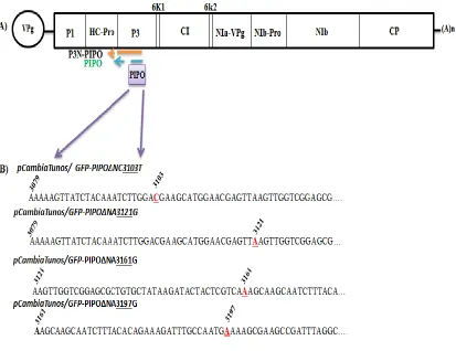
Figure 2.1 Schematic representation of the TuMV genome. The viral RNA is
symbolized by the horizontal line, linked to a single VPg at 5ʹ end and to a poly (A) tail
at the 3ʹ end. Two polyproteins are processed by three different proteinases into 11
mature proteins. Of them, P3N-PIPO, resulting from the translation of the 5ʹ sequence
of TuMV P3 cistron is followed by a small open reading frame protein embedded in the
cistron (A). Different PIPO mutants were created in cDNA, an infectious clone of TuMV
(pCambiaTunos/GFP) which is GFP fused between P1 and HC-Pro. Red letters within
2.4 Plasmid construction for PIPO mutants
All PIPO mutant constructs were obtained by overlapping PCR using Phusion™
High-Fidelity DNA Polymerase® (Fermentas). Mutations were introduced into the full-length
cDNA infectious clone of TuMV (pCambiaTunosGFP) strain UK1 (Gene bank accession
NC-002509) (Cotton et al., 2009). Mutagenic primers for specific positions are shown in
Table 2.1. All of the primers in this study were designed using Lasergene 10 software. In
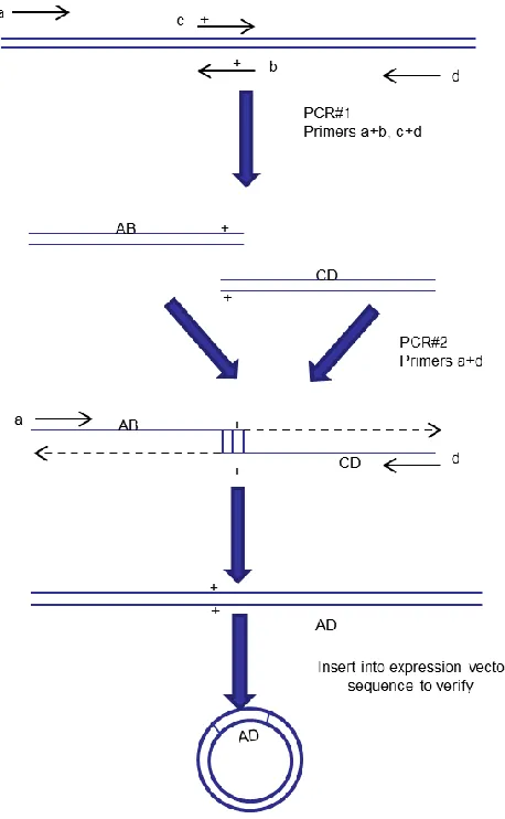
the first step, two pairs of primers were used to generate DNA fragments with
overlapping ends containing the desired mutation. These two DNA fragments were then
mixed and annealed to get hybrid duplexes. In the second step, the resulting hybrids were
then extended and amplified to yield recombined PCR products that could be directly
cloned into an appropriate plasmid vector following restricted digestion (Tomlinson,
1987) (Fig. 2.1). The amplified products were purified from agarose gels by QIAquick
Gel Extraction Kit (Qiagen). The PCR product was ligated into the pCR™-Blunt vector
(Invitrogen) and then transformed by heat shock to E.Coli DH5α strain according to the
manufacturer’s manual (Invitrogen). Single colonies were used to inoculate 3 mL of LB
medium with appropriate antibiotics and grown overnight at 37 ºC. Plasmid extraction
was performed for each culture by QIAprep Spin Miniprep Kit (Qiagen). Digestion was
performed with appropriate restriction enzymes SnaBІ and KpnІ (NEB). The desired
fragment was inserted back into the corresponding restriction sites of the parental
infectious clone (pCambiaTunosGFP). The modified plasmid was confirmed by DNA
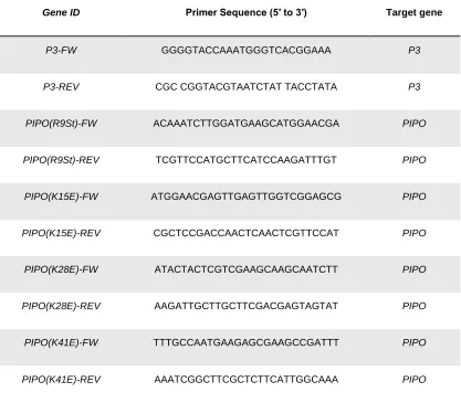
Table 2.1 Mutagenic Primers for PIPO mutants and plasmid construction.
Gene ID Primer Sequence (5′ to 3′) Target gene
P3-FW GGGGTACCAAATGGGTCACGGAAA P3
P3-REV CGC CGGTACGTAATCTAT TACCTATA P3
PIPO(R9St)-FW ACAAATCTTGGATGAAGCATGGAACGA PIPO
PIPO(R9St)-REV TCGTTCCATGCTTCATCCAAGATTTGT PIPO
PIPO(K15E)-FW ATGGAACGAGTTGAGTTGGTCGGAGCG PIPO
PIPO(K15E)-REV CGCTCCGACCAACTCAACTCGTTCCAT PIPO
PIPO(K28E)-FW ATACTACTCGTCGAAGCAAGCAATCTT PIPO
PIPO(K28E)-REV AAGATTGCTTGCTTCGACGAGTAGTAT PIPO
PIPO(K41E)-FW TTTGCCAATGAAGAGCGAAGCCGATTT PIPO
Figure 2.2 Schematic representation of overlapping PCR. Overlapping PCR was used
to create specific nucleotide mutations or generate chimeric gene products. The AB
2.5 Transient expression in Nicotiana benthamiana
Plant expression vectors were transformed by electroporation into Agrobacterium
tumefaciens strain GV3101. For agroinfiltration, agrobacterial cultures were grown
overnight in LB medium containing appropriate antibiotics. Cells were collected by
centrifugation at 4500 rpm for 3 min at room temperature (RT), and the pellet
resuspended in an infiltration buffer (500 µM MgCl 2 and MES buffer containing 200
mM acetosyringone) and 2-3 hours incubation at room temperature performed. For the
cell-to-cell movement assay the culture was diluted to an optical density of 0.01–0.001 at
600 nm (OD600). Fully expanded leaves of four-week-old Nicotiana benthamiana plants were agroinfiltrated with diluted agrobacterial cultures and the agroinfiltrated plants were
maintained under normal growth conditions in a growth room for 3 weeks.
2.6 Cell-to-cell movement assay
To determine whether P3N-PIPO is a necessary factor for cell-to-cell movement of
TuMV, transient expression of the PIPO mutant in Nicotinia benthamiana leaf cells was
used and inoculated leaves were observed under a Leica TCS SP2 inverted confocal
microscope with an Argon ion laser in various time intervals. The intervals were 3, 6, and
9 days post inoculation (dpi). Plants tissue inoculated with various PIPO mutants were
imaged at room temperature. Distal leaves were examined for systemic infection by GFP
localization under UV light and followed by RT-PCR analysis for viral detection after 14
2.7 Confocal microscopy
For confocal microscopy, YFP was excited at 514 nm and captured at 525-650 nm. CFP
was excited at 458 nm and captured at 465–525 nm. GFP was excited at 488 nm and the
emitted light was captured at 500 to 555 nm. Images were captured digitally and handled
using the Leica LCS software.
2.8 Protoplast isolation and transfection assay
Mesophyll protoplasts were prepared from using leaves from four-week-old Nicotiana
benthamiana by the Tape-Arabidopsis Sandwich method (Wu et al., 2009). The leaves
were incubated in 1–1.5% cellulase Onozuka R-10 and 0.2–0.4% macerozyme R-10 in
0.4 M D-sorbitol, 20 mM KCl and 20 mM MES, pH 5.7 for 2 hours at room temperature.
Protoplasts were washed in W5 buffer (154 mM NaCl, 125 mM CaCl2, 5 mM KCl and 2
mM MES, pH 5.7) and resuspended in MMg (0.4 M Mannitol, 15 mM MgCl2, 4 mM
MES, PH at 5.7) buffer at a concentration of 105 cells/mL. Approximately 5 × 10⁴ protoplasts in 0.2 mL of MMg solution were mixed with approximately 30 (20-40) μg of
plasmid DNA at room temperature. An equal volume of a freshly-prepared solution of
40% (w/v) PEG with 0.1 M CaCl2 and 0.2 M mannitol was added, and the mixture was
incubated at room temperature for 5 min. After incubation, 3 mL of W5 buffer was added
Protoplasts were imaged at room temperature after 48 hours post-transfection, using the
confocal microscope described above (Vijayapalani et al., 2012).
2.9 RNA extraction and Quantitative reverse transcription –PCR
To verify the level of replication of the introduced mutations in vivo, total RNA was
extracted from virus-infected TuMV PIPO mutant’s protoplasts using the RNeasy Plant
Minikit (Qiagen) and treated with RNase-free DNaseI. One microgram of RNA
pretreated with DNaseI was used as a template for first strand cDNA synthesis. RT
reactions were performed with the SuperScriptIII First-Strand Synthesis System for
RT-PCR kit (Invitrogen) according to the manufacturer’s instructions. QRT-RT-PCR was
performed using SsoFast EvaGreen Supermix (Bio-Rad) in the CFX96 real-time PCR
system (Bio-Rad) following the manufacturer’s instructions. Relative amounts of all
mRNAs were calculated from threshold cycle values. The Nicotiana benthamiana actin
reference gene was used for normalization. Primers CP-F and CP-R were used for TuMV
detection (Wei et al., 2013). Data were statistically evaluated by the unpaired Student’s
t-test.
2.10 P3N-PIPO artificial frame shift for subcellular localization
Previous high-resolution ultra-structural studies of TuMV revealed that CI protein
colocalizes with P3N-PIPO protein and formed conical structures which anchored to and
extended through PD (Wei et al., 2010). To investigate whether P3N-PIPO mutants retain
their ability for targeting TuMV CI in PD, agrobacterium-mediated transient
fragment, which encodes a PIPO-YFP fusion in-frame with the N-terminus of P3 to
generate P3N-PIPO-YFP, was constructed. In order to do that, two nucleotides in the
G2A6 motif in the TuMV infectious clone was deleted by mutagenic overlap PCR to
create an artificial frame shift (Fig. 2.3).
Figure 2.3 Schematic representation of TuMV P3N-PIPO reading frame. The TuMV
P3N-PIPO fragment was amplified by artificial frame shift with the introduction of two
nucleotides deletion in G2A6 motif of PIPO protein by overlap PCR. The modified
fragment was introduced to appropriate destination vectors, which was used for
2.11 Gateway cloning
Gateway technology (Invitrogen) was used to generate all expression vectors for
subcellular localization, bimolecular complementation assay (BIFC) and yeast two hybrid
screening (Y2H). Gene sequences were amplified by PCR using Phusion® DNA
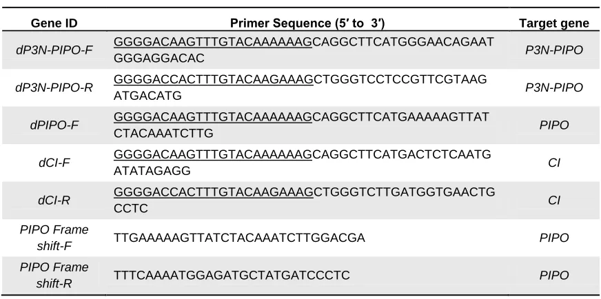
polymerase (NEB) and primers containing the recombinant sites (Table 2.1). The
resulting DNA fragments were separated on 1% agarose gels and then purified using the
QIAquick Gel Extraction Kit. The purified fragment was transferred by recombination
into the entry vector pDONR221 (Invitrogen) using BP Clonase® enzyme mixtures
(Invitrogen) that were set up and incubated overnight at 25 ºC following the supplier’s
recommendations. Then pDONOR vectors were verified by sequencing (Wei and Wang,
2008). For BIFC and Y2H, the afore-mentioned pDONR221 constructs were ligated with
a (35S-YN) gateway or (35S-YC) gateway vector and also a modified (pGADT7)
gateway or (pGBKT7) gateway vector respectively (Lu et al., 2010; Xiong and Wang,
2013). For subcellular localization in Nicotiana benthamiana plant cells P3N-PIPO
mutants and CI modified Gateway fragment ligated with (pEarly101) or (pEarly102)
vector to produce P3N-PIPO mutants which were tagged with yellow fluorescence
Table 2.2 List of primers used for gateway cloning. Gateway sequences were
underlined.
Gene ID Primer Sequence (5′ to 3′) Target gene
dP3N-PIPO-F GGGGACAAGTTTGTACAAAAAAGCAGGCTTCATGGGAACAGAAT
GGGAGGACAC P3N-PIPO
dP3N-PIPO-R GGGGACCACTTTGTACAAGAAAGCTGGGTCCTCCGTTCGTAAG
ATGACATG P3N-PIPO
dPIPO-F GGGGACAAGTTTGTACAAAAAAGCAGGCTTCATGAAAAAGTTAT
CTACAAATCTTG PIPO
dCI-F GGGGACAAGTTTGTACAAAAAAGCAGGCTTCATGACTCTCAATG
ATATAGAGG CI
dCI-R GGGGACCACTTTGTACAAGAAAGCTGGGTCTTGATGGTGAACTG
CCTC CI
PIPO Frame
shift-F TTGAAAAAGTTATCTACAAATCTTGGACGA PIPO
PIPO Frame
2.12 Yeast related methods
2.12.1 Yeast strain and culture
The yeast strain used in this work was the AH109 strain, a derivative of strain PJ69-2A
that includes the ADE2 and HIS3 markers (James et al., 1996). Yeast cells were grown at
30 °C in either YPD (1% bacto-yeast extract, 2% bacto-peptone and 2% glucose) or in a
synthetic dextrose (SD) medium (0.67% bacto-yeast nitrogen base without amino acids,
2% glucose) supplemented with necessary nutrients. To make SD or YPD plates, 2% agar
was added to the liquid medium. The SD or YPD media were autoclaved, and stored in
4°C. Yeast cells were stored on SD plates sealed with Parafilm® at 4 °C for up to several
months. For long-term storage, yeast cells were grown in an appropriate liquid medium at
30 °C overnight, then 0.6 mL of the culture was added to 0.4 mL of 50% sterile glycerol
and stored at -80°C.
2.12.2 Yeast two hybrid screening
Yeast strain AH109 was grown at 28 °C in minimal synthetic defined (SD) base liquid
medium [0.17% yeast nitrogen base without amino acids, 2% dextrose] mixed with
appropriate drop out (DO) supplement powder. For SD solid medium, SD was
supplemented with 1.5% w/v agar. Y2H assays were performed following the Clontech®
yeast protocol handbook. In brief, yeast cells were made competent with the LiAC
method (Xiong and Wang, 2013) and transformed cells were plated onto a selective
medium lacking tryptophan and leucine (SD-Trp-Leu) and a selective medium lacking
2.13Statistical analysis
All statistical analyses were performed using Microsoft Excel spreadsheet software.
Significant differences between the amount of the genomic RNA of mutants relative to
that of the wild type virus in the qRT-PCR assay were analyzed using unpaired Student’s
Chapter 3
3 Results
3.1 Knock-out of the P3N-PIPO expression
To test if P3N-PIPO is essential for TuMV cell-to-cell movement, a P3N-PIPO knock-out
mutant was created by the introduction of a single stop codon into the PIPO frame
without affecting the amino acid sequence of other viral proteins including P3 (Fig.
2.1B). The wild type infectious clone pCambiaTunos/GFP was used as a positive control
and the viral replication defective mutant pCambiaTunos/GFP-NIb-GDD (Fellers et al.,
1998) was created as a negative control. In the replication-defective mutant, the core
motif GDD of the viral RNA-dependent RNA polymerase was mutated to abolish viral
replication. The wild type virus, PIPO knock-out and the GDD mutants were
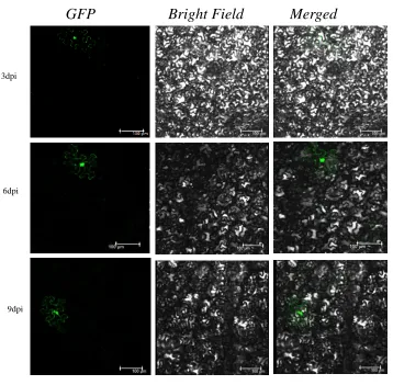
agroinfiltrated into Nicotinia benthamiana leaf cells. It was found that the PIPO
knock-out mutant (GFP-PIPO (R9Stop)) was contained in a single cell only and lost the ability
to move to the neighboring cells, even after 9 days post agroinfiltration (dpa) (Fig. 3.1B).
In contrast, the leaves agroinfiltrated by the wild type clone (pCambiaTunos/GFP)
showed a group of cells of virus infection at 3 dpa that gradually expanded to a large area
of virus infection (Fig. 3.1A). As expected, the replication-defective mutant was only
found in isolated single cells of Nicotinia benthamiana leaves which were agroinfiltrated
by the GDD mutant (Fig. 3.1C). The GFP fluorescence derived from the
replication-defective GDD mutant came from translation of the non-replicative transcript of the
TuMV cDNA (tagged by GFP) under the control of 35S promoter. To monitor if these
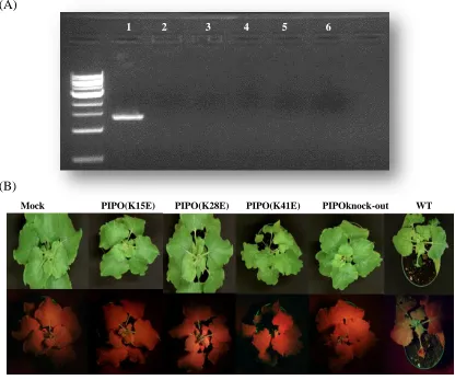
viruses can establish systemic infection, newly emerging leaves (distal - not infiltrated)
agroinfiltration. Only the wild type clone produced strong green fluorescence signal. This
did not occur in other mutants (Fig. 3.2B). Therefore, both PIPO knock-out and GDD
mutants cannot move systemically. Consistently, no viral symptoms were evident in
plants inoculated with these two mutants 14 dpa (Fig. 3.2B). RT-PCR was conducted to
detect the virus in the newly emerging leaves of these plants. No virus was detectable in
GFP Bright Field Merged
3dpi
6dpi
9dpi
3dpi
6dpi
9dpi
GFP Bright Field Merged
3dpi
6dpi
9dpi
C) GFP/NIb-GDD-TuMV
Figure 3.1 Cell-to-cell movement of TuMV requires P3N-PIPO. (A)
pCambiaTunos/GFP-TuMV, (B) GFP-PIPO(R9Stop), and (C) GFP/NIb-GDD
(negative control) were introduced into Nicotiana benthamiana leaves cells via
agroinfiltration, respectively. Infiltrated leaves were examined by confocal
(A)
(B)
Figure 3.2 Detection of TuMV accumulation in upper newly emerging leaves of
Nicotinana benthamiana plants inoculated with PIPO mutants. A) Specific TuMV
primers were used to amplify a P3N-PIPO cDNA fragment derived from viral RNA
isolated from upper newly emerging leaves 14 days post agroinoculation.
pCambiaTunos/GFP-TuMV (Lane 1) and TuMV PIPO substitution mutants respectively
GFP-PIPO(K15E), GFP-PIPO(K28E), GFP-PIPO(k41E), PIPOknock-out (2, 3, 4 and 5)
and mock (Lane 6). B) Nicotinia benthamiana leaves were infected with
pCambiaTunos/GFP-TuMV and TuMV PIPO mutants 14 days post inoculation under
1 2 3 4 5 6
Mock PIPO(K15E) PIPO(K28E) PIPO(K41E) PIPOknock-out WT
3.2 Synonymous mutations in P3N-PIPO
The substitution mutation in PIPO also was performed by site-directed mutagenesis.
These mutations were synonymous with respect to the polyprotein frame. They resulted
in replacement of a negatively charged amino acid with positively charged residue. A
total of three mutants were derived through these substitutions: PIPO(K15E),
PIPO(K28E), and PIPO(K41E) (Fig. 3.3). Transient expression of PIPO mutants in
Nicotinia benthamiana leaf cells was subsequently performed and agroinfiltrated leaves
cells were observed by confocal microscopy in different time intervals (3, 6, and 9 days
post agroinfiltration) (Fig. 3.3). Moreover, RT-PCR results of newly emerging leaves
(not inoculated ones) and GFP localization assay under UV light showed the same GFP
fluorescence pattern as the knock-out mutant and no indication of viral symptoms was
Figure 3.3 TuMV P3N-PIPO substitution mutants impaired intercellular
movement. (A) pCambiaTunos/TuMV, (B) PIPO(K15E), (C)
GFP-PIPO(K28E), (D) GFP-PIPO(K41E), and (E) GFP-NIb-GDD-TuMV(negative control)
A) TuMV-GFP (Positive Ctrl)
B) GFP-PIPO(K15E)
C) GFP-PIPO(K28E)
D) GFP-PIPO(K41E)
A) B)
GFP Bright Field Merged
3.3 Protoplast transfection and quantitative RT-PCR assay
Since three PIPO substitutions mutants [PIPO(K15E), PIPO(K28E), PIPO(K41E)] and
PIPO knock-out mutant lost ability to move between cells, it is necessary to determine if
these mutations affect viral replication (Fig. 3.4). The wild type virus and all the five
mutants including three substitution mutants, PIPO knock-out mutant and the
replication-defective GDD mutant were subjected to the replication assay in protoplasts. The strong
green fluorescence signal, similar to the wild type, was observed of transfected protoplast
by PIPO mutants. Weak GFP signals were also evident for the GDD mutant due to
transcription of the viral cDNA directed by the constitutive 35S promoter. Quantitative
RT-PCR was applied to compare the TuMV transcript levels of different PIPO mutants in
protoplast. For this purpose, total RNA was extracted from transfected protoplast 22
hours post transfection and followed by real time PCR with specific TuMV primers. The
result showed that knock-out or substitution mutations of P3N-PIPO did not significantly
Figure 3.4 Replicaiton assay of TuMV P3NPIPO mutants in Nicotiana benthamiana
mesophyll protoplasts. (A) pCambiaTunos/TuMV, (B) PIPO knock-out, (C)
GFP-PIPO(K15E), (D) GFP-PIPO(K28E), (E) GFP-PIPO(K41E) were transfetcted into the
A)TuMV-GFP
B)GFP-PIPOknock-out
C)GFP-PIPO(K15E)
D)GFP-PIPO(K28E)
GFP Bright Field Merged
Figure 3.5 RT-PCR quantification of PIPO mutants RNA in protoplast. RNA was
extracted from transfected protoplast after 22 hours. The TuMV RNA levels were
normalized to the amount of actin transcript in each sample and then further to that of
wild type virus (pCambiaTunos/GFP-TuMV (WT). The values SEs were calculated from
three biological replicates. Statistical significance was analyzed by the unpaired Students
t-test and (P<0.05)*.
0 0.2 0.4 0.6 0.8 1 1.2
F)Mock
*
WT PIPOknock-out PIPO(K15E) PIPO(K28E) PIPO(K41E) GDD
3.4 P3N-PIPO mutants lost their ability to target the TuMV CI protein
To examine why P3N-PIPO mutations affect cell-to-cell movement, I determined the
subcellular localization of TuMV P3N-PIPO and further tested if TuMV P3N-PIPO
mutants still have ability to direct the targeting of CI to PD. Consistent with the
distribution of TEV P3N-PIPO, TuMV P3N-PIPO-YFP was also located at PD in
Nicotiana benthamiana epidermal cells (Fig. 3.6 B). When expressed alone, the TuMV
CI protein formed aggregates in the cytoplasm (Fig. 3.6 A). However, when TuMV CI
and P3N-PIPO were coexpressed, both of them targeted the PD (Fig. 3.7A). This is in
agreement with the previous finding that TEV P3N-PIPO modulates the PD-localization
of TEV CI (Wei et al. 2010). However, coexpression of the TuMV PIPO knock-out
mutant (which is P3N, N-terminal portion of P3) and TuMV CI failed to direct CI to PD
and both proteins were present in the cytoplasm as aggregates (Fig. 3.7 B). These data
suggest that PIPO is essential for PD-localization of P3N-PIPO and for directing CI to
PD. Coexpression of TuMV CI with P3N-PIPO substitution mutants revealed that these
P3N-PIPO mutants lost the ability to target the CI protein to PD (Fig. 3.8 A, B and C). To
examine if these P3N-PIPO mutants remain to be PD-located proteins like P3N-PIPO,
they were coexpressed with the PD marker PDLP1 (PD-located protein 1). The results
showed that the P3N-PIPO mutants still maintained their ability for PD localization (Fig.
CFP Bright field Merged
YFP Bright field Merged
A) CI-CFP
B) P3N-PIPO-YFP
Figure 3.6 Subcellular localization of TuMV CI and P3N-PIPO proteins in
Nicotiana benthamiana leaf cells. Transient expression of CI-CFP (A) and
P3N-PIPO-YFP (B) was examined by confocal microscopy. Images were taken 48 hrs post
A) P3N-PIPO-YFP+CI-CFP
B) PIPOknock-out-YFP+ CI-CFP
CFP YFP Merged
Figure 3.7 Subcellular localization of TuMV CI in Nicotiana benthamiana leaf
cells. Transient coexpression of CI-CFP with P3N-PIPO-YFP (wild type) (A) or with
the PIPO knock-out mutant (B) was examined by confocal microscopy. Images were
taken 48 hrs post agroinfiltration. The arrows indicated the aggregates of both proteins
CFP YFP Merged A)PIPO(K15E)-YFP+CI- CFP
B)PIPO(K28E)-YFP+CI-CFP
C)PIPO(K41E)-YFP+CI-CFP
Figure 3.8 Subcellular localization substitutions of PIPO mutants in
Nicotiana benthamiana leaf cells. Transient coexpression of CI-CFP with the
PIPO(k15E)-YFP (A), PIPO(K28E)-YFP (B), PIPO(K41E)-YFP (C) was
observed under a confocal microscope. Images were taken 48 hrs post
Figure 3.9 P3N-PIPO substitution mutants keep their ability for PD localization in
Nicotiana benthamiana leaf cells. Transient coexpression of PDLP-CFP (PD marker)
with P3NPIPO-YFP wild type (A), PIPO(K15E)-YFP (B), PIPO(K28E)-YFP (C), and
PIPO(K41E)-YFP (D) was monitored by confocal microscopy. Images were taken 48 hrs A) PIPO-YFP+PDLP1-CFP
CFP YFP Merged
B) PIPO(K15E)-YFP+ PDLP1-CFP
C) PIPO(K28E)-YFP+ PDLP1-CFP