ISSN (Print) : 2320 – 3765 ISSN (Online): 2278 – 8875
I
nternational
J
ournal of
A
dvanced
R
esearch in
E
lectrical,
E
lectronics and
I
nstrumentation
E
ngineering
(An ISO 3297: 2007 Certified Organization)
Vol. 5, Issue 6, June 2016
Assessment of Human Sperm Head
Morphology for Assisted Reproduction
Techniques using Open Source Software
Raghavendra. Maggavi1, Sanjay. Pujari2, Amar.Herekar3
Research Scholar [ECE], Visvesvaraya Technological University, Belagavi, India1
Professor, Dept. of ECE, Angadi Institute of Technology and Management, Belagavi, India2
Assistant Professor, Dept. of ECE, Maratha Mandal Engineering College, Belagavi, India3
ABSTRACT: Infertility affects about 15% of the population and going by the trends seen by infertility clinics worldwide, it is on the rise from its present about 15% of population to about 20% or even more. Head size and head shape defects are important criteria in the determination of a spermatozoon’s morphological normality or abnormality and able to find out the sperm cell is alive or dead. In the present study, we have developed a freely available sperm head morphology analyzer plug-in for open source software. Described the systems functionality and confirmed its validity with respect to the commercial softwares.
KEYWORDS:Microscopic Image, Spermatozoon, WHO, IMAGEJ, Medea LAB CASA.
I.INTRODUCTION
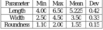
When a couple had undergone the painful experience of miscarriage, and has been investigated thoroughly, have been placed under “Unexplained” category, the options for counselling, planning of treatment and prognosis all becomes troublesome for the treating consultants. Hence, increased research in to the factors governing pregnancy loss becomes validated. Over the years it has been noticed that we had paid relatively less attention to male partner or Sperm proper in contrast to the female factors. Now that we have known almost every possible female factor contributing to the pregnancy loss it’s time we shift our serious attention on to the sperm. The set of four morphometric parameters was obtained. The definitions of these parameters are illustrated in the fig.1. Since the heads of human spermatozoa may not be bilaterally symmetric, head length (L) and width (W) must be carefully and objectively defined [1]. We measured length as the distance between the midpoint of the insertion of the flageller midpiece with the head (fig.1, point a) and the point farthest (b) from it. Head width was then defined as the length of the longest line perpendicular to the line ab and intersecting the sides of the sperm head (fig.1). Sperm head circumference (C) was directly obtained with the digitizer by tracing the border of the head, and in so doing projected area (A) was computed automatically. In characterizing the shape of the head the aspect ratio (length / width) also was determined. The definition of a morphologically normal head spermatozoon as proposed [2] is as given in the table 1.
Table. 1 Normal Head spermotoza (WHO) Parameter Min Max Mean Dev
Fig1.Structure of Human Sperm Head
Though this criteria has been widely adopted by World Health Organization [3] for the Examination and Processing of Human Semen and recommended for human semen morphology assessment, this criteria is still of great scientific controversy, due to lack of accurate clinical validation. The sperm morphological analysis is through manual observe on at least 200 spermatozoa in a microscope. However, with the same criteria, we usually evaluators. In order to get more accurate and consisted morphological evaluation results, better methodology is needed, which is more objective, precise.
II.MATERIALS AND METHODS
Block diagram of the proposed system is shown in fig.2.1. Major steps are explained below. Image Collection:
Papanicolaou stained [4, 13] sperm sample where imaged on to OLYMPUS BX41 [5] microscope attached with digital microscope camera ProgRes® CT3 [6] under 40x magnifications in bright field mode.
Image Analysis:
Sperm cells were displayed on the monitor at equivalent brightness and all the cells which did not present any overlap with debris or other cells were considered for analysis. From each sample heads were captured and analysed using the ImageJ open source software [7] using custom macros. After treatment of the images some of the cells had to be discarded because of defective binarization as observed by incorrect correspondence between the original image and its mask. Each sperm head was measured for four primary parameters [head area (A), head length (L), head width (W), head roundness (R).
Procedure described below.
1. If the image is in RGB form, it is transformed to a gray scale image.
2. Then filtered using un sharp mask filter to increase the intensity of the sperm cells
3. Thresholding is done on the image such that shape of the all sperm cells will be appeared.
4. Heads of the sperm cells will be extracted using head extraction algorithm which is the part of the custom macro.
ISSN (Print) : 2320 – 3765 ISSN (Online): 2278 – 8875
I
nternational
J
ournal of
A
dvanced
R
esearch in
E
lectrical,
E
lectronics and
I
nstrumentation
E
ngineering
(An ISO 3297: 2007 Certified Organization)
Vol. 5, Issue 6, June 2016
Fig. 2 Proposed System
III.RESULTS AND ANALYSIS
The figures [3-5] show analysis on sperm sample which is taken from system microscope first RGB image is converted to 8 bit gray scale. Then head extraction filter is applied on the gray image after that thresholding is made on the image to differentiate sperm cells with respect to background. Next particle analysis is achieved to remove the noise and produce a mask on all sperm cells available in the image. Finally using ROI manager labeling and available in the image. Finally using ROI manager labeling and Measurement is carried out on each sperm cell. Using ImageJ.
Fig. 4 Output of head extraction filter
ISSN (Print) : 2320 – 3765 ISSN (Online): 2278 – 8875
I
nternational
J
ournal of
A
dvanced
R
esearch in
E
lectrical,
E
lectronics and
I
nstrumentation
E
ngineering
(An ISO 3297: 2007 Certified Organization)
Vol. 5, Issue 6, June 2016
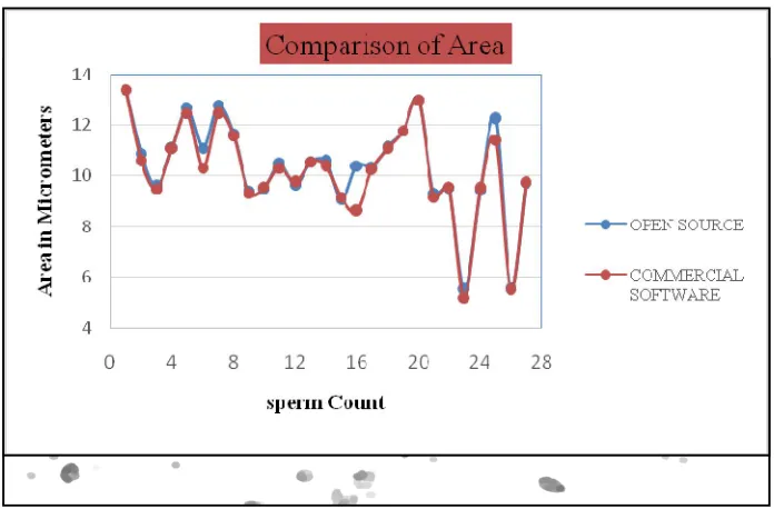
Fig.6 Comparission of sperm head area obtained from open source and commercial software
Fig.8 Comparission of sperm head roundness obtained from open source and commercial software
Fig.9 Comparission of sperm head width obtained from open source and commercial software
The above figures [6- 9] shows measurement on four morphometric parameters for the 27 sperm cells from the sample which are suitable for the analysis. And results using open source software checked for accuracy. Difference in Mean values of length (+ 0.33µm), width (- 0.75µm), area (+ 0.44µm) and roundness (+ 0.06µm) obtained when compared with commercial software MedeaLAB CASA [8].
IV.CONCLUSIONS AND FUTURE WORK
In the present study, we have developed a freely available sperm head morphology analyzer plug-in for open source software. Described the systems functionality and confirmed its validity with respect to the commercial softwares such as Sperm-Class Analyzer [9], Sperm Morphometry Module of ISAS [10, 13], and the Metrix Oval Head Morphology software component of the Hamilton-Thorne CEROS system [11]. Out of nine morphological indices four are automatically measured in the present study (Length, Width, Area and Roundness) [12]. Remaining five morphological indices to be measured automatically in the future work
ISSN (Print) : 2320 – 3765 ISSN (Online): 2278 – 8875
I
nternational
J
ournal of
A
dvanced
R
esearch in
E
lectrical,
E
lectronics and
I
nstrumentation
E
ngineering
(An ISO 3297: 2007 Certified Organization)
Vol. 5, Issue 6, June 2016
[1] Raghavendra. Maggavi, Sanjay. Pujari, Amar. Herekar. “Analysis of human sperm head morphology using open- source software”. International Journal of Engineering Science and Computing.Vol.6 ; pp.5597-5602, 2016 [2] Katz DF, Overstreet JW, Samuels SJ, Niswander PW, Bloom TD, et al. “Morphometric analysis of spermatozoa
in the assessment of human male fertility”. Asian Journal of Andrology;Vol.7:pp.203-10,1986
[3] Menkveld, Cas AG Holleboom and Johann PT Rhemrev, “Measurement and significance of sperm morphology,” Asian Journal of Andrology.Vol.13, pp.59-68, 2011
[4] World Health Organization, WHO laboratory manual for the examination and processing of human semen.5th ed, 2010.
[5] Maree L, du Plessis SS, Menkveld R, van der Horst G. “Morphometric dimensions of the human perm head depend on the staining method used”. Asian Journal of Human Reproduction; Vol.25: pp.1369-82, 2010
[6] http://www.olympusamerica.com [7] http://www.progres-camera.de [8] http://www.imagej.nih.gov [9] http://www.medealab.de
[10] Maroto-Morales A, Ramón M, Garcia-Alvarez O, Soler AJ, Esteso MC, Martinez-Pastor F, Pérez-Guzmán MD, Garde JJ. “Characterization human sperm head morphometry using the Sperm-Class Analyzer”. Asian Journal of Theriogenology;Vol.73:pp.437-48, 2010
[11] Tuset VM, Trippel EA, de Monserrat J. “Sperm morphology and its influence on swimming speed in Atlantic cod”. Journal Application of Ichthyol;Vol.24:pp.398 -405, 2008
[12] Rijsselaere T, Van Soom A, Hoflack G, Maes D, de Kruif A. “Automated sperm morphometry and morphology analysis of canine semen by the Hamilton-Thorne analyzer”. Asian Journal of Theriogenology;Vol.62:pp.1292-306, 2004
[13] Ferreira T, Rasband W. “The ImageJ User GuideVersion1.44”.Available at:http:llimagej.nih.govlijldocsluser-guide.pdf. Accessed February 2011.
[14] Giuseppe Bellastella, Trevor G. Cooper, Marina Battaglia, Anda Ströse, Inma Torres, Barbara Hellenkemper, Carles Soler, Antonio A. Sinisi “Dimensions of human ejaculated spermatozoa in Papanicolaou stained seminal and swim-up smears obtained from the Integrated Semen Analysis System (ISAS®)”Asian Journal of Andrology Vol.12: pp.871-879, 2010
[15] Menkveld R. “Clinical significance of the low normal sperm morphology value as proposed in the fifth edition of the WHO Laboratory Manual for the Examination and Processing of Human Semen”. Asian Journal of Andrology; Vol.12:pp. 47-58, 2010
[16] Auger J. “Assessing human sperm morphology: top models, underdogs or biometrics?.”Asian Journal of Andrology; Vol.12: pp.36-46, 2010
[17] Menkveld R, Stander FSH, Kotze T JvW , Kruger TF, van Zyl JA. “The evaluation of morphological characteristics of human spermatozoa according to stricter criteria”. Asian Journal of Human Reproduction;Vol.5: pp.586-92, 1990
[18] Meschede D, Keck C, Zander M, Cooper TG, Yeung CH, et al. “Influence of three different preparation techniques on the results of human sperm morphology analysis”. International journal of Andrology; Vol.16:pp.362-9. 1993
[19] Garrett C, Baker HW. “A new fully automated system for the morphometric analysis of human sperm heads”. International Journal of Fertility Sterility; Vol.63:pp.1306-17,1995
[20] Davis, R.O. “Automated sperm morphometry and morphology”. In Course XI. Contemporary Methods of Sperm, Oocyte and Embryo Morphology Assessment. 26th Annual Postgraduate Course of the American Fertility Society, Montreal, Canada. pp.61-67, 1993
[21] Coetzee, K. and Kruger, T.F. “Automated sperm morphology analysis”. International journal of Assisted Reproduction Review-7, pp.109-113, 1997.
[22] Coetzee, K., Kruger, T.F. and Lombard, C.J. “Predictive value of normal sperm morphology: a structured literature review”. Asian Journal of Human Reproduction Update, Vol.4,pp.73-82, 1998
[23] Garrett, C. and Baker, H.W.G.: “A new fully automated system for the morphometric analysis of human sperm heads”. International Journal of Fertility Sterility. Vol.63,pp.1306-1317, 1995
[24] Davis, R.P., Bain, D.E., Siemers, R.J. et al. “Accuracy and precision of the Cell Form Human automated sperm morphometry instrument”. International Journal of Fertility Sterility.Vol.58,pp.763-769, 1992
outcome”. Asian Journal of Human Reproduction.pp.1136-1140, 1992a
[27] Davis, R.O. and Gravance, C.G. “Standardization of specimen preparation, staining, and sampling methods improves automated sperm-head morphometry analysis”. International Journal of Fertility Sterility.Vol.59,pp.412-417, 1993
[28] Kruger, T.F. “Computer-assisted sperm analysis systems: morphometric aspects”. Asian Journal of Human Reproduction. Vol.10,pp.46-52,1995
[29] Menkveld, R. and Kruger, T.F. “Advantages of strict (Tygerberg) criteria for evaluation of sperm morphology”. International Journal of Andrology., Suppl. Vol.2, pp.36-42, 1995
[30] Kruger, T.F., Acosta, A.A., Simmons, K.F. et al. “New method of evaluating sperm morphology with predictive value for human in vitro fertilization”. Asian Journal of Urology, Vol.3, pp.248-251, 1987
[31] Kruger, T.F., Du Toit, T.C., Franken, D.R. et al. “A new computerized method of reading sperm morphology (strict criteria) is as efficient as technician reading”. International Journal of Fertility Sterility. Vol.59, pp.202-209. 1993
[32] Kruger, T.F., Du Toit, T.C., Franken, D.R. et al. “Sperm morphology: assessing the agreement between the manual method (strict criteria) and the sperm morphology analyzer IVOS”. International Journal of Fertility Sterility.Vol.63,pp.134-142, 1995
[33] Lacquet, F.A., Kruger, T.F., Du Toit, T.C. et al. “Slide preparation and staining procedures for reliable results using computerized morphology (IVOS). International Journal of Archive Andrology, Vol. 36, pp.133-153. 1996 [34] Liu, D.Y. and Baker, H.W.G. “Relationship between human sperm acrosin, acrosomes, morphology and
fertilization in vitro”. Asian Journal of Human Reproduction. Vol.5, pp.298-303.1990
[35] Kruger, T.F., Menkveld, R., Stander, F.S.H. et al. “Sperm morphologic features as a prognostic factor in in-vitro fertilization”. International Journal of Fertility Sterility, Vol. pp.46,1118-1123.1986
[36] Teifoory N,Moradi M H and Nafisi V R “A new method for sperm segmentation in microscopic image” Proc. 11th Iranian Biomedical Engineering Conference Amirkabir University of Technology. 2002
[37] Nafisi, Moradi and Naser-Esfahani “A template matching algorithm for sperm tracking and classification.” Physiological Measurement. 2005
[38] Sedighi S, Moradi M H and Nafisi “Compare several methods for sperm segmentation in microscopic image” Master project, Islamic Republic of Azad University of Technology. 2004
[39] Wijchman, J., Wolf, B.D., Graafe, R. and Arts, Variation in semen parameters derived from computer- aided semen analysis, within donors and between donors”. International Journal of Journal of Andrology, Vol.22, pp.773-780. 2001
[40] Verstegen, J., Iguer-Ouada, M. and Onclin, K. “Computer assisted semen analyzers in Andrology research and veterinary practice”. International Journal of Theriogenology, Vol.57, pp.149-179. 2001
[41] Linneberg, C., Salamon, P., Svarer, C. and Hansen, L. “Towards semen quality assessment using neural networks”. Proceedings of IEEE Neural Networks for Signal Processing IV, Ermioni, 6-8 September 1994, pp.509- 517. 1994
[42] Ostermeier, G., Sargeant, G., Yandell, T. and Parrish, J. “Measurement of bovine sperm nuclear shape using Fourier harmonic amplitudes”. International Journal of Andrology, Vol.22, pp.584-594. 2001
[43] Otsu, N. “A threshold selection method from gray- level histograms”. IEEE Transactions on Systems, Man and Cybernetics, Vol. 9, pp.62-66. 1979
[44] Biehl, W.M., Pasma, P., Pijl, M., Sánchez, L. and Petkov, N. “Classification of boar sperm head images using learning vector quantization”. Proceedings of the European Symposium on Artificial Neural Networks, pp.545-550. 2006
[45] Alegre, E., Biehl, M., Petkov, N. and Sánchez, L. “Automatic classification of the acrosome status of boar spermatozoa using digital image processing and LVQ”. Computers in Biology and Medicine, Vol.38, pp.461-468. 2008
[46] Nowshiravan Rahatabad, F., Moradi, M.H. and Nafisi, V.R. “A multi steps algorithm for sperm segmentation in microscopic image”. Proceedings of the World Academy of Science, Engineering and Technology, Vol.12, pp.43-45. 2005
ISSN (Print) : 2320 – 3765 ISSN (Online): 2278 – 8875
I
nternational
J
ournal of
A
dvanced
R
esearch in
E
lectrical,
E
lectronics and
I
nstrumentation
E
ngineering
(An ISO 3297: 2007 Certified Organization)
Vol. 5, Issue 6, June 2016
[48] Park, K., Yi, W. and Paick, J. “Segmentation of sperms using the strategic Hough transform”. Annals of Biomedical Engineering, Vol.25, pp.294-302. 1997
[49] Carrillo, H., Villarreal, J., Sotaquira, M., Goelkel, M. and Gutierrez, R. “A computer aided tool for the assessment of human sperm morphology”. Proceedings of the 7th IEEE International Conference on Bioinformatics and Bioengineering, pp.1152-1157.2007
[50] Carrillo, H., Villarreal, J., Sotaquira, M., Goelkel, M. and Gutierrez, R. “Spermatozoon segmentation towards an objective analysis of human sperm morphology”. Proceedings of the 5th International Symposium on image and Signal Processing and Analysis, pp.522-527. 2005
[51] Abbiramy V.S. and Shanthi, V. “Spermatozoa segmentation and morphological parameter analysis based detection of teratozoospermia”. International Journal of Computer Applications, Vol.3, pp.19-23. 2010
[52] Wang, Z., Bovik, A.C., Sheikh, H.R. and Simoncelli, E.P. “Image quality assessment: From error measurement to structural similarity”. IEEE Transaction on Image Processing, Vol.13, pp.1-14. 2004