i
Osteogenic Effects of Pigment Epithelium
Derived Factor on Mesenchymal Stem Cells
Mina Elahy
College of Health and Biomedicine Victoria University
Submitted in fulfillment of the requirements of the degree of doctor of philosophy
ii
Abstract
The field of bone tissue engineering has expanded in the recent decade to meet the
increasing need to replace bone tissue in skeletal disease, congenital malformation,
trauma, and tumours. Stem cell encapsulation has become a promising method in the
future of this field. Alginate is a natural polymer that has been used widely for stem cell
transplantation due to its biocompatibility. Pigment epithelium-derived factor (PEDF) is
known for its anti-cancer properties due to its anti-angiogenic and anti-proliferative
properties, particularly against osteosarcoma, a type of primary bone cancer. This
study investigated the osteogenic effect of PEDF on mesenchymal stem cells (MSCs) in
monolayer cell cultures and encapsulated in alginate beads in vitro and in vivo. Stem
cells were isolated from the bone marrow of mouse long bones, and PEDF was used as
an osteogenic supplement to differentiate MSCs to osteoblasts in both monolayers and
in alginate beads (3D structure). Differentiation to osteoblasts was evaluated by
qualitative and quantitative methods such as immunocytochemistry, mineralisation
staining, and immunoblotting for the in vitro part of the study. The in vitro study shows
that PEDF can stimulate MSCs to differentiate into osteoblasts in both monolayers and
alginate beads. Furthermore, alginate beads containing PEDF degraded significantly in
comparison to alginate beads alone, indicating that PEDF could be used as an agent to
modify alginate and make it suitable for stem cells to interact and proliferate inside the
beads. These results then have been taken further to an in vivo ectopic model. The
iii microcomputed tomography (µCT) analysis indicate that PEDF in a physiological dose is
able to induce bone formation in vivo with and without co-encapsulation with MSCs.
These findings can be useful in order to introduce a new biological model for future use
iv
Student declaration
Doctor of Philosophy Declaration
“I, Mina Elahy, declare that the PhD thesis entitled “osteogenic effects of pigment
epithelium derived factor on mesenchymal stem cells” is no more than 100,000 words in
length including quotes and exclusive of tables, figures, appendices, bibliography,
references and footnotes. This thesis contains no material that has been submitted
previously, in whole or in part, for the award of any other academic degree or diploma.
Except where otherwise indicated, this thesis is my own work”.
Signature
v
Preface
The following work presented in this study was carried out in collaboration with other researchers:
(i) Tissue processing, Alcian blue, and H & E staining were performed at the CELL Central
School of Anatomy, Physiology & Human Biology, University of Western Australia, 35
Stirling Highway, Crawley, WA, 6009.
(ii) The micro-CT analyses of mouse limbs were performed in the Department of
vi
Acknowledgement
I would firstly like to acknowledge my family, and in particular my parents Saeid and
Mehri. Thank you for your unconditional love and support, and offering so much
wisdom. I am eternally grateful for everything. My sisters Taraneh and Elahe for their
love, support, and understanding, which helped me tremendously.
To my supervisor Professor Crispin Dass, thank you for introducing me to the field of
bone research and taking my ideas on board and letting me fly higher and bringing me
back to reality again. Thank you for your patience in proof reading and help with
publications. Your advice throughout this process kept me focused on this topic and
without your guidance this study would not have been possible. I am so grateful.
Dr. Swati Baindur-Hudson, Thank you for your time and suggestions. Thank you to
Nikola Popovik and all the staff at the Building 6 -St. Albans campus, Victoria University
and all the staff at the CHIRI Biosciences Research Precinct at Curtin University, who
generously provided time, expertise, and research equipment for the final part of my
study. Thanks are due to Professor Gregory Blatch and Professor Terence McCann for
facilitating my relocation from Melbourne to Perth.
I also want to thank Professor Philip Newsholme, who embodied the figure of a kind
professor and welcomed me to his research group and gave me valuable advices. I am
also indebted to Professor John Mamo, who inspired me to take pride in my research
and encourage my crazy ideas and gave me the opportunity to be part of his research
vii by Associate Professor Michael Doschak. Thank you for your time and generosity.
viii
Abbreviations
ARS alizarin red staining
ALP alkaline phosphatase
RGD arginine-glycine-aspartic acid
ACD asymmetric cell division
Ba2+ barium
BMUs basic multicellular units
BMPs bone morphogenetic proteins
BV bone volume
BSA bovine serum albumin
β- GP β-glycerophosphate
Ca2+ calcium
CaCl2 calcium chloride
CD markers cluster of differentiation markers
Col-I collagen I
CFU-f colony forming unit – fibroblast
CSD critical-size bone defect
DMEM Dulbecco’s modified Eagle medium
DAB 3, 3’-diaminobenzidine
DAPI 6-diamidino-2-phenylindole dihydrochloride
ix EDTA ethylenediamine tetraacetic acid
ECM extracellular matrix
FGF fibroblast growth factor
FBS foetal bovine serum
HSC haematopoietic stem cell
H & E staining haematoxylin and eosin staining
HA hydroxyapatite
ISCT International Society for Cellular Therapy
MMPs matrix metalloproteinase
MMP13 matrix metalloproteinase-13
MMP14 matrix metalloproteinase-14
MMP2 matrix metalloproteinase-2
MT1-MMP membrane type 1 matrix metalloproteinase
MSCs mesenchymal stem cells
NCPs non-collagenous proteins
OCN osteocalcin
OPN osteopontin
PAR protein partitioning-defective protein
PBS phosphate buffered saline
PEDF pigment epithelium-derived factor
x
PLGA polylactic-co-glycolide
PCL polycaprolactone
PGA polyglycolic acid
PHEMA polyhydroxyethylmethacrylate
PLA polylactic acid
PLG polylactic-coglycolide
PMMA polymethylmethacrylate
PTFE polytetrafluoroethylene
RCL reactive centre loop RCL
RT-PCR reverse transcript polymerase chain reaction
(rh) BMP-2 recombinant human bone morphogenetic protein-2
ROP ring–opening polymerisation
Sr2+ Strontium
SC subcutaneously
TV tissue volume
TGF-ß transforming growth factor- beta
xi
List of Publications and Awards
Original papers
1. Elahy M, Dass C.R. Dz13: c-Jun downregulation and tumour cell death. Journal of Chemical Biology and Drug Design, 2011. Dec;78(6):909-12. doi: 10.1111.
2. Elahy M, Baindur-Hudson S, Dass C R. The emerging role of PEDF in stem cell biology. Journal of Biomedicine and Biotechnology, 2012:239091. doi: 10.1155/2012/239091
3. Elahy M, Baindur-Hudson S, Cruzat VF, Newsholme P, Dass C, R. Mechanisms of PEDF mediated protection with respect to ROS damage and inflammation in diabetic retinopathy and neuropathy. Journal of Endocrinology, 2014. Sep; 222(3):R129-39. doi: 10.1530/JOE-14-0065. 4. Elahy M, Doschak M, Hughes J, Baindur-Hudson S, Dass C R. Alginate bead-encapsulated PEDF induces ectopic bone formation in vivo in the absence of co-administered mesenchymal stem cells. Journal Current Drug Target (in Press).
5. Alcantara M; Nemazannikova N; Elahy M; Dass C R. Pigment epithelium-derived factor upregulates collagen I and downregulates matrix metalloproteinase 2 in osteosarcoma cells, and colocalises to collagen I and heat shock protein 47 in foetal and adult bone. Journal of Pharmacy and Pharmacology. 2014 Nov; 66 (11):1586-92. doi: 10.1111/jphp.12289.
6. TanM.L, ShaoP, MoorstM, FriedhuberA M, ElahyM, Indumathy S, DunstanD.E, WeiY, Dass
C R. The potential role of free chitosan in bone trauma and bone cancer management.
Biomaterials. 2014 Sep; 35(27):7828-38. doi: 10.1016
Conference Papers
1. Elahy M, Baindur-Hudson S, Dass C.R. Osteogenic differentiation of mesenchymal stem cells by PEDF in monolayer culture and encapsulated in alginate microbeads Oral presentation 1st PHARMA
conference 2013, Singapore
2. Elahy M, Baindur-Hudson S, Dass C.R. Osteogenic differentiation of mesenchymal stem cells by PEDF in monolayer culture and encapsulated in alginate microbeads. Poster presentation 25th Lorne
xii 3. Elahy M, Baindur-Hudson S, Dass C R. PEDF can differentiate mesenchymal stem cells to osteoblasts in vitro and in vivo. Accepted for Oral presentation, Bone Tech conference 2013, Singapore.
xiii
Table of Contents
Title ... Error! Bookmark not defined.
Abstract ... ii
Student declaration ... iv
Preface ... v
Acknowledgement ... vi
Abbreviations ...viii
List of Publications and Awards ... xi
Table of Figures ... xvi
List of Tables ... xix
1. Literature review ... 21
1.1. Stem cells ... 21
1.1.1. Stem cell definition ... 21
1.1.1.1. Stem cell division ... 22
1.1.2. Murine bone marrow-derived stem cells ... 25
1.2. Bone tissue engineering ... 26
1.2.1. Bone structure and function ... 26
1.2.2. Bone extracellular matrix (ECM) ... 28
1.2.3. Healing, a natural process ... 29
1.2.4. Bone grafts ... 34
1.2.4.1. Autografts ... 35
1.2.4.2. Allografts ... 36
1.2.4.3. Xenograft ... 36
1.2.4.4. Prosthesis ... 37
1.2.4.5. Alternative solution: tissue engineering ... 37
1.2.5. Scaffolds ... 38
1.2.5.1. Scaffold requirements ... 38
1. 2.5.2. Scaffold materials ... 40
1. 2.5.2.1. Synthetic polymers ... 41
1.2.5.2.2. Natural polymers ... 42
1.2.5.2.3. Composites ... 42
1.2.5.2.4. Blends ... 43
1.2.6. Natural polymers: alginates ... 44
1.2.6.1. Structure and sources ... 44
1.2.6.2. Cell encapsulation and interaction ... 46
xiv
1.2.7. Growth factors as osteogenic supplement ... 48
1.2.7.1. Bone morphogenetic proteins (BMPs) ... 48
1.2.7.2. Platelet-rich plasma (PRP) ... 50
1.2.8. PEDF ... 50
1.2.8.1. PEDF and mesenchymal stem cells (MSCs) ... 52
1.3. Aims and Objectives ... 53
2. Material and Methods ... 56
2.1. In vitro study ... 56
2.1.1. Cell culture ... 56
2.1.2. Stem cell isolation and expansion ... 56
2.1.3. Immunocytochemistry ... 58
2.1.4. Flow cytometry analysis ... 58
2.1.5. Tri-lineage differentiation ... 59
2.1.5.1. Adipogenic differentiation ... 59
2.1.5.2. Chondrogenic differentiation ... 60
2.1.5.3. Osteogenic differentiation ... 61
2.1.6. Fabrication of alginate beads with MSC encapsulation ... 61
2.1.7. Viability of MSCs (PEDF and beads) ... 62
2.1.8. Osteogenic differentiation of MSCs ... 62
2.1.9. Fluorescence immunocytochemistry ... 63
2.1.10. Alizarin red staining (ARS) ... 64
2.1.11. von Kossa staining ... 65
2.1.12. Immunoblotting ... 66
2.1.13. Alkaline phosphatase activity ... 67
2.1.14. Statistical analysis ... 67
2.2. In vivo study ... 68
2.2.1. Animals ... 68
2.2.2. Procedure ... 68
2.2.3. Microtomography (µ-CT) and bone volume analysis ... 69
2.2.4. Immunohistochemistry and histological staining ... 70
2.2.4.1. Immunohistochemistry ... 70
2.2.4.2. Alcian Blue staining ... 71
2.2.4.3. Haematoxylin and eosin staining (H & E staining) ... 72
3. Results ... 74
3.1. In vitro studies ... 74
3.1.1. Stem cell isolation, identification, culture, and viability ... 74
3.1.1.1. Immunocytochemistry, FACS analysis and trilineage differentiation ... 75
3.1.1.2. Stem cell viability ... 79
3.1.2. Osteogenic differentiation of MSCs in monolayer culture ... 81
3.1.2.1. Fluorescence immunocytochemistry ... 81
xv
3.1.2.3. Alizarin red staining (ARS) ... 90
3.1.2.4. Immunoblotting ... 93
3.1.2.5. ALP activity ... 95
3.1.3. Osteogenic differentiation of MSCs encapsulated in alginate beads ... 97
3.1.3.1. Alginate bead degradation and cell release ... 97
3.1.3.2. Alizarin red staining ... 99
3.1.3.3. von Kossa staining... 100
3.2. In vivo study ... 103
3.2.1. Macroscopic findings ... 103
3.2.2. Micro-CT ... 105
3.2.3. Haematoxylin and eosin (H & E staining) ... 108
3.2.4. Alcian blue staining ... 112
3.2.5. Immunohistochemistry ... 114
4. Discussion ... 125
4.1. In vitro study ... 125
4.2. In vivo study ... 129
5. Conclusions, limitation and future directions ... 136
5.1 Conclusion ... 136
5.2 Limitation and future directions: ... 137
xvi
Table of Figures
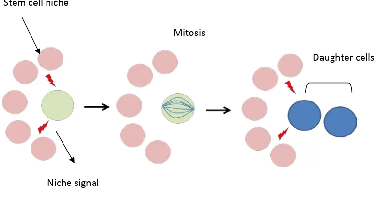
Figure 1. Asymmetric cell division (ACD) ... 24
Figure 2. Differentiation capacity of mesenchymal stem cells into mesenchymal lineage. adapted from Caplan et.al [21] ... 25
Figure 3.The remodelling of compact bone ... 28
Figure 4. Matrix changes during endochondral ossification. ... 30
Figure 5. Alginate structure. ... 45
Figure 6. PEDF is expressed in the epiphyseal cartilage (growth plate) and in the areas of active bone remodelling. ... 54
Figure 7. Morphological features of the isolated mesenchymal stem cells (MSCs) ... 74
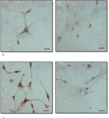
Figure 8. Immunocytochemical identification of harvested mesenchymal stem cells (MSCs) by light microscopy. ... 77
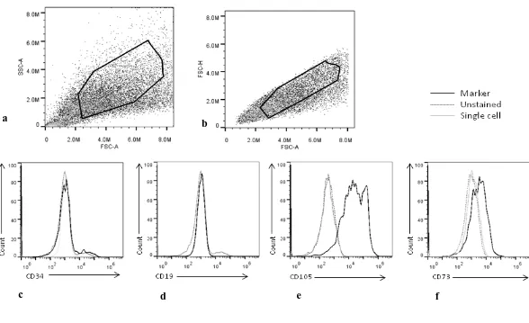
Figure 9.Flow cytometric identification of harvested mesenchymal stem cells (MSCs). ... 78
Figure 10. Differentiation capacity of mesenchymal stem cell. ... 79
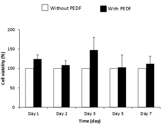
Figure 11. Viability of mesenchymal stem cells (MSCs) in the presence of pigment epithelium-derived factor (PEDF). ... 80
Figure 12. Viability of mesenchymal stem cells (MSCs) in the presence of pigment epithelium-derived factor (PEDF). ... 81
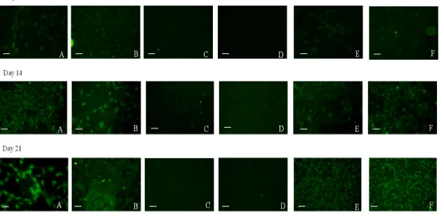
Figure 13. Immunocytochemistry of osteopontin (OPN) marker in monolayer culture of differentiated mesenchymal stem cells (MSCs) under conditions A – F (key below) at days 7, 14, and 21. ... 83
Figure 14. Immunocytochemistry of osteocalcin (OCN) marker in monolayer culture of differentiated mesenchymal stem cells (MSCs) under conditions A – F (key below) at days 7, 14, and 21. ... 84
Figure 15. Immunocytochemistry of collagen I (Col I) in monolayer culture of differentiated mesenchymal stem cells (MSCs) under conditions A – F (key below) at days 7, 14, and 21. ... 85
xvii Figure 17. von Kossa staining results of mesenchymal stem cells (MSCs) grown on normal plate
surface. ... 88
Figure 18. von Kossa staining results of mesenchymal stem cells (MSCs) grown on osteogenic plates. ... 89
Figure 19. The mineral concentration of mesenchymal stem cells (MSCs) grown on normal (non-bone-matrix-coated) plate surface as measured by Alizarin Red ... 91
Figure 20. The mineral concentration of mesenchymal stem cells (MSCs) grown on osteogenic (bone matrix- coated) plate surface as measured by Alizarin Red. ... 92
Figure 21. Immunoblot analysis shows the expression level of four osteogenic markers; alkaline phosphatase (ALP), collagen-1 (Col-1), osteopontin (OPN), and osteocalcin (OCN) at day 14. ... 94
Figure 22. Alkaline phosphatase activity of mesenchymal stem cells (MSCs) in monolayer culture in osteogenic plate at days 7, 14, and 21. ... 96
Figure 23. Time course of mesenchymal stem cell (MSC) growth and development in alginate beads. ... 98
Figure 24. Time course of alginate bead degradation over a period of 21 days in cell culture. .. 99
Figure 25. Mineralisation by released cells from alginate beads in osteogenic plate at day 21 in monolayer culture. ... 100
Figure 26. von Kossa staining results of mesenchymal stem cells (MSCs) released from alginate beads on normal (non-bone-matrix-coated) plate surface. ... 101
Figure 27. von Kossa staining results of mesenchymal stem cells (MSCs) released from alginate beads on osteogenic plate surface. ... 102
Figure 28. Macroscopic observation of beads implanted in Balb/c mice for 35 days. ... 104
Figure 29. Micro-CT images of alginate bead implants. ... 106
Figure 30. Quantitative micro-CT measurements. ... 107
Figure 31. Haematoxylin and eosin (H & E) staining for different groups. ... 110
Figure 32. Haematoxylin and eosin (H & E) staining for different groups. ... 111
Figure 33. Haematoxylin and eosin (H & E) staining for muscle surrounding the implant site for four different groups. ... 112
xviii Figure 35. Alcian blue staining for the four different groups. ... 113 Figure 36. Immunostaining of alginate bead implant paraffin sections for bone marker
osteocalcin (OCN) in the four different groups. ... 115 Figure 37. Immunostaining of alginate bead implant paraffin sections for bone marker
osteopontin (OPN) in the four different groups. ... 116 Figure 38. Immunostaining of alginate bead implant paraffin sections for bone marker alkaline
phosphatase (ALP) in the four different groups. ... 117 Figure 39. Immunostaining of alginate bead implant paraffin sections for bone marker
collagen-I (Col-collagen-I) in the four different groups. ... 118 Figure 40. Immunostaining of alginate bead implant paraffin sections for bone matrix marker
pro- collagen-I (pro-col-I) in four different groups. ... 120 Figure 41. Immunostaining of alginate bead implant paraffin sections for bone matrix marker
heat shock protein 47 (HSP47) in four different groups. ... 121 Figure 42. Immunostaining of alginate bead implant paraffin sections for bone matrix marker
membrane-type matrix metalloproteinase (MT1-MMP) in the four different groups. .... 122 Figure 43. Immunostaining of alginate bead implant paraffin sections for bone matrix marker
xix
List of Tables
xx
Chapter
21
1. Literature review
1.1. Stem cells
1.1.1. Stem cell definition
Stem cells are usually defined as clonogenic, immature, and undifferentiated
cells that are capable of keeping their stemness state during cell division [1]. These cells
have two unique features that are critical for tissue homeostasis; firstly, the
self-renewal property and secondly, the ability to undergo multi-lineage differentiation [2].
The activity of stem cells is at its peak during embryonic development and can generate
all three embryonic germ layers - ectoderm, endoderm, and mesoderm in the
developing embryo. The first successful attempt at isolation of embryonic stem cells
(ESCs) from mice embryos was in 1981 [3, 4], though the first human ESCs were
isolated in 1998 [5]. Adult stem cells are responsible for homeostasis of the tissues in
which they reside. These cells can be found in most tissues throughout the body such
as the brain, bone marrow, liver, and retina in a particular area of the tissue called the
stem cell niche. The stem cell niche is a microenvironment that contains all the cellular
and molecular factors that regulate and support stem cells. In spite of the unique
properties of stem cells (self-renewal and differentiation), adult stem cells can stay
dormant through most of their lifetime and are activated by specific environmental
factors under certain circumstances such as injuries and diseases [6, 7]. Stem cells need
to be held within the niche and this happens via adhesion between stem cells and the
22 ascribed to stem cell niches: 1) perpetuation of quiescence, 2) elevation of cell
numbers, and 3) direction of cell fate and differentiation [8]. Upon division, if a cell is
placed outside the niche, it commits to differentiation depending on the different
microenvironmental stimuli and signalling it encounters in its new environment [6].
1.1.1.1. Stem cell division
Asymmetric cell division (ACD) is the way in which stem cells can maintain their
self-renewal ability and give rise to progeny in other lineages at the same time [9].
Drosophila has been the model for studying ACD [10-13], which revealed two separate
mechanisms-extrinsic and intrinsic- involved in ACD. In the extrinsic mechanism, cell
division takes place in a way that one of the daughter cells maintains access to the
stem cell niche and replaces the divided parent cell, which results in maintaining the
stem cell pool while the other daughter cell is isolated from the stem cell niche. Losing
contact with the signalling molecules within the niche will initiate differentiation
pathways. However, in the intrinsic mechanism, asymmetric inheritance of specific
proteins such as protein partitioning-defective protein (PAR) in Caenorhabditis elegans
[14] and cyclin D2 - a cell cycle regulator in cells- (specifically in mammalian brain cells)
can give differential fate to one of the daughter cells just after cell division [15]. A
23 In bone marrow, there are two known cell populations: the haematopoietic
stem cells (HSC) that can give raise to various blood cells, that is, myeloid and
lymphoid, and a rare population of non-haematopoietic adult stem cells that resides in
the bone marrow as well as most connective tissues of the body. The latter cells have
the potential to differentiate into a variety of mesenchymal tissues such as bone,
cartilage, adipose, and muscle. By placing whole bone marrow in plastic culture dishes,
and removing nonadherent cells after four hours, it was demonstrated for the first time
that bone marrow contains a heterogeneous population of cells [17]. The first adherent
cells resulting from that experiment formed round-shaped colonies containing
fibroblastoids, called a Colony Forming Unit – fibroblast (CFU-f). It was also observed
that these adherent cells became more homogeneous in appearance after being
passaged several times, and could differentiate into other mesenchymal cells such as
bone and adipose cells [17, 18]. Further studies investigated the proliferative ability of
these cells as well as their multipotency and differentiation capacity, not only into the
three mesenchymal lineage cell types but also into other cell types such as neurons and
muscles (Figure 2) [19-22]. The term mesenchymal stem cells (MSCs) was suggested for these cells in 1991 [23], considering the multilineage differentiation ability of these
cells. However, the misconception that using MSCs as a general term for true stem cells
or expanded multipotent progeny has led to the phrase “multipotent mesenchymal
stromal cells (MSCs)” from The International Society for Cellular Therapy (ISCT) [23, 24].
24 to be defined as stem cells: 1) the shape and plastic-adherence capability, 2) the
expression/lack of expression of surface antigen - cluster of differentiation (CD)
markers, and 3) the ability to selectively differentiate into chondrogenic or osteogenic
lineages in response to environmental stimuli [25].
Figure 1. Asymmetric cell division (ACD)
In extrinsic ACD after cell division, one of the cells that remain in the stem cell niche will maintain the stem cell pool whereas the other cell that loses contact with the niche will go through differentiation pathways.
Stem cell niche
Niche signal
Mitosis
25 Figure 2. Differentiation capacity of mesenchymal stem cells into mesenchymal lineage. adapted from Caplan et.al [21]
1.1.2. Murine bone marrow-derived stem cells
The mouse has been considered the model of choice for various types of scientific
research. Nevertheless, difficulties in mouse bone marrow stem cell isolation present
some challenges in investigating the principals of stem cell biology and their
therapeutic applications [26, 27]. MSCs have been isolated from several species such as
human, mouse, rat, dog, baboon, pig, sheep, goat, and rabbit [28]. Although there are a
26 isolation and expansion of MSCs in the mouse is far more difficult. During isolation,
these cells are usually contaminated with haematopoietic cells, which results in a
heterogeneous cell population. Furthermore, expansion of a heterogeneous cell
population can occur. This issue has been addressed in the human [20, 29] and other
species [30, 31] by serial passaging of adherent cells or co-culture with other cells such
as endothelial cells [32, 33]; though this approach has not been very helpful when it
comes to mouse bone marrow-derived MSC isolation as the long term expansion of
mouse MSCs during culture involves challenges due to the tendency of these cells to
lose their proliferative potential or result in highly proliferative MSC populations [34,
35]. However, with certain methods and culture conditions, it is possible to establish a
proliferative and homogeneous population of MSCs with the capacity of tri-lineage
differentiation [36-38]. These MSCs would be a very useful tool toward understanding
and interpreting MSC-based therapies and data for future clinical applications such as
tissue engineering.
1.2. Bone tissue engineering
1.2.1. Bone structure and function
Bones provide mechanical support for muscles and promote movement, as well as
protecting internal organs. The mechanical support and properties that bone provides
27 are comprised of compact (or cortical) bone, which contains ~ 80 – 90 % mineralised
tissue providing the mechanical strength. The ends of long bones are made up primarily
of trabecular (or cancellous) bone. In contrast, only 15 – 25 % of the trabecular bone is
mineralised. Thus, while trabecular bone contributes to the mechanical strength, its
initial function is metabolic, as this bone functions as a supply of calcium and
phosphate ions. Other than the mentioned functions for bone, recently its role in
metabolism came into light. These includes existence of a novel endocrine regulatory
loop in which insulin signalling in the osteoblast controls postnatal bone development
and simultaneously regulates insulin sensitivity and pancreatic insulin secretion to
regulate glucose homeostasis. [39, 40]
In addition, in the adult organism, many bones contain cavities filled with the bone
marrow, and represent the anatomical site for blood cell and platelet production
(haematopoiesis) [41].
Bones are mainly comprised of three different cell types: osteoblasts, osteocytes, and
osteoclasts. Osteoblasts, which derive from MSCs, are cuboidal, post-proliferative cells
with high synthetic activity and are responsible for bone extracellular matrix deposition
and mineralisation. Osteocytes are star-shaped mature osteoblasts and are smaller in
size, and are embedded in a mineralised matrix and are the most abundant cell type in
mature bone. Osteoclasts are multinucleated cells of haematopoietic origin with
28 of osteoblasts and osteoclasts secure bone homeostasis during development and
remodelling throughout a lifetime (Figure 3) [42, 43].
Figure 3.The remodelling of compact bone
Osteoclasts acting together in small group excavate a tunnel through the old bone. Osteoblasts enter the tunnel behind them, line its walls, and begin to form new bone.
1.2.2. Bone extracellular matrix (ECM)
Bone extracellular matrix (ECM) is composed of two main components. The organic
part, which makes 30-40 percent of the tissue, mostly consists of type I collagen fibrils
in a bed of proteoglycan aggregates (mainly biglycan and decorin) and glycoproteins.
Glycoproteins represent the largest proportion of non-collagenous proteins (NCPs) and
include thrombospondin [44], bone sialoprotein [45], alkaline phosphatase [46],
osteonectin [47], osteopontin, and osteocalcin [48], as well as amino acids with high
29 mineral part of the bone is mainly made of calcium phosphate crystals in the form of
hydroxyapatite (HA), which constitutes 60-70 percent of the bone tissue. In addition to
HA, other minerals such as bicarbonate, citrate, magnesium, potassium, and sodium
are also found [49].
1.2.3. Healing, a natural process
Bone is a highly vascularised tissue and, as mentioned before, the balance between the
activities of osteoclasts (bone-resorbing cells) and osteoblasts (bone-forming cells)
leads to a continuous remodelling of the bone, which makes it adaptable to mechanical
stress and helps the tissue to maintain its health and repair potential. Bone
homeostasis occurs via basic multicellular units (BMUs) [50]. Each unit includes a
“cutting cone” of osteoclastic bone reabsorption followed by osteoblasts laying down
new bone in the trail of osteoclasts, and the end result is an osteon [51]. Proper cell
proliferation, differentiation, migration, and remodelling of the extracellular matrix will
lead to the development and regeneration of bone tissue. Cellular condensation
initiates bone formation, where mesenchymal cells spread out, migrate, proliferate,
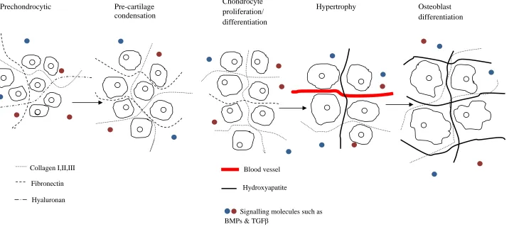
30 Hyaluronan
Collagen I,II,III
Hydroxyapatite Fibronectin
Blood vessel Prechondrocytic Pre-cartilage
condensation
Chondrocyte proliferation/ differentiation
Hypertrophy Osteoblast differentiation
Signalling molecules such as BMPs & TGFβ
Figure 4. Matrix changes during endochondral ossification.
31 There are two mechanisms responsible for bone development: bones can
directly develop from MSCs into osteoblasts, called intramembranous ossification, or at
an early stage where a cartilage template can form, which, after a while, can be
replaced by bone, called endochondral ossification.
Intramembranous ossification occurs during development of flat bones and is
peripheral to the site of the fracture during bone healing. During intramembranous
ossification, cells of the mesenchymal lineage, which are embedded in a membrane of
connective tissue, directly undergo osteogenic differentiation and synthesize the
osteoid (non-mineralised matrix), which eventually mineralise. Stem cell specification
toward the osteogenic lineage is basically regulated by the combined action of three
transcription factors, such as RUNX2, OSX and nuclear β-catenin. Several experimental
studies suggest that RUNX2 directs osteogenesis in the early phases of stem cell
differentiation, while OSX and nuclear β-catenin act downstream consolidating the
transition toward the osteoblastic phenotype [52, 53].
The latter (endochondral ossification) takes place during the development of short and
long bones, the growth of the length of long bones (growth plate), and during the
natural healing of bone fractures. [54, 55]. During this process the mesenchymal
progenitor cells first aggregate into a cartilage template and are then replaced by bone
[55, 56]. Prior to endochondral ossification, migration of pre-chondrocytic
mesenchymal cells occurs (Figure 4). Migration of pre-chondrocytic mesenchymal cells
32 hyaluronan and collagen type-I. These mesenchymal cells produce condensed nodules
following the chondrogenic phase [55, 57].
During the condensation stage, lots of changes occur during cell-matrix
interactions. These changes are mediated by specific molecules including N-cadherin,
fibronectin, syndecans, tenascin, thrombospondins, neural cell adhesion molecule,
focal adhesion kinase, paxillin, and matrix metalloproteinases (MMPs) [57]. The
extracellular space becomes limited due to increased activity of hyaluronidase and
more compact dispersion of collagen types I and III and fibronectin. Afterwards,
pre-chondrocytic cells proliferate and differentiate to osteocytes to provide both
mechanical support and scaffold for the hard osteoid tissue that will form later [58].
Chondrocyte differentiation is defined by cartilage-supporting matrix synthesis, which
includes a variety of collagens such as collagen-II, collagen-IX, and collagen-XI, and
various proteoglycans such as aggrecan. The process of chondrocyte maturation
continues by hypertrophy as the cells secrete hydroxyapatite into the ECM for
mineralisation [59, 60]. During hypertrophy, chondrocytes secrete collagen type-X and
matrix metalloprotease-13 (MMP13), and these events lead to changes in ECM protein
composition. Degradation of the ECM facilitates vascular invasion and recruitment of
chondroclasts, which results in the removal of apoptotic chondrocytes and migration of
new MSCs which later differentiate into osteoblasts that exude bone matrix [60].
Woven bone gradually substitutes cartilaginous tissue. This remodelling process is
33 metalloprotease-14 (MMP14), which also known as membrane type 1 matrix
metalloprotease (MT1-MMP) as collagen I, II, and III are substrates for MMP14
protease function [61-63]. To do so, MMP14 uses matrix metalloprotease-2 (MMP2) as
a substrate and activates it by cleaving its pro-domain, which leads to bone formation
stage [64, 65]. Furthermore a large number of small signalling molecules like cytokines
and growth factors (IL-4, BMPs, TGF-ß and VEGF) further influences bone cell activity.
These factors are produced by hypertrophic chondrocytes during the hypertrophy
stage and thus are part of mechanisms by which bone cells can influence each other’s
behaviour. Some are incorporated into the bone matrix and are released again when
bone matrix is resorbed. This can locally alter behaviour of bone cells. An important
regulatory mechanism is the RANK-RANKL pathway. Osteoblasts produce RANKL that
attaches to receptors on the surface of pre-osteoclasts and signals these cells to
differentiate into mature osteoclasts. In this stage, bone matrix is made of mineralised
and proteinaceous ECM and is called the primary bone formation stage. As the process
continues to its final stage, the secondary bone formation stage, the ECM becomes
more solid and reconstructs itself to weight-tolerable cortical or trabecular bone [66].
Inflammation is the key difference in developmental skeletogenesis versus
regenerative skeletogenesis. During regenerative skeletogenesis, inflammatory cells
secrete necessary growth factors and cytokines to recruit MSCs and initiate bone
formation. However, developmental skeletogenesis is regulated by
34 Usually, bone has the ability to heal small lesions due to its ability to undergo
spontaneous regeneration, so no invasive procedure such as surgery is required and
conventional therapy like casting would be sufficient. However, spontaneous healing
does not apply in cases like non-union or critical-size bone defect (CSD). Non-union is
defined as “a fracture that is over nine months old and has not shown radiographic
signs of progression toward healing for three consecutive months” [69]. In the case of
continuous poor or no healing, the final stage of aseptic non-union would be
pseudoarthrosis. One approach toward management of pseudoarthrosis is grafting,
which sometimes involves not only complete removal of the fracture site but also
tissue around the non-union site [51]. Critical-size bone defect is “the smallest size
intraosseous wound in a particular bone that will not heal spontaneously during the
lifetime of the animal”, and the plan for therapeutic management of CSD is bone
grafting and transplants [51].
1.2.4. Bone grafts
In order to have a successful bone graft, there are certain criteria which have to be
considered. During bone regeneration after the bone graft, three crucial processes
have to take place in order to generate new bone formation: osteogenesis,
35 Osteogenesis is the act of bone formation as a result of osteoid expression by
osteoblasts followed by mineral deposition. Osteoinduction however, is the induction
of differentiation of undifferentiated cells such as stem cells and other osteoprogenitor
cells toward the osteoblast lineage. Osteoconduction is the ability of the graft to
promote and enhance migration and attachment of osteoprogenitor cells and
osteoblasts as well as vessel formation in order to support the new tissue formation. To
have a clinically and physiologically functional bone graft, the graft has to exhibit the
above features.
1.2.4.1. Autografts
Autogenous cancellous bone or autograft is believed to be the ideal graft material as it
is harvested from and implanted into the same individual [70]. These grafts are usually
taken from the iliac crest, rib, fibula, or tibia of the patient. These segments of bone are
highly vascularised and possess osteogenic potential due to the presence of viable
osteoprogenitor cells as well as osteoconduction/induction capacity. Furthermore, as
this graft is from the same patient, it will not transmit disease to the recipient and the
risk of immunoreactions is low. However, cancellous bone taken from the patient is not
readily available in unlimited quantities. The process of harvesting the bone imposes
potential complications and pain to the patient as well as possible complications that
may occur after surgery including inflammation, infection, and donor site morbidity
36 1.2.4.2. Allografts
Allografts are harvested from a human donor other than the patient, the prerequisite
of harvesting being genetic compatibility. The graft specimen has to go through some
processes to eliminate and reduce the risk of immunoreactions and disease transfer
such as freeze-drying, washing, demineralisation, and gamma-irradiation or ethylene
oxide sterilization. However, although these procedures reduce the risk of disease
transmission and immunoreactions, they also lessen the osteogenic potential of these
grafts [73, 74].
1.2.4.3. Xenograft
Unlike autografts and allografts, xenografts are obtained from a non-human donor.
Xenografts are not commonly accepted or used in clinical applications due to ethical
issues and disease transfer. These grafts are usually used as bone void filler to fill burr
hole and craniotomy defects and in smoothing facial skeletal contour abnormalities.
Examples of these grafts include: BioOss [75] an inorganic matrix from cows [76],
porcine organic bone matrix [77], equine [78], and bio coral grafts [79, 80], and Norian
37 1.2.4.4. Prosthesis
The use of a large prosthesis is a common solution to overcome the above-mentioned
complications. Nonetheless, as there is no biological interaction between the
prosthesis and tissue, the prosthesis may simply give in and the revision surgery may be
required in order to fix or replace it [82, 83]. This becomes particularly problematic in
growing recipients [84]. Other concerns such as risk of allergy and toxicity [85] raise the
new idea of using alternative materials, and consequently, open a new horizon of tissue
engineering.
1.2.4.5. Alternative solution: tissue engineering
The search for alternative approaches toward bone repair and replacement of
the conventional methods has resulted in the new field of tissue engineering. This field
has evolved rapidly over the last 15 years. Scaffolds with designed microstructures give
sound structural support and adequate mass transport of nutrients and oxygen to
facilitate tissue regeneration. There are numerous reports on various tissues grown in
vitro including bone [74], cartilage [86], main bronchus [87], and blood vessels [88]. The
achievements in the area of tissue engineering in various disciplines are due to the rapid
advancement in knowledge of stem cell biology and increased understanding of their
response to environmental cues [89]. The unique ability of MSCs to sense and react to
38 makes them an excellent candidate for use in tissue engineering. However, one cannot
disregard the need of a suitable vehicle for stem cell delivery. Hence, lots of effort has
been put into fabrication of an optimal scaffold with the necessary characteristics for
stem cell delivery [66].
1.2.5. Scaffolds
Similar to scaffolds in the field of construction, the scaffold for tissue engineering and
regenerative medicine needs to be strong, reliable, and able to endure the
environmental conditions for the specific period of time. Moreover, the scaffold should
be removable without damaging or affecting the newly formed or repaired structure
[90]. In tissue regeneration, the scaffold is a three-dimensional (3D) construct that acts
as a template for cell adhesion, proliferation, differentiation, and extracellular matrix
formation to provide a suitable environment for the newly regenerated tissue.
1.2.5.1. Scaffold requirements
For successful bone tissue engineering, a suitable environment in which osteogenic
cells are able to migrate, differentiate, and proliferate is necessary. The scaffold, as a
three-dimensional structure, can accommodate stem cells in this context and promote
new bone formation as well as provide mechanical support during bone regeneration
39 to proliferate and differentiate, a few considerations need to be taken into account
(Table 1). The biocompatibility of the material that the scaffold is being made of is
important to avoid an adverse immunological reaction it may cause in the body.
Biodegradability is another important characteristic of the scaffold, which means that
the scaffold should be able to be degraded inside the body naturally at an appropriate
time and controllable rate. In addition, the degradation products should not be toxic
and must be metabolised naturally in the body [92]. Furthermore, porosity and
permeability of the scaffold is essential for high yield of cell seeding (in vitro) and
proper infiltration (in vivo), nutrient transport, tissue ingrowth, and vascularisation
[93]. The mechanical stability of the scaffold is another important, desirable feature
[92]. The construct should mimic the native bone environment and structure to make
the scaffold ideally osteoconductive, osteoinductive, and osseointegrative of stem cells
40 Table 1. Summary of scaffold features and their effect on bone regeneration.
Scaffold characterisation Biological effect
Biomaterial and biocompatibility - Cell proliferation and differentiation - Appropriate for in vivo implantation
Geometry and architecture - Encourage three dimensional growth of the cell - Controlling the growing tissue morphology - Support cell proliferation
Porosity - Encouraging cell differentiation, recruitment, aggregation, and vascularisation
Mechanical properties - Support mechanical loading
Degradation rate - Make space for new tissue ingrowth - Allow the extracellular matrix to remodel
Biochemical stimuli - Embody proper growth factors and cytokines for cell function enhancement
1. 2.5.2. Scaffold materials
A variety of materials including metals, ceramics, polymers (natural and synthetic), and
their blends have been used for the replacement and repair of damaged bone tissues.
Metals and ceramics have two major disadvantages for tissue engineering applications:
they are non-biodegradable, and their processability is limited [95]. Synthetic and
natural polymer scaffold applications have been comprehensively examined in the lab
and clinic in order to replace bone tissue [96-99]. The polymer materials are more
41 desirable properties such as biocompatibility and degradation, mechanical properties,
and microstructure [100, 101].
1. 2.5.2.1. Synthetic polymers
Synthetic polymers, both organic and inorganic materials, are used in a wide variety of
biomedical applications. The family of saturated aliphatic polymers including polylactic
acid (PLA), polyglycolic acid (PGA), poly (lactic-coglycolide) (PLG), and their blends, are
the conventional materials that have been used in bone tissue engineering. These
polymers have a high molecular weight and are usually polymerised via a condensation
reaction. The other method of polymerisation is ring–opening polymerisation (ROP),
which has been used in the polylactic group. The main core of all aliphatic polymers is
the same, the only difference being the substituent group that accounts for a variation
in molecular weight and degradation rate [14, 15].
There are other biodegradable synthetic polymers that are being investigated in
the field of tissue engineering. These polymers include polycaprolactone (PCL),
polyanhydrides, polyphosphazenes, and bioactive glass. The ability to attach to bone
and soft tissue makes these materials good candidates for bone graft material. These
polymers are able to form a layer of hydroxyapatite in the margin of bone and allow
the graft material to integrate the implant to live tissue [92, 102]. Synthetic
non-degradable polymers include alloplastic, polymethylmethacrylate (PMMA),
42 104]. To overcome the undesirable feature of non-degradability in these useful
materials, and induce controlled degradation and improve biocompatibility, one can
combine them with degradable and natural polymers.
1.2.5.2.2. Natural polymers
The first biomaterials with biodegradability features that had been considered for use
in bone tissue engineering clinically were natural polymers. Collagen [105-107], fibrin
[108-111], silk [112-114], hyaluronic acid [115, 116], chitosan [117, 118], and alginate
[91, 106, 109, 119-122] are used in bone and cartilage tissue engineering applications.
These materials are able to enhance cell attachment and proliferation in biological
systems due to their better interactions with cells compared to other polymers such as
synthetic ones. Natural polymers have a highly organised structure and also possess
extracellular ligands that may bind to cell receptors. However, limited supply and low
weight-bearing capacity, high cost, and difficulty in processing them for clinical
applications are some disadvantages of natural polymers [71, 118].
1.2.5.2.3. Composites
Composite materials consist of two or more separate materials which can benefit from
all the positive feature of each to provide a scaffold with better features. The
43 made of hyaluronic acid and organic collagen. HA belongs to the mineral part of the
living bone, and is required for better osteoconductivity. In polymer/ceramic
composites, HA can be used as the ceramic part while collagen, gelatin, chitosan, chitin,
elastin, poly methymethacrylate, polypropylene fumarate, polyphosphazenes, and poly
hydroxybutyrate, PCL, PLA, PLG, poly anhydride, and polyorthoester, each can serve as
the polymer component of the composite. In such constructs, the living bone matrix
can spontaneously integrate into the HA layer. The degradation pattern of the polymer
may be affected by the bioactive phase in the polymer composite. Changes in
degradation kinetics are related to a pH-buffering effect at the surface of the polymer
due to rapid proton exchange for alkali in the ceramic, which changes the rate of
polymer degradation in pH-dependent cases (acidic degradation) [123-125]. In
composite constructs, degradation rates may change due to ceramic accessibility in the
composite structure, which leads to water absorption and hydrophilicity of the
hydrophobic polymer structure [125]. An improved environment for cell seeding,
survival, growth, differentiation, load-bearing, and other mechanical properties are
some of the beneficial features of composite material for tissue engineering [126].
1.2.5.2.4. Blends
A blend is a category of polymeric material which is manufactured by a combination of
synthetic–natural, natural–natural, and synthetic–synthetic polymers. The purpose of
blending these materials is to benefit from idiosyncratic advantages of each material,
44 production cost of synthetic material, biocompatibility, and controlled biodegradability
of the natural polymers to match the cell growth rate [127]. PLA, PLG, and PCL as
synthetic polymers and gelatin, elastin, chitosan, starch and alginate as natural
polymers have been widely used in manufacturing blend materials [128, 129].
1.2.6. Natural polymers: alginates
Due to certain interesting chemical and physical properties of alginates, they have been
used in a broad range of applications in different fields. In the food and beverage
industry, alginates are used as thickeners and stabilisers. They also are used for yeast
encapsulation in the ethanol production industry. Alginates are also applicable in other
industries such as paper and paint production, ceramic shaping, sewage water
treatment and purification [130-133]. Alginates have also been used in the healthcare
and pharmaceutical industries as matrices for cell encapsulation and transplantation
since 1980 when the first successful encapsulation of islet cells was achieved [134].
1.2.6.1. Structure and sources
Alginate refers to a family of polyanionic copolymers of 1-4 linear linkages of
ß-D-mannuronic acid (M) and α-L-gluronic acid (G). The various compositions of these
isomers results in at least three distinctive conformations of the hexopyranose ring
45 subsequently affect the behaviour of the encapsulated cells. For example, a high G
content leads to more stable gel compared to the high M content, which can affect the
final purpose of encapsulation (cell proliferation and growth, or metabolism and
secretory activity) [135, 136]. These polysaccharides were initially isolated from brown
algae, namely Laminaria hyperborea, Ascophyllum nodosum, and Macrocystis pyrifera
and bacteria such as Azotobacter and Pseudomonas. However, isolation from bacteria
is not cost-effective, so the material is sourced from brown algae [132]. (Figure 5)
Figure 5. Alginate structure.
46 1.2.6.2. Cell encapsulation and interaction
Sodium alginate is soluble in an aqueous solution, which is called a hydrogel. Ionic
interaction with certain divalent cations such as calcium (Ca2+), strontium (Sr2+),and
barium (Ba2+) at room temperatureresults in the gelation of sodium alginate and
consequent formation of a three dimensional structure. Mechanical properties
including mechanical stability, viscosity, and elastic modulus of alginates are the key
factors that affect the interaction of cell-gel during cell encapsulation [137, 138]. As
there are no receptors for alginate polymers in mammalian cells, this polymer acts as a
practically inert environment and, consequently, the adhesion and proliferation of cells
within alginate is not affected by it [139]. To improve the cell-gel interaction, some
adhesion molecules such as laminin [140], collagen [141], gelatin [91], and fibrin [109]
have been supplemented into alginate during cell encapsulation. However, coupling
using such materials is challenging to control and may lead to non-specific cell
interactions. Hence, coupling of short chain amino acids was introduced to improve the
cell-gel interaction. The fibronectin-derived adhesion peptide arginine-glycine-aspartic
acid (RGD) and its subtypes are commonly used to improve the cell adhesion properties
of alginate; considering that RGD-receptors have been identified and well characterized
in mammalian cells [142]. Furthermore, the manipulation of alginate composition with
the above-mentioned elements could affect and improve the degradation rate and
47 The mechanism that is responsible for nutrition supply and waste removal from
encapsulated cells is simple diffusion because of the absence of a vasculature system.
As a result, the size of the sphere is an important limiting factor in mass transport in
encapsulated cells [143]. Encapsulation can be achieved by using simple methods such
as a syringe or pipette [133, 144] or more precise methods including atomisation and
emulsification. However, the resultant spheres obtained from the different methods
would vary in size, which eventually will affect the survival and proliferation of
encapsulated cells. It has been shown that spheres with a diameter of 0.9-1 mm
displayed a cell layer approximately 0.2 mm thick at the periphery, while cells in the
centre of the microcapsules were dead, which indicates insufficient nutrition transport
to the centre. However, this problem can be resolved by reducing the size of the
spheres [143, 145].
1.2.6.3. Stem cell encapsulation in alginate for bone regeneration
Encapsulation of bone marrow-derived stem cells in alginate for the purpose of bone
tissue formation has been examined extensively and has shown promising results as an
alternative solution for bone healing. However, to use alginate encapsulation clinically,
a few strategies have been introduced. One approach is to encapsulate the
48 with osteogenic supplements in order to initiate and support osteogenic differentiation
[148-150].
1.2.7. Growth factors as osteogenic supplement
In recent years, growth factors such as bone morphogenetic proteins (BMPs) have been
used extensively in bone tissue engineering. These are the main signalling molecules
for growth, proliferation, and differentiation of bone progenitor cells, and deliver
certain advantages that make them effective and suitable for use in pre-clinical and
clinical settings [151].
1.2.7.1. Bone morphogenetic proteins (BMPs)
Bone morphogenetic proteins belong to the superfamily of transforming growth
factor-beta (TGF-ß) and are structurally related. To date more than twenty BMPs have been
identified. Several BMPs such as -2, -4, -6, -7, and -9 have shown direct effects on bone
formation through bone morphogenic cascades, which result in proliferation and
differentiation of osteoprogenitor cells and MSCs, and final bone formation [151, 152].
Furthermore, two BMPs are available commercially for use in clinical treatment -
recombinant human (rh) BMP-2 (Infuse™) [153] and rhBMP-7 (OP-1™) [154]. These
BMPs are being used widely as substitutes for autografts or in combination with grafts
49 The use of BMPs as an alternative for grafts has several advantages such as reducing
morbidity- and surgery-related complications and also overcoming the limitation of
supply as BMPs can be made using recombinant DNA technology [155]. However, the
application of BMPs also has certain limitations. As BMPs works in a dose-dependent
manner, the concentration of BMPs needs to reach a threshold level in order to initiate
bone formation. Additionally, there is a rapid systemic clearance half-life for rh-BMP-2
(7-16 minutes) and 10-15 hours for rhBMP-7, which necessitates the need to use
supraphysiological dosesin order to rectify the rapid wash-out in order to achieve a
satisfying response [148]. Moreover, other limitations such as insufficient
responsiveness of cells, possible inhibitory effect of high doses of BMPs on other
tissues, and possible side-effects have come to light [156]. Furthermore, recent
contradicting reports have been published about no positive effect of BMP-2 on
fracture healing, as well as increasing debate with regards to its side effects such as
osteolysis, implant reposition, loss of alignment, urogenital, bladder retention, and
bone overgrowth into the spinal canal, which are the most common complications that
have reported when rhBMP-2 is used compared with other graft methods [157-172].
The production cost of recombinant protein is another reason to search for other
50 1.2.7.2. Platelet-rich plasma (PRP)
The concentration of platelet in plasma is almost 1x10⁶ platelets in 5 ml of plasma,
which contains a 3- to 5-fold increase in growth factor concentration. The main role of
platelets is in wound healing. The first reaction after wounding and bleeding is
activation of platelets by contact with collagen and subsequent release of the growth
factors to initiate the healing process [173]. The known cytokines in platelet are mostly
involved in cell proliferation, chemotaxis, cell differentiation, and angiogenesis, which
make it a good candidate to be used in bone repair [174, 175]. The majority of the
growth factors that have been considered to be used as a supplement for osteogenic
differentiation are somehow derivatives of PRP such as: platelet-derived growth factor
(PDGF) [176], vascular endothelial growth factor (VEGF) [177], and fibroblast growth
factor (FGF) [178]. While it has been shown that using PRP and its derivatives can
support bone formation in different animal models [179-183], not all the studies are
supportive of using PRP as a reliable and efficient clinical therapy in bone complications
[184-186].
1.2.8. PEDF
Pigment epithelium-derived factor (PEDF) is a glycoprotein that belongs to the
superfamily of serpin protease inhibitor proteins without inhibitory function, encoded
51 evolution [187]. It is a protein of 418 amino acids, with a size of 50kDa and is widely
expressed in most bodily tissues [188]. The highest amount of expression has been
observed in the eye, foetal and adult liver, adult testis, ovaries, placenta, and the
pancreas. [189]. A significant reduction in the expression of PEDF is found in senescent
(aging) cells [190]. PEDF was originally isolated from the conditioned medium of
cultured human foetal retinal pigment epithelium cells [189]. It is an extracellular
protein which shows the typical secondary and tertiary structure of a serpin and binds
to collagen-1 and heparin. The α-sheet is the dominant feature of the secondary
structure and comprises the core structural domain of the protein, being closely
involved in dynamic movements that are part of serpin function [191]. The existence of
a reactive centre loop (RCL) is another feature of serpins, and it is a proteinase
recognition site and a critical component of the function of serpins [189]. PEDF
contains an RCL structure but the function of this is still unknown [189].
While PEDF is increasingly becoming known for its anti-cancer properties, it is a
pluripotent molecule with neurotrophic qualities, as well as having anti-angiogenic,
anti-proliferative, pro-differentiation, neuroprotective, and anti-inflammatory roles
[192-194]. Recent studies have demonstrated that PEDF supports the survival and
proliferation of neural, retinal, and embryonic stem cell populations [195]. PEDF is also
detected in areas of endochondral ossification and active bone remodelling [196]. As
mentioned before, endochondral ossification is one of the main processes that MSCs
52 1.2.8.1. PEDF and mesenchymal stem cells (MSCs)
PEDF is one of the most abundant proteins identified in murine MSC
(mMSC)-conditioned medium [197]. Immunofluorescent staining has shown a high level of PEDF
in the rough endoplasmic reticulum/Golgi areas [197]. PEDF is also found to be located
near the plasma membrane and in the extracellular space, giving PEDF the ability to
bind to collagen and proteoglycans in the extracellular matrix.
During differentiation of MSCs to osteoblasts, the expression of several genes
begins or is elevated and this includes PEDF. It has been shown that a high level of PEDF
is expressed in osteoblasts during the early stages of bone development, and to a lesser
extent in osteoclasts [198, 199]. Osteoblasts and possibly osteoclasts are able to
synthesise and release PEDF, and this protein has a critical role in normal and abnormal
bone angiogenesis [198, 200]. In developing bones, blood vessel growth is localised and
MSCs may play a role as pericytes in support of newly formed blood vessels, which is a
very active process during endochondral ossification [201, 202]. In locations such as the
long bone growth plate, blood vessels selectively invade the region between
hypertrophic chondrocytes and newly formed bone matrix. These newly-formed
vessels allow migration of osteoblasts, which leads to new bone matrix deposition and
bone elongation. PEDF is expressed in the epiphyseal cartilage and in the areas of
active bone remodelling in the primary spongiosa and periosteum of metaphyseal bone
[196]. There is a gradual decrease in the intensity of PEDF expression as chondrocytes
53 PEDF varies in bone (Figure 6); a high level of expression was observed in the germinal
zone, followed by a decrease in expression in the proliferation, maturation, and final
hypertrophic zones [196, 198].
1.3. Aims and Objectives
The hypothesis of the current study is that PEDF is intrinsically involved in
osteoblast differentiation and bone formation.
The main objective of this thesis was to investigate the effect of PEDF as an
osteogenic supplement for MSCs in an alginate bead scaffold for bone tissue formation
in vitro and in vivo.
In order to examine the hypothesis, a series of in vitro experiments were carried
out. The stemness of the isolated cells from mouse bone marrow first examined using
specific surface markers and other necessary assays such as tri-lineage differentiation.
The osteogenic potential of PEDF then examined in normal and osteogenic plates in
mono layer and capsulated in alginate and confirmed via several qualitative and
quantitative methods including immunofluorescent, immunoblottig, von kossa , alizarin
Red staining and enzyme activity assay. The study further taken to animal model to test
54 Epiphyseal plate
Bone marrow cavity
(Mesenchymal stem cell source)
Germinal zone (containing stem cells) or resting zone (R)
Proliferative zone (P)
Maturation zone (M)
Upper and lower hypertrophic zone (UH, LH)
Ossification zone (OZ) PEDF expression zone
Figure 6. PEDF is expressed in the epiphyseal cartilage (growth plate) and in the areas of active bone remodelling.
The highest level of expression is observed in the proliferative zone, maturation zone, and upper hypertrophic zone respectively.
55
Chapter
56
2. Material and Methods
2.1. In vitro study
2.1.1. Cell culture
All the cell culture and in vitro assays were carried out in Greiner culture flasks or
plates (96-well, 23-well, 12-well and 6-well) at 37°C and 5% CO2 unless otherwise
specified.
All cell culture reagents and chemicals were purchased from Sigma-Aldrich (St Louis,
MO, USA) unless otherwise stated. Dulbecco’s modified Eagle medium (DMEM), with
low glucose, supplemented with 1% penicillin/streptomycin and 10% heat-inactivated
foetal bovine serum (FBS) is referred to as complete media. Each experiment was
repeated twice and carried out with 4 replicates (n=4) unless otherwise stated.
2.1.2. Stem cell isolation and expansion
Wild-type 6 weeks Balb/c mice were sourced from the Monash Animal Research
Platform, Clayton- VIC, Australia. Animal ethics approval was granted prior to
commencing the study from the Victoria University Animal Experimentation Ethics
Committee (AEEC 16/10).
Mesenchymal stem cells were isolated from the marrow resident in mice long bones
57 by cervical dislocation and the hind limbs were removed and kept on ice in complete
media, while removing all the muscles and connective tissue from bone by scraping and
also during cell collection. After removing the growth plates, bone marrow was flushed
out with complete media using a 27 gauge syringe. The cell suspension was then
filtered through a 100 mm mesh filter (Millipore) to remove any bone or muscle tissue
and then cultured in a 150 cm culture flask in 20 ml of complete medium. Every 8
hours, the nonadherent cells were washed then removed with phosphate buffered
saline (PBS) and fresh media added for the next 72 hours. The adherent cells were then
washed with PBS and the fresh media added every 3 days for the next 3-4 weeks until
cells became 80-90% confluent. Cells were split and passaged every 10-14 days
following media removal and washed with 10 ml PBS. Cells were detached following
incubation with 2 ml of 0.25% trypsin/1 mM ethylenediaminetetraacetic acid (EDTA)
for 2 minutes at room temperature and grown until passage 4 for the derivation of a
pure stem cell population and to increase the cell number. All experiments were
performed within 10 passages. As mentioned before, the culture media contained 10%
FBS which contains PEDF. Unsupplemented medium, that is one without FBS would
leads to senescence and inhibits cell proliferation and ultimately leads to apoptosis.
However, considering that all the cells were treated with the same basic media the
58 2.1.3. Immunocytochemistry
Stemness of the isolated cells is indicated by the expression of CD73 and CD105 as well
as a lack of expression of hematopoietic markers, CD34 and CD19 [25, 69, 203].
Immunocytochemical analysis was performed to characterise the surface antigen
expression of CD34, CD19, CD73, and CD105 of the isolated cells according to a
previously described protocol, with some modifications [204]. Cells were blocked after
permeabilisation using 0.3% saponin following by a peroxidase block (Dako,
Melbourne, Australia) for 5 min, with 2% serum (Dako, Melbourne, Australia)
corresponding to the appropriate antibodies for 30 minutes. Cells were then incubated
overnight with anti-CD34, -CD19, -CD73, and -CD105 (Santa Cruz) in 1:500 dilution, and
were subsequently probed with a biotinylated secondary antibody (Dako, Melbourne,
Australia) at a 1:2000 dilution followed by Vectastain ABC kit (Vector Laboratories,
Burlingame, CA, USA) according to the manufacturer’s instructions and
3,3’-diaminobenzidine (DAB) staining. Haematoxylin staining was used to visualise the
nucleus. Microscopic imaging was performed using a Zeiss Axioplan 2 microscope
(Zeiss-Australia).
2.1.4. Flow cytometry analysis
The expression of surface markers was also evaluated using an Attune® acoustic
focusing cytometer (Thermofisher Scientific, NY, USA). Cells were detached with