Available online on 15.11.2018 at http://jddtonline.info
Journal of Drug Delivery and Therapeutics
Open Access to Pharmaceutical and Medical Research© 2011-18, publisher and licensee JDDT, This is an Open Access article which permits unrestricted non-commercial use, provided the original work is properly cited
Open Access
Research Article
Preparation and Characterization of Solid Lipid Nanoparticles Loaded with
Cisplatin
Indrayani D. Raut*1, Arehalli S. Manjappa2, Shrinivas K.Mohite1, Rajendra C. Doijad3
1 Rajarambapu College of Pharmacy, Kasegaon (India) 2 Tatyasaheb Kore College of Pharmacy, Warananagar (India) 3 KIMSDU’s, Krishna Institute of Pharmacy, Karad (India)
ABSTRACT
Cisplatin (Cis diaminedichloro platinum) is the first platinum drug to be used as an anticancer drug, and it is widely used in the treatment of testicular, head, neck, ovarian and lung cancer. The use of Cisplatin is limited due to its intrinsic and acquired resistance and severe side effects such as chronic neurotoxicity and nephrotoxicity. The colloidal carriers such as emulsion, liposomes, polymeric nanoparticles have been extensively studied to overcome above limitations. The solid lipid nanoparticles (SLNs), amongst other colloidal carriers, were found to be an ideal carrier for lipophillic drug for better stability and release retardation. Cisplatin loaded solid lipid nanoparticles was prepared by microemulsion technique. Stearic acid was used as lipid. The other excipients were used as DPPG, Soya lecithin and Poloxamer P407 and acidic buffer pH4. Probe sonication was used for 10 min at 79 Amplitude. Cisplatin SLNs Batch C13 showed particle size of 119.23±1.52 nm, Zeta potential of -37.33±2.47 mV, % Entrapment efficiency of 90.2 ± 2.1 %., % Drug loading capacity of 1.62 ± 1.34 %., The TEM study of optimized Cisplatin SLN illustrated the spherical shape of nanoparticles. Total release amount of Cisplatin was 82.62± 2.04 % after 48 hrs. The formulation performed kinetics study followed Peppas plot equation The SLNs of Cisplatin met all the requirements of a colloidal drug delivery system. They had particle size in nanosize; their size distribution was narrow and all the particles were in spherical shape and stable. Keywords: Cisplatin, Solid Lipid nanoparticles, zeta potential, Particle size, Transmission electron Microscopy.
Article Info:Received 01 Oct, 2018; Review Completed 31 Oct 2018; Accepted 02 Nov 2018; Available online 15 Nov 2018 Cite this article as:
Raut ID, Manjappa AS, Mohite SK,Doijad RC, Preparation and Characterization of Solid Lipid Nanoparticles Loaded with Cisplatin, Journal of Drug Delivery and Therapeutics. 2018; 8(6):125-131 DOI: http://dx.doi.org/10.22270/jddt.v8i6.2033
*Address for Correspondence:
Indrayani D. Raut, Assistant Professor, Department of Pharmaceutics, Rajarambapu College of Pharmacy, Kasegaon, District - Sangli - 415 404, M.H., India
INTRODUCTION
Cisplatin is a Antineoplastic agent used in the treatment of different malignancies. It is widely used in the treatment of testicular, head, neck, ovarian and lung cancer. The use of Cisplatin is limited due to its intrinsic and acquired resistance and severe side effects such as chronic neurotoxicity and nephrotoxicity.1 Cisplatin has limited bioavailability due to its poor aqueous solubility and it also shows harmful effects on various organs. Furthermore, the intravenous administration of Cisplatin produces adverse effects like renal failure, ototoxicity, loss of hearing, cerebral blindness, tubular necrosis and papilloedema. All these effects limit the amount of drug given to the patients. The colloidal carriers such as emulsion, liposomes, and polymeric nanoparticles have been extensively studied to overcome above limitations. The solid lipid nanoparticles (SLNs), amongst other colloidal carriers, were found to be
an ideal carrier for lipophillic drug for better stability and release retardation. Further, the bioavailability of poorly soluble drug can be increased by incorporating them into SLNs.2
Targeting the drug to the desired site of action would not only improve the therapeutic efficiency but also enable a reduction of the amount of drug which must be administered to achieve a therapeutic response, thus minimizing unwanted toxic effects. Lipid-based particulate delivery systems, like liposomes, micelles, nanocapsules, and solid lipid nanoparticles have been developed especially to solubilize poorly water-soluble and lipophillic drugs. These lipid-based systems have the advantage that comprises bio-derived and/or biocompatible lipids that often result in lower toxicity.2 This study is aimed to develop and characterize the Cisplatin loaded SLNs which are colloidal drug delivery in sustained manner with high degree of specificity.
MATERIAL AND METHOD Material
Cisplatin (Khandelwal Laboratories Pvt. Ltd. Thane), DPPG ( Lipoid Germany) Stearic Acid , Poloxamer P-407, Soya Lecithin, citric acid, Potassium dihydrogen phosphate,
Disodium hydrogen phosphate, sodium chloride ( Loba chemie Pvt. Ltd, Mumbai )
Method of preparation of Solid Lipid Nanoparticles
Cisplatin loaded solid lipid nanoparticles were prepared by Microemulsion technique. In this technique Stearic acid melt at 800 C. and the DPPG was melted at 1220 C and mixed with melted Stearic acid. To this mixture soya lecithin and Poloxamer P407 were added and stirred for 5 min. Cisplatin was dissolved in acidic buffer pH 4 and incubated at 370 C for 30 min. This solution was heated at 80 0C. This aqueous phase was added to melted lipid phase. This solution was kept on probe sonicator for a period of 10 min at 79 amplitude. Finally o/w emulsion was formed. This microemulsion was carefully added drop wise into 10 ml ice cold acidic buffer pH 6,5 and 4 present in beaker with a continuous stirring at 2000 rpm on magnetic stirrer. The SLN dispersion was stirred for 3hr after addition of microemulsion.5,6
Table 1: Formulation of Cisplatin SLNs
Sr. No. Ingredients C11 C12 C13
1. Cisplatin 10 mg 10 mg 10 mg
2. Stearic acid 33.31 mg 33.31 mg 33.31 mg 3. Soya lecithin 500 mg 500 mg 500 mg 4. Poloxamer 407 28.50 mg 28.50 mg 28.50 mg
5. DPPG 15 mg 15 mg 15 mg
6. Acidic Buffer upto 20 ml (pH 6) 20 ml (pH 5) 20 ml (pH 4)
Characterization of Cisplatin Solid Lipid Nanoparticles:
1. Particle Size Analysis:
The particle size and polydispersity index (PDI) of prepared solid lipid nanoparticle were determined using a Malvern Zetasizer ZS90 (Nanoseries Malvern Instrument). Each sample was diluted 10 times with distilled water to avoid multiscattering phenomena and is placed in disposable sizing cuvette. The ploydispersibility index was studied to determine the narrowness of particle size distribution. The size analysis of sample consisted of 3 measurements and results were expressed as mean size ± Std. Deviation.5
2. Zeta Potential:
The zeta potential of prepared Cisplatin solid lipid nanoparticles was performed using Zeta meter. Zeta potential is as scientific term for electrokinetic potential in colloidal dispersion. Zeta potential is a key indicator of the stability of colloidal dispersions. The zeta potential of prepared Cisplatin solid lipid nanoparticles was determined using a Malvern Zetasizer ZS90 (Nanoseries Malvern Instrument). The pre concentrated Cisplatin SLNs 1ml sample was diluted 10 times with distilled water in 10 ml volumetric flask. From this solution 1 ml is placed in disposable zeta potential cuvette. 7
3 Determination of % Entrapment efficiency:
Entrapment efficiency of Cisplatin loaded solid lipid nanoparticles was estimated by centrifugation method. In this method prepared solid lipid nanoparticles were placed in cooling centrifugation tube and centrifuged at 15000 rpm for 45 min at 60C. The pellets were dissolved in 5 ml of
methanol and evaporated using heating Mentle. Then 1 ml of OPDA and 2 ml of Phosphate buffer pH 7.4 were added. The mixture was then heated for 10 min and volume was adjusted with Dimethyl formamide upto 10 ml. Entrapped Cisplatin was determined using UV spectrophotometer at 705 nm. The samples from supernatant were diluted before going for absorbance measurement. Encapsulation efficiency is also expressed as percent of drug trapped and was calculated using equation.8
%EE= Total amount of drug incorporated in SLN / initial weight of drug X 100.
4. Determination of% Drug Loading capacity:
Loading capacity (LC %) can be calculated by dividing the amount of total entrapped drug by the total nanoparticle weight. In drug delivery, yield, given as a percent, is a reflection of the amount of drug delivered per amount encapsulated. Loading capacity helps you to deal with nanoparticles after their separation from the medium and to know their drug content. It is calculated using the following equation8
%LC = [Entrapped Drug/Amount of drug + Lipid+ Excipient] * 100
5. Surface Morphology:
nanoparticulate carriers system can be efficiently evaluated.9
6. In-Vitro Drug Release:
In-vitro drug release of prepared cisplatin solid lipid
nanoparticles was performed by using Dialysis bag.The release of Cisplatin from the solid lipid nanoparticles was measured in triplicate in Phosphate Buffer Saline pH 7.4. 2 mg equivalent cisplatin SLN were taken in a dialysis bag and both the end of dialysis bag were closed and placed in beaker containing 75ml PBS pH 7.4. This dialysis bag was arranged in such a way that it just touches the surface of buffer solution. The whole set up was placed on a magnetic stirrer which was rotated at 50rpm. Temperature of buffer was maintained at 37±10C. 2 ml aliquots of release medium were withdrawn at a time interval of 0.5, 1, 2, 4, 8, 12, 24 and 48 hrs. To maintain the sink condition, same volume of dissolution media was added. The aliquots were centrifuged at 3000rpm for 5 to 10 min. at room temperature. Supernatant was diluted with 2 ml phosphate buffer PH 7.4 and 1 ml OPDA solution. This solution was heated at 100∘C for 15 min in order to get light green
colored solution and volume was adjusted with Dimethyl Formamide to 10 ml. This was estimated by UV Visible spectrophotometer at 704 nm. The concentration of drug was calculated from straight line equation obtained from standard curve of Cisplatin.10
The data obtained from In-vitro release was subjected to kinetic study. 10
7. Stability study of Optimized Cisplatin SLNs: The
purpose of stability testing is to provide information on how the quality of drug product varies with time under the
influence of variety of environmental factors such as temperature, humidity and light and to establish a shelf life for drug product at recommended storage conditions.10
Procedure: From the different SLN batches, Formulation
C13 was tested for stability studies. Formulation C13 was divided into 3 sample sets and stored at 40± 10C, 250±20C and 60% ± 5% RH, 400±2 0C and 75%± 5 % RH.
After one month, particle size, entrapment efficiency of selected formulations was determined.
RESULT AND DISCUSSION
1. Particle size Analysis of Cisplatin SLNs:
The particle size is the most important property for in vivo integrity and biological fate of nanoparticles. Development of carriers with appropriate size plays an important role in the field of drug delivery. It is stated that nanoparticles in the range of 100-200nm have the highest potential to extend the circulation time in the blood stream, because they are small enough to avoid selective uptake in the liver. The vasculature of cancer cells is leaky unlike the normal cells. Small size of nanoparticles allows them to permeate through this leaky vasculature and to be retained in the tumour cells. This phenomenon improves the intracellular accumulation and localization of SLNs in tumor without affecting normal cells. All the batches produced nanoparticles in the size range between 100-200 nm. Amongst them the particle size of batch C13 was found to be 119 nm and was, therefore, optimized and selected for
In-vitro drug release. Out of three graphs of particle size
of each batch of Cisplatin SLNs one less particle size of graph showed in Figure 1,2,3.
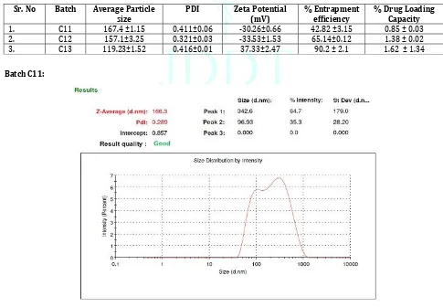
Table 2: Characterization of Cisplatin SLNs
Sr. No Batch Average Particle
size PDI Zeta Potential (mV) % Entrapment efficiency % Drug Loading Capacity
1. C11 167.4 ±1.15 0.411±0.06 -30.26±0.66 42.82 ±3.15 0.85 ± 0.03 2. C12 157.1±3.25 0.321±0.03 -33.53±1.53 65.14±0.12 1.38 ± 0.02 3. C13 119.23±1.52 0.416±0.01 37.33±2.47 90.2 ± 2.1 1.62 ± 1.34
Batch C11:
Batch C12:
Figure 2: Particle Size and PDI of Batch C12 Cisplatin SLNs Batch C13:
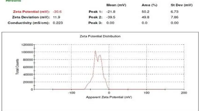
Figure 3: Particle Size and PDI of Batch C13 Cisplatin SLNs 2. Determination of Zeta potential:
The zeta potentials of all the batches were greater than 30mV with negative charge indicating the stability of the formulations. The highest stability as measured by the zeta potential was shown by batch C13 which was formulated at pH 4. Moreover, the anionic lipid DPPG might have added to the negative charge thereby improving the stability of formulation. Out of the three graphs of Zeta Potential of Each batch of Cisplatin SLNs, one graph which is high zeta potential value showed in Figure 4,5and 6.
Batch C11:
Batch C12
Figure 5: Zeta Potential of Batch C12 Cisplatin SLN Batch C13
Figure 6: Zeta Potential of Batch C13 Cisplatin SLN
3. % Entrapment Efficiency:
Drug entrapment efficiency is an important property in drug loaded nanocarriers and directly affects the therapeutic effect of system. The entrapment efficiencies have been shown in table no. 2. The results show that the amount of drug entrapped was more due to the DPPG, soya lecithin and three times Stearic acid increased acidic buffer pH 4.The entrapment efficiency of pH 5 and 6 batches showed less entrapment than pH 4 batch C13.The effect of acidic pH buffer on the % Entrapment efficiency. The effect on entrapment was observed in batch C13 i.e. 90.2 ± 2.1 %. The higher the encapsulation capacity of nanoparticles the larger the amount of drug released at the tumor site.
4. % Drug Loading Capacity:
The % Drug loading capacity of Cisplatin C13 was found to be 1.62 ± 1.34 %. It was shows a good result.
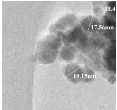
5. Surface Morphology:
The TEM study of optimized Cisplatin SLN illustrated the spherical shape of nanoparticles. The TEM image of the nanoparticles is depicted in Figure No.7.It is evident from
the image that the particles were spherical in shape with narrow size distribution and no aggregation. Particles of optimized Cisplatin SLN formulation were of nanosize. The surfaces of particles are smooth moreover their integrity was found to be intact.
6. In-vitro drug release of Cisplatin Solid lipid Nanoparticles:
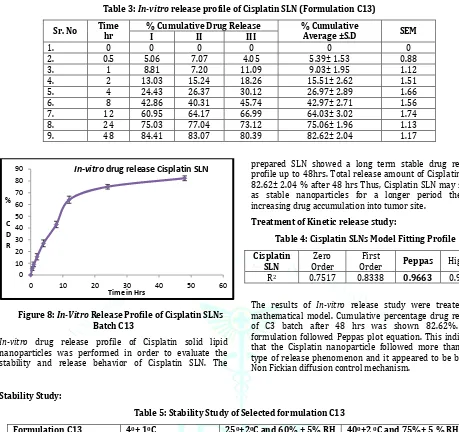
Table 3: In-vitro release profile of Cisplatin SLN (Formulation C13)
Sr. No Time hr % Cumulative Drug Release % Cumulative Average ±S.D SEM
I II III
1. 0 0 0 0 0 0
2. 0.5 5.06 7.07 4.05 5.39± 1.53 0.88
3. 1 8.81 7.20 11.09 9.03± 1.95 1.12
4. 2 13.03 15.24 18.26 15.51± 2.62 1.51
5. 4 24.43 26.37 30.12 26.97± 2.89 1.66
6. 8 42.86 40.31 45.74 42.97± 2.71 1.56
7. 12 60.95 64.17 66.99 64.03± 3.02 1.74
8. 24 75.03 77.04 73.12 75.06± 1.96 1.13
9. 48 84.41 83.07 80.39 82.62± 2.04 1.17
Figure 8: In-Vitro Release Profile of Cisplatin SLNs Batch C13
In-vitro drug release profile of Cisplatin solid lipid
nanoparticles was performed in order to evaluate the stability and release behavior of Cisplatin SLN. The
prepared SLN showed a long term stable drug release profile up to 48hrs. Total release amount of Cisplatin was 82.62± 2.04 % after 48 hrs Thus, Cisplatin SLN may serve as stable nanoparticles for a longer period thereby increasing drug accumulation into tumor site.
Treatment of Kinetic release study:
Table 4: Cisplatin SLNs Model Fitting Profile Cisplatin
SLN Order Zero Order First Peppas Higuchi R2 0.7517 0.8338 0.9663 0.9298
The results of In-vitro release study were treated by mathematical model. Cumulative percentage drug release of C3 batch after 48 hrs was shown 82.62%. The formulation followed Peppas plot equation. This indicates that the Cisplatin nanoparticle followed more than one type of release phenomenon and it appeared to be by the Non Fickian diffusion control mechanism.
Stability Study:
Table 5: Stability Study of Selected formulation C13
Formulation C13 40± 10C 250±20C and 60% ± 5% RH 400±2 0C and 75%± 5 % RH
% Entrapment efficiency 89.04±1.26 87.32±1.58 86.25±2.22
Particle Size 120.2±2.24 122.5±1.65 123.4±3.02
Stability study of the prepared Cisplatin solid lipid nanoparticles C13 were carried out, by storing the formulation C13 at 40± 10C, 250±20C and 60% ± 5% RH, and 400±2 0C and 75%± 5 % RH in humidity control oven for month. The %Entrapment efficiency and Particle size
were carried out. The results % Entrapment efficiency and Particle size at different storage condition are shown in Table 5.The comparing this data with the previous data of C13 it was observed that there is slight increase in Particle size and little Decrease in the % Entrapment efficiency.
Figure 9: Particle size of Cisplatin SLNs of batch C13 after stability 0
10 20 30 40 50 60 70 80 90
0 10 20 30 40 50 60
%
C D R
Time in Hrs
CONCLUSION
The Cisplatin loaded solid lipid nanoparticles met all the requirements of a colloidal drug delivery system. They had particle size in nanosize; their size distribution was narrow and all the particles were in spherical shape. The percent entrapment efficiency and percent drug loading capacities were in the acceptable limits. Moreover, the In-vitro
release pattern revealed that they served the purpose of being a sustained drug delivery system
ACKNOWLEGEMENT
Authors are grateful to Khandelwal Laboratories Pvt. Ltd. Thane for providing the gift sample of Cisplatin. We are also thankful to the Management of Rajarambapu college of Pharmacy, Kasegaon for providing the necessary facilities to carry out this work.
CONFLICT OF INTEREST
The authors declare no conflict of interest.
REFERENCES
1. Nabuhiro N, Souichiro O, Horacio C, Miyamoto M, Novel Cisplatin- incorporated polymeric micells can eradicate solid tumors in mice. Cancer Res 2003; 63:8977-83.
2. Mukherjee S, Roy S, Thakur RS, Solid Lipid Nanoparticles: A Modern Formulation Approach in Drug Delivery System, Indian Journal of Pharmaceutical Sciences, 2009; 71(4):349-358.
3. Reddy R.N., Sharif A, Solid Lipid Nanoparticles: An Advanced Drug Delivery system, 2013; 4(1):161-171.
4. Sathali AH, Ekambaram P, Priyanka K. Solid Lipid Nanoparticles: A Review. Scientific Reviews and Chemical. Communication2012; 2(1):80-102.
5. Doijad RC, Manvi FV, Ghodhwani DM, Joseph R, Deshmukh NV, Formulation and Targetting efficiency of cisplatin Engineered solid lipid nanoparticles, Indian journal of pharmaceutical sciences, 2008; 203-207.
6. Bende Kruijff, Gea Speelmans, Wendy H.H.M. Sips, Ruud J. H. Grisel, Rutger,. M. Staffhorst, Anne Marie J. Fichtinger-Schepman, Jan Reedijk, The interaction of the anti-cancer drug cisplatin with phospholipids is specific for negatively charged phospholipids and takes place at low chloride ion concentration , Biochimica et Biophysica Acta(BBA)-Biomembranes,1996; 1284(1):60-66.
7. Pandita D, Ahuja A, Velpandian T, Lather V, Dutta T, Khar RK, Characterization and In-vitro assessment of paclitaxel loaded lipid nanoparticles formulated using modified solvent injection technique, Pharmazie, 2009; 64: 5
8. Singh S, Dobhal AK, Jain A, Pandit JK, Formulation and Evaluation of Solid Lipid Nanoparticles of a water soluble drug: Zidovudine, Chem. Pharm, Bulletin, 2010; 58/(5):650-655.
9. DaWei C, Yi Fan L, LiXiang R, XiuLi Z, Solid lipid nanoparticles for enhancing Vinpocetine’s oral bioavailability, Journal of controlled release, 2006; 114:53-59.