STUDIES ON POLYHEDRAL NIOSOMES
A thesis presented by
Parinya Arunothayanun, B.Pharm.
in partial fulfilment o f the requirements for the degree of
Doctor o f Philosophy
of
the University o f London
October 1998
ProQuest Number: 10104872
All rights reserved
INFORMATION TO ALL USERS
The quality of this reproduction is dependent upon the quality of the copy submitted.
In the unlikely event that the author did not send a complete manuscript and there are missing pages, these will be noted. Also, if material had to be removed,
a note will indicate the deletion.
uest.
ProQuest 10104872
Published by ProQuest LLC(2016). Copyright of the Dissertation is held by the Author.
All rights reserved.
This work is protected against unauthorized copying under Title 17, United States Code. Microform Edition © ProQuest LLC.
ProQuest LLC
789 East Eisenhower Parkway P.O. Box 1346
ZMZ'XA^SA/iA^
u ^ a i L ^ a i m
^avaniarv mmvnnaai'd
The most exciting phrase to hear in
science,
the one that
heralds new discoveries, is not 'Eureka!' (I found it!) but
'That's funny .. . '
ACKNOW LEDGEM ENTS
The last three years in the School of Pharmacy have been one of the most fantastic times of my life. I would like to thank the following people who have been taking part in both my studies and contributing to my joyous experiences.
First and foremost, I would like to express my sincere gratitude to my supervisor, Professor Alexander T. Florence, for his superb guidance and encouragement during my time of study. I will always remember your excellent supervision (also with aspects of fun) which has been in the kindest, most open-minded and friendly way.
I wish to convey special thanks to Dr Ijeoma F. Uchegbu for invaluable techniques, consolation and friendship. Dr Duncan Q. M. Craig is gratefully acknowledged for his support and advice in thermal studies. I am deeply indebted to Dr John A. Turton for his superb supervision during the in vivo study. I would like to give my appreciation to Professor Gregory Gregoriadis for being kind to me during my stay in CDDR.
My thanks also go to Mr Dave McCarthy for his friendship and excellent microscopic work. Miss Clair Liu for her HSDSC introduction, Mr Thongchai Sooksawate for his help with micropipette techniques, Mr Douglas Banning for advice in shear flow studies, Ms Annie Cavanagh for immediate help with slides and posters, M r W ilfred Baldeo for technical advice in HPLC, and Mr Steve Coppard for his help during in vivo studies. Miss Marie- Sophie Bernard, Mr Bipin Shah, Mr Sandeep Kiri, Miss Jayna Patel were also appreciated for fruitful collaborations.
The Government Pharmaceutical Organisation, Thailand is gratefully acknowledged for financial support.
For friendship, a big big thank go to Sasitorn, Jittima, Ruedeekorn, Sudax, Sakthi, Gill, Yvonne, Dejana, Ibrahim,Victoria, Roghieh, Dejana, Brenda, Steve, Kent, Malini, Begona, Mia, Jean Christophe, Kirsten and all my friends in CDDR. Affection from all Thai friends in London is greatly admired.
There were also times when I experienced down-hearted moments. It would never have been possible for me to be here and now without tremendous support from my family (and their long distance calls), and all my friends. I would like to thank Prapassom, Pomtip and, especially, Itsaraet for being such good friends of mine all the ways.
ABSTRA CT
Niosomes prepared from non-ionic surfactants were studied and characterised with regard to their physicochemical and biological properties. Hexadecyl diglycerol ether (C1 6G2) and
a series of poly(oxyethylene) alkyl ethers form, with an equimolar amount of cholesterol, a mixture of largely spherical and tubular niosomes. In the absence of cholesterol, they form polyhedral stmctures below their phase transition temperature (Tm) and they transform into spheres on heating above Tm. Various properties, namely vesicle shape, size, encapsulation efficiency, membrane permeability, thermal behaviour, viscosity, and osmotic activity were investigated using in addition, as a comparator, sorbitan monostearate (Span 60) niosomes. The role of membrane composition was in particular an issue.
Rheological studies showed that viscosity of niosomes can be affected by a number of factors including vesicle shape. The values of intrinsic viscosity were used in attempt to predict the hydration and the volume fraction of the vesicle dispersions.
Polyhedral niosomes formed by CJ6G2 are less osmotically active, and more permeable than
their spherical/ tubular counterparts, formed with cholesterol. The release profiles of 5(6)- carboxyfluorescein from both types of niosomes are also different and can be affected by the content of the hydrophilic surfactant poly(24) oxyethylene cholesteryl ether (Solulan C24) in the membranes.
The high encapsulation efficiency of luteinising hormone releasing hormone (LHRH) in C1 6G2 niosomes was achieved when these are prepared with the remote loading method
(exploiting either pH or (NH4)2S0 4 gradients). In vitro studies showed that spherical/
tubular C,6G2 niosomes are more stable than their polyhedral counterparts in rat plasma and
muscle homogenate. Following intramuscular injection in rats, ^^^I-LHRH solution was cleared from the site of injection within 2 h, whilst both polyhedral and spherical/ tubular CjgG2 niosomes act as a depot and release ^^^I-LHRH over 25 h and 49 h, respectively.
TABLE OF CONTENTS
A C K N O W LE D G E M E N TS....iv
A B S T R A C T .... v
L IS T OF F IG U R E S...xiii
L IS T OF T A B L E S...xviii
A B B R E V IA T IO N S...x i x
CHAPTER 1
INTRODUCTION TO VESICULAR SYSTEMS
...
1
1.1. DEFINITIONS ... 1
1.2. FORMATION OF V E S IC L E S ... 2
1.3. SOME PHYSICOCHEMICAL ASPECTS OF V E S IC L E S ... 4
1.3.1. Thermal behaviour of vesicle m em b ran es... 4
1.3.2. Vesicle shape transform ation... 5
1.3.3. Osmotic b e h av io u r... 9
1.3.4. Rheological p ro p e rtie s... 10
1.4. NON-SPHERICAL V E S IC L E S ... 11
1.5. APPLICATIONS OF VESICULAR SYSTEMS ... 16
1.5.1. Medical applications ... 16
1.5.2. Non-medical applications ... 19
OUTLINE OF WORK IN T H E SIS... 20
CHAPTER 2
THE IMPORTANCE OF MEMBRANE COMPOSITION TO THE
PHYSICOCHEMICAL PROPERTIES OF V E SIC LE S
...
21
MATERIALS AND METHODS ... 22
2.2. MATERIALS ... 22
METHODS... 25
2.3. GENERAL METHODS FOR PREPARATION OF N IO S O M E S ... 25
2.4. EFFECT OF MEMBRANE COMPOSITION ON VESICLE SHAPE ... 26
2.4.1. Cryo-scanning electron microscopy (Cryo-SEM) ... 26
2.4.2. Temperature-induced vesicle shape transform ation... 26
2.4.3. An investigation into phase transition behaviour of polyhedral niosomes.26 2.4.4. Transfer of cholesterol from spherical/ tubular niosomes to polyhedral niosomes ... 27
2.5. EFFEC T OF M EM BR A N E CO M PO SITIO N ON V ESIC LE SIZE AND ENCAPSULATION E F F IC IE N C Y ... 27
2.5.1. Size of unsonicated niosomes ... 27
2.5.2. Size stability of sonicated C1 6G2 niosomes ... 27
2.5.3. Transmission electron microscopy (T E M )...
28
2.5.4. Separation of unencapsulated material and determination of encapsulation e ffic ie n c y ... 28
2.6. MEMBRANE PERMEABILITY OF NIOSOMES ... 29
2.6.1. Release of CF from Span 60 niosomes ... 29
2.6.2. Release of CF from niosomes ... 29
RESULTS AND DISCUSSION ... 30
2.7. EFFECT OF MEMBRANE COMPOSITION ON VESICLE SHAPE ... 30
2.7.1. Temperature-induced vesicle shape transform ation... 35
2.7.2. Phase transition behaviour of polyhedral and spherical/tubular niosomes...39
2.7.3. Cholesterol transfer: observation through shape tran sfo rm atio n 46 2.8. EFFECT OF MEMBRANE COMPOSITION ON VESICLE SIZE AND
ENTRAPMENT EFFIC IEN C Y ... 50
2.8.1. Span 60 niosomes ... 50
2.8.2. Cj6G2 niosomes ... 51
2.8.2.a. Growth in size o f sonicated polyhedral niosomes . . . . 52
2.9. EFFECT OF MEMBRANE COMPOSITION ON MEMBRANE PERMEABILITY...55
2.9.1. Multi-component n io s o m e s ... 55
2.9.2. Release of CF from C1 6G2 niosomes ... 57
2.10. CO N C LU SIO N S... 58
CHAPTER 3
VISCOSITY OF NIOSOME D ISPE R SIO N S
...
60
3.1. IN TRO D U CTIO N... 60
3.1.1. Rheology of colloidal dispersions ... 61
3.1.2. Problems in the determination of volume fraction and hydration values of vesicle d isp ersio n s... 64
MATERIALS AND METHODS ... 66
3.2. MATERIALS ... 66
METHODS... 67
3.3. VISCOMETRIC STUDIES OF SOLULAN C24 MICELLAR SOLUTIONS . . . 67
3.4. VISCOMETRIC STUDIES OF SPAN 60 NIOSOME D ISPER SIO N S... 67
3.4.1. Effect of vesicle concentration, size and membrane c o m p o sitio n 67 3.4.2. Effect of electrolytes ... 68
3.4.3. Effect of preparation methods and lam ellarity... 68
3.5. VISCOMETRIC STUDIES OF C ^ A NIOSOME D ISPER SIO N S... 69
RESULTS AND DISCUSSION ... 69
3.6. VISCOMETRIC STUDIES OF SOLULAN C24 MICELLAR SOLUTIONS . . . 69
3.7. VISCOMETRIC STUDIES OF SPAN 60 N IO SO M E S... 70
3.7.1. Effect of vesicle concentration... 70
3.7. La. Estimations o f volume fraction and h y d r a tio n 73 3.7.2. Effect of vesicle size ... 77
3.7.3. Effect of vesicle membrane com position ... 77
3.7.3.a. Hydration ... 78
3.7.4. Effect of electrolytes ... 79
3.7.5. Effect of preparation methods ... 81
3.8. VISCOMETRIC STUDIES OF C ^ A N IO SO M E S... 82
3.8.1. Effect of vesicle shape and tem perature... 82
3.9. C O N C LU SIO N S... 85
Chapter 4
OSMOTIC BEHAVIOUR OF POLYHEDRAL NIOSOMES
...
86
4.1. IN TRODUCTION ... 86
MATERIALS AND METHODS
... 874.2. MATERIALS ... 87
METHODS... 88
4.3. OSMOTIC ACTIVITY OF POLYHEDRAL NIOSOMES ... 88
4.4. THE RELEASE O EC E FROM POLYHEDRAL N IO S O M E S ... 88
4.4.1. The influence of osmotic gradient on CF re le ase ... 88
4.4. La. Effect ofN aC l on the phase transition behaviour . . . . 89
4.6. THE RELEASE OF CF FROM POLYHEDRAL N IO S O M E S ... 94
4.6.1. The influence of osmotic gradient on CF re le ase ... 94
4.6.La. The effect o f co-entrapped NaCl on CF r e le a s e 98 4.6.1.b.Effect ofN aC l on the phase transition behaviour o f polyhedral niosomes ... 103
4.6.2. The effect of pH on CF release ... 106
4.7. C O N C L U SIO N ... 108
CHAPTER 5
IN VITRO/ IN VIVO EVALUATION OF LUTEINISING HORMONE
RELEASING HORMONE (LHRH) LOADED NIOSOMES
...
109
5.1. IN TRO D U CTIO N ... 109
5.1.1. L H R H ... 109
5.1.2. Approaches to the delivery of LHRH ... I l l 5.1.3. In vitro/ in vivo evaluation of LHRH loaded Cj^Gj n io so m e s... 112
MATERIALS AND METHODS
... 1135.2. MATERIALS ... 113
METHODS... 113
5.3. PREPARATION OF LHRH LOADED POLYHEDRAL N IO S O M E S 113 5.4. ANIMAL H U SB A N D R Y ... 115
5 .5 ./A V7TR0 E V A L U A T IO N ... 115
5.5.1. Preparation of plasma ... 115
5.5.2. Preparation of 5% muscle h o m o g e n a te ... 116
5.5.3. Evaluation of niosomes in v itr o... 116
5 .6 ./VV7V0 E V A L U A T IO N ... 116
5.8. W WTRO STABILITY S T U D Y ... 120
5.9. IN VIVO CLEARANCE S T U D Y ... 122
5.10. CO N C LU SIO N S... 127
CHAPTER 6
MANIPULATION OF NIOSOMES: APPROACHES TO BIOMIMETIC
PULSED DELIVERY AND FABRICATION OF MICROSTRUCTURES
128 6.1. IN TRO D U CTIO N ... 1286.2. A BIOMIMETIC APPROACH TO PULSATILE DELIVERY ... 129
MATERIALS AND METHODS ...
1316.2.1. Materials ... 131
METHODS...
1316.2.2. Preparation of niosomes filled micropipettes ... 131
6.2.3. Preparation of LHRH loaded n io so m e s... 132
6.2.4. Extrusion of microspheres and niosomes from m icropipettes... 132
6.2.5. Preparation of niosome filled c a p illa rie s... 133
6.2.6. RESULTS AND DISCUSSION ... 133
6.3. MICROFABRICATION OF SURFACTANT STRUCTURES... 137
MATERIALS AND METHODS ...
1376.3.1. Materials ... 137
6.3.4. RESULTS AND DISCUSSION ... 138
6.4. C O N C LU SIO N S... 146
CHAPTER 7
CONCLUSIONS AND FUTURE W O R K
... 1477.1. C O N C LU SIO N S... 147
7.2. FUTURE W O R K ... 150
B IB L IO G R A P H Y... 152
LIST OF FIGURES
Chapter 1
Figure 1.1: Schematic representation of a vesicle ... 1
Figure 1.2: Schematic representation of lipid bilayer polymorphic phases... 4
Figure 1.3: Micropipette suction of liquid phase and gel p h a s e ... 5
Figure 1.4: Fusion of two v e s ic le s ... 6
Figure 1.5: Three spherical vesicles connected by a tether ... 7
Figure 1.6: Schematic representing the mechanisms of budding/ fission processes 7 Figure 1.7: Conformai diffusion of a toroidal vesicle with holes... 8
Figure 1.8a: Helical structures of p-Methoxy benzylidene-p-n-butylaniline and 10% cholesteryl nonanoate... 12
8b: Helical structures of comeal lens of beetles...12
9a: Schematic representation of scroll formation from planar bilayers 13 9b: Hollow cylinder formed from a diacetylenic phospholipid... 13
I Oa: Optical micrograph of C , discomes... 14
10b: Confocal laser scanning micrograph of a polyhedral vesicle... 14
II a: Electron micrograph of a helical bundle of vesicles...15
11b: Electron micrograph of geodesic niosomes... 15
lie : Optical micrograph of vesicle-enclosed tubules...15
1 Id: Electron micrograph of vesosomes ... 15 Figure
Figure
Figure
Figure
Figure
Figure
Figure
Figure
Figure 2.2: Molecular structures of cholesterol and Solulan C24 ... 25
Figure 2.3a: Phase contrast micrograph of Ci^EOg polyhedral niosomes ... 31
Figure 2.3b: Photomicrograph of CF loaded C1 6G2 polyhedral niosomes... 31
Figure 2.4: Photomicrograph of spherical n io s o m e s ... 32
Figure 2.5a: Cryo-scanning electron micrograph of C1 6G2 polyhedral niosomes . . 33
Figure 2.5b: Cryo-scanning electron micrograph of C1 6G2 spherical niosomes. . . . 33
Figure 2. 5c&d : Cryo-scanning electron micrographs of tubular niosomes ... 34
Figure 2.6: Shape transformation of Ci^EG, polyhedral niosomes ... 36
Figure 2.7: Shape transformation of a CJ6G2 polyhedral vesicle ... 37
Figure 2.8: “Budding o f f ’ of CigG2 spherical v e sic le s... 38
Figure 2.9: HSDSC trace of polyhedral n io s o m e s ... 41
Figure 2.10: Endothermie transitions of niosomes on increasing cholesterol content.42 Figure 2.1 la: Endothermie transitions of polyhedral niosomes on increasing Solulan C24 content ... 44
Figure 2.1 lb: Exothermic transitions of C1 6G2 polyhedral niosomes on increasing Solulan C24 content... 44
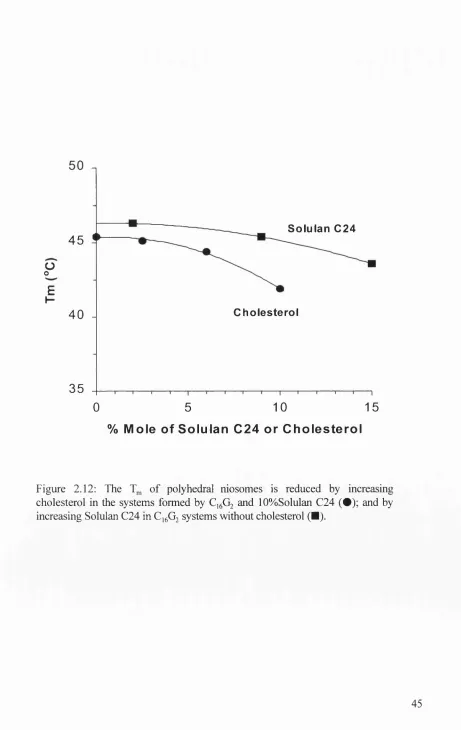
Figure 2.12: Transition temperatures of C1 6G2 niosomes on increasing cholesterol and Solulan C24 content... 45
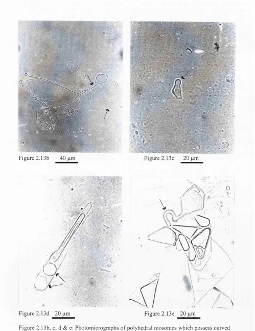
Figure 2.13a: Photomicrograph of a mixture of C1 6G2 polyhedral niosomes and spherical niosomes before incubation... 47
Figure 2.13b-e:Photomicrographs of polyhedral niosomes after incubation at 55°C with spherical n io so m es... 48
Figure 2 .14b: Photomicrographs of a mixture of Cj^EOg polyhedral niosomes and
unsonicated spherical niosomes after incubation at 55°C 49
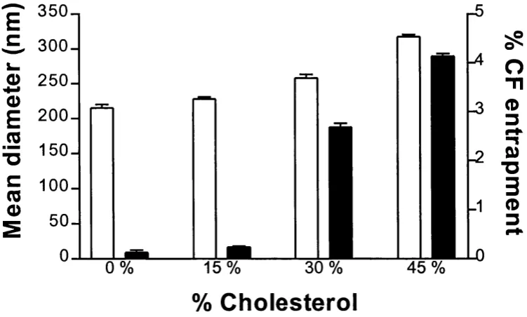
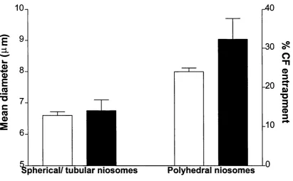
Figure 2.15: The size and % entrapment of sonicated CF loaded Span 60 niosom es... .50 Figure 2.16: The size and % entrapment of CF loaded unsonicated C1 6G2 niosomes 51
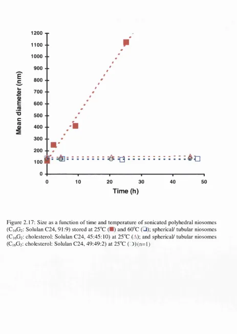
Figure 2.17: Size as a function of time and temperature of sonicated C1 6G2 niosomes.53
Figure 2.18a&b: Transmission electron micrographs of unsonicated C1 6G2 polyhedral
niosomes... 54
Figure 2.18c&d: Transmission electron micrographs of sonicated CJ6G2 polyhedral
niosomes ... 54
Figure 2.19a: Release profiles of CF from the five systems: CF solution; a mixture
of CF solution and empty Span 60 niosomes; CF loaded Span 60
niosomes with 15%; 30%; and 45% cholesterol ... 56
Figure 2 .19b: Release profile of CF from a multi-component niosome dispersion and
the predicted release p ro f ile ... 56
Figure 2.20: Release profile of CF from CjgG2 niosomes ... 57
Figure 2.21: Schematic representing factors affecting shape and molecular
conformation of polyhedral n io s o m e s ... 59
Chapter 3
Figure 3.1: Reduced specific viscosity plots of Solulan C24 micellar solutions . . 70
Figure 3.2: Relative viscosity of Span 60 niosomes with 1 & 10% Solulan C24. . 71
Figure 3.5: Reduced specific viscosity plots of 278 nm-Span 60 niosomes,
assuming (p = C, 1.5C, 2C, and 3C... 76
Figure 3.6: Relative viscosity of 270 nm-Span 60 niosomes formed with 10%
Solulan C24 or 10% dicetyl phosphate in water and in 0.5 M NaCl . . 80
Figure 3.7: Relative viscosity of sonicated hand-shaken vesicles and sonicated
reversed phase evaporation vesicles ... 82
Figure 3.8: Relative viscosity of C1 6G2 niosomes at different temperature... 83
Chapter 4
Figure 4.1 : The reduction in niosome mean diameter in NaCl so lu tio n s... 92
Figure 4.2: The reduction in mean niosome diameter in glucose s o lu tio n ... 92
Figure 4.3: The calculated water efflux from niosomes in NaCl solutions... 93
Figure 4.4: The cumulative CF released from niosomes in NaCl solutions over a
5 h period ... 95
Figure 4.5: The cumulative CF released from niosomes 5 h after dispersion in
NaCl solutions... 96
Figure 4.6: Schematic representing osmotic behaviour of C1 6G2 niosomes... 97
Figure 4.7: The release of CF from C1 6G2 niosomes into isotonic media (2 M NaCl)
and hypotonic media (water)... 100
Figure 4.8: Release of CF from Cj^G2 polyhedral niosomes with 2 or 9% Solulan
C24 into hypotonic media (w a te r)... 101
Figure 4.9: Photomicrographs of Ci^G2 polyhedral niosomes with 9% Solulan C24
encapsulating CF prepared in 2.0 M NaCl and then dialysed against
Figure 4.10: Photomicrographs of CigG2 polyhedral niosomes with 2% Solulan C24
encapsulating CF prepared in 2.0 M NaCl and then dialysed against
2 M NaCl and water... 102
Figure 4.11: HSDSC traces of 2% Solulan C24-Polyhedral niosomes ... 104
Figure 4.12: HSDSC traces of 9% Solulan C24-Polyhedral niosomes in 2M NaCl.. 105
Figure 4.13: HSDSC traces of 15% Solulan C24-Polyhedral niosomes in 2M NaCl..106
Figure 4.14: M olecular structure of 5(6)-carboxyfluorescein ... 107
Figure 4.15: Release of CF from C ,6G2 niosomes at pH 5 and pH 8... 107
Chapter 5
Figure 5.1: M olecular structure of LHRH ... 110
Figure 5.2: Entrapment of LHRH acetate into polyhedral n io s o m e s ... 119
Figure 5.3: Release profile of ‘^^I-LHRH from niosomes in plasma and in 5%
muscle h o m o g e n a te ... 121
Figure 5.4: ‘^‘^I-LHRH remaining at the intramuscular injection site ... 123
Figure 5.5: ^^^I-LHRH detected in blood after intramuscular injection ... 124
Chapter 6
Figure 6.1 : Electron micrograph of a synapse... 129
Figure 6.2a: Schematic representing the releasing of active agents from a neuron. 130
Figure 6.2b: Schematic representing a suggested biomimetic delivery device... 130
Figure 6.5;
Figure 6.6a:
Figure 6.6b:
Figure 6.7:
Figure 6.8:
Figure 6.9:
Figure 6.10:
Figure 6.11:
Figure 6.12:
Figure 6.13:
Release profile of LHRH acetate obtained by pulsatile expulsion . . . 134
Extrusion of single polystyrene m icro sp h eres... 136
Extrusion of single n io so m e s... 136
Electron micrographs of a silica capillary tube loaded with niosomes. .. 136
Extrusion of a 30 pm-spherical vesicle... 140
Extrusion of a 30 pm-polyhedral v e s ic le ... 140
Extrusion of three consecutive large polyhedral niosomes ... 141
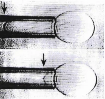
Straight rods obtained by extruding polyhedral n io s o m e s ... 142
Suction of an oligolamellar spherical vesicle ... 144
Suction of a polyhedral vesicle ... 145
LIST OF TABLES
Table 2.1: Materials used ... 23Table 2.2: Phase transition temperatures and shape transformation temperature of polyhedral niosomes ... 40
Table 3.1 : Intrinsic viscosity of niosomes... 73
Table 3.2: Intrinsic viscosity and hydration value of niosomes at various range of assumed volume fractions... 76
Table 4.1 : The increase in mean diameter of niosomes encapsulating 2 M NaCl after being dispersed in water for 5 h ... 93
ABBREVIATIONS
C^EO^ polyoxyethylene 2 cetyl ether
C„EO, polyoxyethylene 5 cetyl ether
hexadecyl diglycerol ether
C.6G3 hexadecyl triglycerol ether
C.sEO^ polyoxyethylene 2 stearyl ether
C,«EO, polyoxyethylene 5 stearyl ether
CF 5(6)-carboxyfluorescein
GPP Critical packing parameter
DCP Dicetyl phosphate
DPPC l,2-Dipalmitoyl-sn-glycero-3-phosphocholine
DRV Dehydration-rehydration vesicles
DSC Differential scanning calorimetry
DSPC l,2-Distearoyl-sn-glycero-3-phosphocholine
HPLC High performance liquid chromatography
HS Hand-shaking
HSDSC High sensitivity differential scanning calorimetry
I.D. Inner diameter
LHRH Luteinizing hormone releasing hormone
MW Molecular weight
O.D. Outer diameter
PBS Phosphate buffered saline
PCS Photon correlation spectroscopy
PEG Polyethylene glycol
REV Reverse-phase evaporation vesicles
SDS Sodium dodecyl sulphate
SEM Scanning electron microscopy
TBS Tris buffered saline
CHAPTER 1
INTRODUCTION TO VESICULAR SYSTEMS
1.1. DEFINITIONS
Certain amphiphilic molecules self-assembl on dispersing in aqueous media to form sealed
vesicles which are composed of bilayer membranes enclosing an aqueous core (Figure 1,1).
_w"CCÛQQÛrw-Figure 1.1: Schematic representation of a vesicle formed by a bilayer of amphiphilic molecules.
Vesicular dispersions formed by phospholipids, so called “liposomes”, have been used
widely as cell models and drug delivery systems (Bangham et a i, 1965). The term
“niosome” was initially used by L’Oreal, France (Vanlerberghe et a i, 1973), and refers to vesicles formed mainly by self-assembly of synthetic non-ionie surfactants with an optional
addition of charged amphiphiles and cholesterol (Florence & Baillie, 1989). However, other
names such as non-phospholipid liposomes or non-ionic liposomes have also been used since
they are considered as analogues of liposomes.
Niosomes offer a versatile alternative to liposomes as their surfaces, which can
subsequently elucidate the vesicle behaviour in vivo, can be modified by the choice o f a wide range o f surfactants with differing structure. Due to the fact that both liposomes and
niosomes are vesicles comprising bilayers, their physicochemical properties have very
much in common. This chapter reviews general knowledge on some aspects o f vesicular
systems relevant to the studies in this thesis.
1.2. FORMATION OF VESICLES
Amphiphilic molecules aggregate in aqueous solution to give a number o f forms such as
micelles, inverted micelles, spherical micelles, planar bilayers, and curved bilayers
(vesicles). However, in non-polar organic media, “inverted” or “inverse” vesicles can also
be formed (Kunieda & Rajagopalan, 1996). A critical packing parameter (GPP) of
amphiphilic molecules, defined as
CPP = v / l , a , (1.1)
has been derived to predict the structure o f surfactant assemblies, where v = hydrocarbon
chain volume, 1^, = hydrocarbon critical chain length, = hydrophilic head group area
(Israelachvili et a l, 1980; Israelachvili, 1991). A surfactant with a GPP value o f 0.5-1 is predicted to form flexible bilayers which can aggregate into closed vesicles. However, the
GPP calculated for some vesicle-forming amphiphiles do not fall in the suggested
solvophobic forces, van der Waals forces, electrostatic interactions, entropy effects, and
hydrogen bonds, that determine the shape o f supramolecular assemblies (Gmner, 1987;
Fuhrhop & Koning, 1994).
Apart from phospholipids, the various chemical structures o f synthetic surfactants that form
vesicles on hydration have been reviewed by Ozer et a l, (1991), Florence (1993), and Uchegbu & Florence (1995). Formation o f vesicles can also be induced by an interaction
between positively and negatively charged amphiphiles e.g. in the mixture o f sodium
dodecyl sulfate and dodecyltrimethylammonium bromide (Kamenka et a l, 1992). It is noteworthy that vesicles can also be formed by monolayer membranes in the case of
bolaform amphiphiles which carry two hydrophilic head groups, one on each end o f the
hydrophobic chain (Fuhrhop & Koning, 1994).
Various preparation methods to obtain vesicle dispersions with required optimum size and
lamellarity have been reviewed (New, 1990), providing the concentration o f amphiphiles
exceeds the critical bilayer concentrations (CBC), the value depending on amphiphile; for
example with distearoyl phosphatidylcholine, the CBC is on the order o f lCt’° M (Hristova
& Needham, 1995). Vesicles can be prepared simply by hydrating dried film o f lipids or
surfactants in round bottom flasks, the so called “hand-shaking method”. Other techniques
have also been developed to avoid the use o f organic solvents e.g. formation o f vesicles in
gas streams (Talsma et a l, 1994), inducing micelle-vesicle transitions by removal of surfactants from lipids containing mixed micelles via dialysis (New, 1990), or by removing the water-interacting head group o f surfactant via an enzymatic process (Chopineau et al,
1.3. SOM E PH YSICOCHEM ICAL ASPECTS OF VESICLES
1.3.1. Therm al Behaviour of Vesicle M embranes
The physicochemical properties o f the bilayer membranes can be diverse depending on the
com position o f the membranes which determines the polymorphic phase o f the bilayers.
Figure 1.2 shows some examples o f bilayer geometries assumed by lipid-water systems.
Bilayers undergo changes in physical state with temperature. Above the phase transition
temperature (T^) the bilayer exists as a two-dimentional liquid. Below this temperature the
bilayer is a two-dimenfional solid and the lipid molecules are tightly packed in a crystal
lattice (Gruner, 1987).
m m m
M n m ii
/ / / / / / / #
WD5IHII
illUÜMill
U M S H I M H
m m m
itm tm m
ininnrminit
n n u B i
iluuiiiiiiuuii
ummuhi
<a) L q (b) (c)
Figure 1.2: A schematic representation of lipid bilayer polymorphic phases. The Lp and Lp. phases the chains are in a gel (frozen) state, whereas in the phase is a liquid crystalline (melted chain) bilayer phase with fluid acyl chains.
The value o f T^ o f bilayers varies with the composition of membranes and reflects various
properties. For example, the lipids with longer hydrophobic chains form membranes which
have higher phase transition temperatures and are less permeable to entrapped solute
compared to those formed by lipids with shorter chain lengths (Buckton et al, 1992). The
mechanochem ical properties o f membranes can be different depending on their
A ddition o f cholesterol into the gel state membranes has been found to increase the
mobility o f amphiphiles, this therefore increasing the overall membrane fluidity and
decreasing the phase transition temperature and enthalpy (New, 1990; Taylor & Morris,
1995).
Figure 1.3: Videomicrographs of vesicles formed by stearoyloleoyl p h o s p h a tid y l c h o lin e and p a lm ito y lo le o y l p h o s p h a tid y l ethanolamine (40:60) as the temperature is decreased: (a) liquid phase vesicle at 20°C; (b) gel phase vesicle at 13°C showing 20% reduction in vesicle membrane area when experimenting with equal suction. From Needham & Zhelev (1996).
1.3.2. Vesicle Shape Transform ation
Vesicles can exhibit a variety o f morphologies and shape transformations, as observed in
biomembranes, since the molecules in the bilayers can move laterally along the membranes.
Membrane fluctuations can be induced by various factors such as temperature, the bilayer
asymmetry, or applied external forces (Lipowsky, 1995).
Cytomimetic behaviour has been widely observed. Fusion o f vesicles (Figure 1.4) involves
aggregation o f vesicles with close apposition o f bilayers following a destabilising process
leading to merging o f the bilayers and mixing o f the contents o f each vesicle. These
processes can be induced by a number o f factors including temperature (Allen et ai, 1990),
fusogenic molecules such as polyethylene glycols (Yamazaki & Ito, 1990), cations or
anions (Hope et al, 1983; Allen et al, 1990; Mosharraf et al, 1995; Menger & Lee, 1995)
which allow a fusion process through hydrophobic or charge interactions, depending on the
nature and charge o f the lipids (Israelachvili, 1991 ; Bernsdorff et a/., 1996).
Figure 1.4: Fusion of two didodecyldimethylammonium bromide vesicles taking place over a few seconds after injecting sodium acetate in the vicinity o f the vesicles (bar^25 pm). From Menger & Gabrielson (1995).
The healing process takes place if small defects are created due to the fluidity o f the
membranes; for example, following exposure to low concentrations o f sodium c ho late,
didodecyldimethylammonium bromide vesicles were observed to break open and reseal into
intact vesicles (Menger & Lee, 1995). However, it was found that membrane defects
created by osmotic shrinkage can re-anneal in the liquid state, but not in the gel state
(Disalvo c /a/., 1996).
Membranes are sensitive to the change in temperature, especially near their phase transition
temperature. Dimyristoylphosphatidylglycerol vesicles were reported to develop extended
1996). The events take place when the surface area-to-volume ratio o f the vesicles is
increased due to the increase in excess area and/or decrease in volume, as shown in figure
1.6.
Figure 1.5: A single spherical egg phosphatidylcholine vesicle loaded with fluorescent dextran was transformed, by application o f hyperosmotic glucose solution, into these three spheres connected by a narrow tether. Bar=l 0 pm. From Mathivet et al.
(1996).
Fleating >► T h e r m a l e x p a n s i v i t y o f the m e m b r a n e s is h i g he r t han that o f e n t ra p p e d wa te r
V
)|^Excess ^ s urface area
^ n c l o s e d ’ v o l u m e
A
A r e a f V o l u m e
^ B u d d i n g / fission
« E v a p o r a t i o n o f w a t e r . W a t e r e f fl ux e g. o pe n the c o n t a i n e r ► f t o m vesi cl es # I n c r e a s i n g m e d i u m o sm ol a r i t y
e.g. a d d i t i o n o f suc r ose
Conventional spherical vesicles can exhibit conformai diffusion, induced, for example , by
temperature changes. The shapes o f the vesicles change continuously and randomly, and
the
finally reach an equilibrium non-spherical topology of which^elastic curvature energy of the
fluid membranes is minimized under certain physical constraints; for example , genus 1
(vesicles with one hole) or genus 2 (vesicles with 2 holes) vesicles (Lipowsky, 1995;
Michalet & Bensimon, 1995) (Figure 1.7).
Figure 1.7: Examples of conformai diffusion of a toroidal vesicle with holes: all shapes have the same bending energy, surface area, enclosed volume, and total mean curvature. From Lipowsky (1995).
Shape transformations have also been studied by exposing the vesicles to various physical which
agitations. For example, cylindrical vesicles^formed with dimyristoyl phosphatidyl choline
can be split into a number o f smaller vesicles when manipulated by optical tweezers
(Lipowsky, 1995). Theoretical studies o f the axisymmetric vesicles flowing down narrow
capillaries showed that a vesicle experiences a shape transformation starting from a sphero-
cylinder shape and ending in a bell shape on increasing o f pressure gradient, as observed
in red blood cells (Bruinsma, 1996). Effect of gravity was also studied theoretically which
physical stress (Evans & Kwok, 1982; Lasic & Needham, 1995; Needham & Zhelev, 1996).
For example, Menger & Gabrielson (1995) found that an oligolamellar giant vesicle can be
ruptured layer-by-layer, which results in a smaller vesicle using sharp micropipette.
1.3.3. Osmotic Behaviour
Cells posses semi-permeable membranes and respond to osmotic gradients (Yoneda, 1973).
Their volume and/or turgor pressure are controlled by modulating cytoplasmic osmolality
in response to changes in environmental osmolality by means o f diffusion or transporter
proteins. It is well known that lipid vesicles are osmotically sensitive (Bangham et al, 1967). Osmotic stress can affect properties o f vesicles in many aspects from inducing
membrane undulation (Menger & Lee, 1995) to change in vesicle size (Sun et a l, 1986), shape (Bemdl et a l, 1990) and membrane permeability (Iga et a l, 1989).
Under hypertonic gradients, osmotic shrinkage causes changes in the interlamellar distance
in multilamellar vesicles (Viera et al, 1996). Previous studies found that osmotic shrinkage can cause defects at the membrane surface, an event which is likely to occur when the
bilayer is in the gel state (Disalvo et a l, 1996). Water outflow can also induce topological changes o f vesicles, as discussed in section 1.3.2.
Changes in vesicle volume and area due to osmotic stress lead to changes in the elastic
modulus o f bilayers, their morphology, and to subsequent vesicle lysis (Senisterra et a l, 1988; Mui et a l, 1993). In hypotonic media, an excess o f water influx beyond a critical point, depending on the system, produces an expansion o f the bilayer resulting in changes
subjected to hypotonic solution, mast cell vesicles o f mice can increase in diameter up to
73% (Brodwick et a l, 1992) compared to 152% found in inner medullary collecting duct cells o f rats (Grünewald et a l, 1993). The latter was found to regulate the volume by releasing sorbitol, an intracellular osmolyte, to the external medium. The same response is
also found in the secretion o f bioactive substances, e.g. hormone is released from the
intracellular vesicles o f endocrine cells following cell swelling induced hy medium
hypo-osmolarity or permeant molecules (Inukai et a l, 1992).
1.3.4. Rheological properties
The rheological characteristics o f vesicular suspensions have not been widely studied. A
limited amount o f work has been published on the rheology o f dilute vesicle dispersions
(Florence, 1993; Muzzalupo et a l, 1996). Some have concentrated on the viscoelastic nature o f systems (Smeulders et a l, 1990; Hoffmann et al, 1994 & 1995 ; Seki & Komura,
1995) or on their flow properties at high concentration (Kato et a l, 1983; Sakai et al, 1997), both key topics.
Vesicles, particularly if multilamellar, might be compared, theoretically, with rigid sphere
systems e.g. latex (Saunders, 1961; Wang, 1970), silica dispersions (Van der W erff et a l, 1989), or systems such as microemulsions (Matsumoto & Sherman, 1969; Attwood et al, 1974; Baker et a l, 1984), or emulsions (Sherman, 1968; Kita et a l, 1977). Multilamellar vesicles have been considered as rigid spherical particles in previous studies by Hoffmann
There are a number o f rheological studies considering a vesicle dispersion as a model
for an erythrocyte suspension (Bruinsma, 1996). One may consider red blood cells to be
analogous to large uni- or pauci-lamellar niosomes in the blood; both are comprised o f
simple bilayers without any complex internal structures, although the high concentration
o f red blood cells in the vessels and their distinctive shape make it unlikely that their
behaviour can be compared directly to that o f dilute suspensions o f spherical vesicles.
The effect o f vesicle addition on the viscosity o f topical gel preparations has been studied
(Bonté et a/., 1994). Rheological measurements are useful in monitoring the transformation o f vesicles into micelles (Hassan et a l, 1996). The viscosity o f vesicle dispersions is also important for their in vivo behaviour. One example is the work performed by Sakai et a l (1997) in which the surface o f vesicles entrapping hemoglobin was modified with l,2-dipahnitoyl-5«-glycero-3-phosphatidylethanolamine-N-
[poly(ethylene glycol)]. This was found to prevent the aggregation o f vesicles, improve
flow properties on mixing with blood and albumin, and to provide a better blood flow and
O2 supply profile in vivo compared to unmodified vesicles.
1.4. NON-SPHERICAL VESICLES
In living systems, although most cells appear to be spherical in shape, other structures,
which include some liquid crystalline assemblies such as helical structures found in
DNA, or in the visual systems, also exist. Many attempts have been put forward to build
simpler models for such structures found in the living systems, using synthetic materials
(Figure 1.8) which also lead to a large number o f applications. Amphiphilic molecules
can swell on hydration to form bilayers which, considered as a precursor, evolve to
Figure 1.8: Helical structures o f (a) the liquid crystals formed by p-Methoxybenzylidene-p- n-butylaniline and 10% cholesteryl nonanoate appear to be similar to those found in (b) corneal lens o f beetles (bar^lO pm). From Brown & Wolken (1979).
T ubular or cylindrical vesicles are commonly found coexisting with spherical vesicles as
a result from the swelling process o f lecithin and synthetic analogues. They can also be
formed by fusion o f spherical vesicles; for example, addition o f calcium chloride to
phosphatidylserine vesicles leads to vesicle aggregation, fusion, and subsequent formation
o f multilayered cylinders (Fuhrhop & Helfrich, 1993). Spherical vesicles formed by
didodecyldim ethyl ammonium hydroxide transform into tubular vesicles when their
hydrophilic groups are titrated with HBr. As a result from bilayer asymmetry, with
hydroxide interior and bromide exterior, the smaller head group area o f the bromide
surfactant allows vesicles to swell to a point where they can no longer pack spherically and
prefer tubular structures (Kachar et al, 1984). Figure 1.9a shows plausible ultrastructures
which can be formed following the scrolling of bilayers. Diacetylenic lecithins undergo an
intermediate helical ribbon bilayers, due to the packing defects in closed spheres, to form
hollow tubules so called “microcylinders” (Figure 1.9). It has also been shown that tubular
Figure 1.9: (a) Schematic representation o f scroll formation from planar bilayers. From Fuhrhop & Flelfrich (1993). (b) An example o f hollow cylinder formed from a diacetylenic phospholipid. The tubules have a diameter o f ca. 0.5 pm with the wall being made up of helically wrapped bilayers. From Schnur & Shashidhar (1994).
Disc-shaped structures, with sizes in the nanometre range, were observed with poly-15-
oxyethylene glyceryl-a,a’-dioctadecylethers (Okahata et al, 1981), and some glycolipids
(Zariff et al, 1994). Large disc-shaped vesicles o f 15-100 pm, so called discomes (Figure 1.1 Oa), can be obtained by cooling the mixed micellar liquid o f (hexadecyl diglycerol
ether): cholesterol: Solulan C24 (poly-24-oxyethylene cholesteryl ether) (50:20:30)
(Uchegbu & Florence, 1995). It was suggested that a heterogenous distribution o f the
different surfactants in the bilayer is responsible for the formation o f such structures. This
was evidenced by the formation o f discomes induced by solubilisation o f C,6G2:
cholesterol: dicetyl phosphate (69: 29: 2) and (47.5: 47.5: 5) vesicles with the soluble
surfactant Solulan C24 (Uchegbu et al, 1992) and octylglucoside (Seras et al, 1992),
w m m
Figure 1.10: Some examples of non-spherical vesicles from the mixed surfactant system, C,^G2: Solulan C24. (a) An optical micrograph of discomes obtained from the 50:50 mixture
(bar=20 pm), (b) Confocal laser scanning micrograph in pseudocolours of a polyhedral vesicle obtained from the 91:9 mixture (bar=5 pm). From Uchegbu et al. (1996).
Polyhedral niosomes (Figure 1.10b) were found in the low-cholesterol region of the Cj^Gj-
cholesterol- Solulan C24 phase diagram (Uchegbu el at., 1996). They possess straight edges and
flattened contours with their membranes in the gel state (Lp) at room temperature (Uchegbu et
al., 1997). The release of encapsulated carboxyfluorescein and nicotinamide adenine dinucleotide
has also been found to be thermo-responsive although the membrane are still in gel phase at
studied temperature. Extruded and sonicated dipalmitoyl phosphatidyl choline vesicles (diameter
<200 nm) were also observed to possess such faceted structures by Cryo-TEM when vitrified
below the T^, but appear to be smooth, spherical vesicles above the T^ (Andersson et ai, 1995).
It was explained that in small vesicles with high membrane curvature the tilted bilayers Lp. cannot
be packed into close spheres when the bilayer changegfrom the into the Lp. phase. This
packing problem leads to the formation of a number of flat bilayer areas, joined together by small
highly curved edges.
were, it was speculated, bundles of tubular vesicles associated into vesicle helices (Sakthivel
et a i, 1998) (Figure 1.11a). Sternberg et a l (1995) reported the formation of multiple
vesicle structures, so called geodesic structures (Figure 1.11b), which are aggregates of
small spherical niosomes packed in a disordered face-centred cubic lattice and which appear
to be similar to the structures found in certain bacterial membranes. Interestingly, the bola-
amphiphiles C„[G^ ] 2 (n=10, m=2 and 3), dicarboxylic amides with two oligoglycine head
groups, were found to form vesicle-enclosing tubules (Figure 1.11c) (Shimizu e t a l, 1996).
It was also reported that such structures do not form with €^[0 2 ] 2 or by replacement of the
glycine with pro line or N-methylglycine. The multicompartment vesicles, so called
vesosomes (Figure 1.1 Id), were constructed by encapsulating vesicle aggregates within large
bilayer vesicles using a molecular-recognition process mediated by a biotin-streptavidin
complex (Walker et a l, 1997).
100 nm
•V.
Figure 1.11: Some examples of complex vesicles, (a) A helical bundle of vesicles formed by a lipophihc polyamide dendrimers (Sakthivel et a l, 1998). (b) A geodesic structure formed by aggregation of sorbitan mono-oleate: cholesterol: dicetyl phosphate (19:19:2) niosomes (Sternberg et a i, 1995). (c) Uniform tubular structures which enclose a number o f spherical vesicles formed by C,q[G3 ] 2 (Shimizu e t a l, 1996). (d) A vesosomes formed by wrapping the
biotin-streptavidin-induced vesicle aggregates with a larger streptavidin-incorporated bilayer membrane (Walker et a i, 1997).
1.5. APPLICATIONS OF VESICULAR SYSTEMS
1.5.1. M edical Applications
Vesicular systems have been widely used as drug carriers which rely on the slow release
o f drugs from the systems to achieve sustained activity and reduced toxicity o f drugs
(Baillie, 1988; Florence & Baillie, 1989; Uchegbu et a l, 1995). They have also been used successfully as immunoadjuvants for a broad range o f antigens from short peptides to
particulates, and both humoral and cell-mediated immunity are induced (Hassan et a l, 1996). Use o f radiopaque loading vesicles which accumulate in specific organs have been
found useful in organ imaging (Erdogan et a l, 1996). Apart from parenteral delivery of vesicles, they have also been studied for other routes including transdermal (Schreier &
Bouwstra, 1996) and ophthalmic routes (Durrani et a l, 1992). Vesicles have also been used orally to deliver peptide and protein drugs (Yoshida et a l, 1992), or as immunoadj uvants (Jackson et a l, 1990), since they can be uptaken by M-cells o f Peyer’s patches and translocated to the lymphatics (Childers et a l, 1990).
Vesicles have been so far considered as a biocompatible system although there are concerns
over toxicity o f amphiphiles used, a concern which depends on their chemical structures
(Riess et a l, 1991; Holland et a l, 1992; Mahe et a l, 1992; Lasic & Barenholz, 1996). Studies in cell culture showed that the toxicity o f poly oxy ethylene cholesteryl ethers are due
to their free and micellar forms, being greatly reduced when the surfactant is incorporated
exert cytolytic activity following intravenous administration in rabbits (Yoshida & Nakae,
1986). Non-ionic surfactants are generally biodegradable in vivo and show extremely low toxicity when administered by the subcutaneous and intramuscular routes to rats (Brewer,
1994).
There is a need to optimise the properties o f vesicles e.g. size, membrane composition,
surface characteristics, in order to obtain the required encapsulation efficiency and drug
delivery pattern in vivo. Vesicles with larger size have higher volume to entrap hydrophilic drugs (Iga et a l, 1989). Addition o f cholesterol into the vesicle membranes also increases entrapment efficiency due to packing effects on bilayers and the consequent reduction o f
free motion o f the encapsulated drugs inside the vesicles (Taylor et al, 1990). The presence o f charged amphiphiles in the multilamellar vesicles increases the interlamellar distance
which, in turn, enlarges the aqueous regions between closed bilayers, hence the higher
entrapment (Panico et a l, 1997). Charged vesicles also enhance the incorporationof the drug which possesses opposite charge by the electrostatic interaction (Taylor et a l, 1990); for example, cationic lipids are used to increase the entrapment o f DNA.
W hilst large vesicles are preferred when the drug loaded vesicles are administered
intramuscularly, as they are not easily drained into the lymphatic systems, the use o f small
vesicles are required for intravenous injection. However, there is a need for an optimum
size due to the size dependent affinity o f complement in the recognition o f vesicles which
can promote the uptake into the reticuloendothelial systems. For example, Harashima et a l (1995) studied the intravenous fate o f liposomes, with a size o f 0.2-1 pm, and found higher
are rapidly removed from the circulation (Ghosh & Bachhawat, 1995).
M odification o f the vesicle surfaces with hydrophilic polymers, such as derivatives of
polyethylene glycol (PEG), glucuronide, or monosialoganglioside, provides “stealth”
systems which prevent opsonisation and ensures the vesicle longevity in the blood stream
(Oku & Namba, 1994). The presence o f a fluorinated core in the membranes, as in vesicles
formed with fluorinated phosphatidyl cholines, was found to enhance the retention o f drug
in vivo. This is due to their lipophobic character which reduces the adsorption and anchoring or lipophilic plasma proteins on the membranes, therefore inhibiting the
recognition and uptake by the macrophages (Santaella et al., 1993). Coated polymers can also provide a barrier to drug release which results in a prolonged release profile (Durrani
etal., 1992).
The use o f vesicle for drug delivery purposes is not limited just to sustaining the release of
drug from the vesicles but also to controlling the release o f drug when required. Such
release o f drug can be triggered when subjected to certain stimuli. For example, near the
phase transition temperatures, the vesicle membranes become highly leaky to water-soluble
contents due to disorder at the boundaries between solid and fluid domains in the lipids.
The concept o f temperature-sensitive vesicles exploits such knowledge by choosing the
proper combination o f lipids, which provides vesicle membranes with the phase transition
temperature near the temperature o f tumors or the applied temperature in local
target the drug loaded vesicles to specific sites or organs. One simple approach is an
addition o f antibodies recognizing antigens on tumour cell surfaces into the vesicle
membranes which leads to the high concentration o f drug at the tumour and thus reduces
toxicity due to the presence o f drug in other organs (Gabizon & Papahadjopoulos, 1988).
Vesicular dispersions have been formulated combined with other systems which can
improve delivery profiles; for example, vesicle-in-water-in-oil (v/w/o) system (Yoshioka
& Florence, 1994), drug and hydrogel co-entrapped in vesicles (Monshipouri & Rudolph,
1995), and vesicles loaded into polysulfone capillary fibers as an intraocular implant
(Rahimy (3/., 1994).
1.5.2. Non-medical applications
Composed o f bilayers, vesicles offer a particular valuable model for cells and cell
membranes which serve the need to understand various behaviours o f the living cells. They
have been used to study the cytomimetic responses (see section 1.3), the spontaneous
interbilayer transfer o f lipids (Bittman, 1993; Suzuki et a l, 1995), the mechanisms o f cell damage by toxic compounds e.g. asbestos (Erdogdu & Hasirci, 1994), or to reconstruct
membrane proteins for pharmacological studies (Tank & Miller, 1983).
Vesicles have been used as catalytic substrates which offer suitable conditions for various
chemical and biotransforming reaction, i.e. large surface area, hydrophilic/hydrophobic
boundary, and their hydrophilic compartments (Lasic & Barenholz, 1996). They have been
widely used in the food industries firom immobilizing enzymes in cheese ripening processes
1995). Cylindrical vesicles formed by diacetylenic lecithins (Figure 1.9) which can be
polymerized and metal-coated also offer a number o f applications such as microcathodes
and anti-corrosive paints (Neitchev et a l, 1986; Schnur & Shashidhar, 1994).
OUTLINE OF W ORK IN THESIS
Physicochemical properties o f niosomes formed mainly by sorbitan monostearate (Span
60), hexadecyl diglycerol ether (C1 6G2), and a series o f poly(oxyethylene) alkyl ethers are
studied. In Chapter 2, the effect o f vesicle membrane composition on their morphology,
thermal behaviour, and the basic properties needed for the drug delivery such as mean size,
entrapment efficiency, and membrane permeability are investigated. These are followed by
studies to understand the flow properties o f vesicles in Chapter 3, and their osmotic
behaviour when challenged to a range o f osmotic gradients in Chapter 4. In vivo studies in rats are presented in Chapter 5, to determine the ability o f the niosomes as a depot at the
site o f injection for delivery o f a model peptide drug, luteinizing hormone releasing
hormone (LHRH). An attempt to achieve a pulsatile delivery pattern is shown in Chapter
6, where the application o f micropipette technique is used as a model device for delivery
o f single niosomes. Such technique is also used for fabrication o f microstructures from pre
formed niosomes. Conclusions and suggestions for future work are then presented in
CHAPTER 2
THE
IM PO R TA N C E
OE
M EM BRAN E
COMPOSITION TO THE PHYSICOCHEMICAL
PROPERTIES OE VESICLES
2.1. INTRODUCTION
In order to formulate a vesicle dispersion, the lipid or surfactant which forms the vesicle
membranes are considered as the main excipients. Optimum properties o f vesicles such as
size, shape, surface characteristics, and encapsulation efficacy, which lead to a required
behaviour in vivo, can be obtained by using the proper compositions o f such excipients
along with suitable preparation methods.
Earlier studies in our laboratory showed that vesicles can form various structures on varying
the amount o f a hexadecyl diglycerol ether (Figure 2 .1), cholesterol and a poly (24)
oxyethylene cholesteryl ether (Solulan C24) (Figure 2.2) (Uchegbu, 1996). In this chapter,
we show the effect o f membrane composition on the general properties o f vesicles,
including shape, size, entrapment efficacy and membrane permeability. Niosomes formed
by C]6G2 have been further investigated along with those formed by Span 60 (sorbitan
m onostearate) (Figure 2.1), a non-ionic surfactant commonly used to form vesicles. The
microscopy, transmission electron microscopy (TEM), and cryo-scanning electron
microscopy (cryo-SEM), which reveal a variety o f vesicle structures i.e. polyhedral,
spherical, and tubular forms, depending on membrane composition, temperature, and
preparation methods. The ability o f a series o f poly (oxyethylene) surfactants and some
phospholipids (Figure 2.1) to form vesicles with various shapes in the same fashion as that
found in were also studied. The phase behaviour o f niosomal membranes was
investigated using high sensitivity differential scanning calorimetry. The interaction
between bilayer membranes o f polyhedral niosomes and spherical niosomes was also
studied in order to exploit the polyhedral structures as a tool to determine transfer o f
membrane components. Laser diffraction and photon correlation spectroscopy were used
for measuring mean vesicle diameter. The encapsulation efficiency o f vesicles and their
membrane permeability were studied using a fluorescence marker, 5 (6)-carboxyfluorescein
(CF). The ability o f cholesterol to reduce membrane permeability was also exploited in an
established multi-component system which contains a cocktail o f vesicles with various
cholesterol contents.
MATERIALS AND METHODS
2.2. MATERIALS
The materials used in the various experiments in this chapter are as given in Table 2.1. All
reagents and chemicals were o f analytical grade, unless otherwise stated. All materials were
Table 2.1: Materials used
M ATERIAL SOURCE
5(6)-carboxyfluorescein (CF) Sigma Chemical Company (UK)
CigEO; (polyoxyethylene 2 cetyl ether, Brij 52) Sigma Chemical Company (UK)
Cj^EOj (polyoxyethylene 5 cetyl ether) Sigma Chemical Company (UK)
Ci^Gj (hexadecyl diglycerol ether) Donated by L’Oreal (France)
CjgEOj (polyoxyethylene 2 stearyl ether, Brij 72) Sigma Chemical Company (UK)
C,gEOg (polyoxyethylene 5 stearyl ether) Sigma Chemical Company (UK)
Chloroform (HPLC grade) Rathburn Chemicals Ltd (UK)
Cholesterol Sigma Chemical Company (UK)
1,2-Dipalmitoyl-sn-glycero-3-phosphocholine (DPPC) Lipoid GmbH (Germany)
1,2-Distearoyl-sn-glycero-3-phosphocholine (DSPC) Lipoid GmbH (Germany)
Disodium hydrogen phosphate BDH Laboratory Supplies (UK)
Hydrochloric acid BDH Laboratory Supplies (UK)
Isopropanol BDH Laboratory Supplies (UK)
Phosphotungstic acid TAAB Laboratories equipment
(UK)
Potassium chloride BDH Laboratory Supplies (UK)
Sodium dihydrogen phosphate BDH Laboratory Supplies (UK)
Sodium chloride BDH Laboratory Supplies (UK)
Sodium hydroxide BDH Laboratory Supplies (UK)
Solulan C24 (poly-24-oxyethylene cholesteryl ether) Donated by Ellis & Everald (UK)
Span 60 (sorbitan monostearate) Sigma Chemical Company (UK)
Trizma HCl (tris(hydroxymethyl) aminomethane HCl) Sigma Chemical Company (UK)
D-alpha tocopheryl polyethylene glycol 1000
succinate (TPGS)
Eastman Chemical Company
B
C
OH
D
-C H ^ O ^ H
G—P —G
G—P —G
Figure 2.2: M olecular structures o f (A) cholesterol and (B) Solulan C24
METHODS
2.3. GENERAL METHODS FOR PREPARATION OF NIOSOMES
Lipid/surfactant mixtures were dissolved in chloroform, and the solvent was removed under
reduced pressure at 60°C, in a round bottom flask. Residual organic solvent was removed
by drying the lipid film under a stream o f nitrogen for 15 min. The obtained lipid/ surfactant
film was then hydrated, with either water, 5mM CF in phosphate buffered saline (PBS;
NaCl 140mM, Na2HP0 4 0.18mM, NaH2PO4.2H20 3.2mM, KCl 2.7mM, pH adjusted with
IM N aO H ), or in Tris buffered saline (TBS; Trizma H61 lOmM, NaCl 140mM, pH adjusted
with IM N aO H ) (generally at pH 7.4, unless otherwise stated), at 60°C for 1 h with constant
shaking on a mechanical shaker. The dispersions were then probe sonicated if small
vesicles were required. Niosome dispersions, with a final lipid/surfactant concentration o f
experiments.
2.4. EFFECT OF MEMBRANE COMPOSITION ON VESICLE SHAPE
2.4.1. Cryo-scanning electron microscopy (Cryo-SEM)
Polyhedral and spherical/ tubular niosomes prepared from Cholesterol, Solulan C24
in water were plunge frozen in Liquid Nitrogen Slush, transferred to the Philips XL20
scanning electron microscope and surface moisture removed by sublimation (at -40°C for
10 min). Samples were transferred back to a cryo-work station, gold coated at -180°C and
photographed by SEM.
2.4.2. Temperature-induced vesicle shape transformation
Changes in morphology o f niosomes were observed using a LINKAM system (ECS 196)
with temperature control fitted to a Nikon Microphot FXA light microscope.
2.4.3. An investigation into phase transition behaviour o f polyhedral niosomes
The phase behaviour o f niosomes with various membrane compositions were studied by
using a high sensitivity differential scanning calorimeter (HSDSC) (MicroDSC, Setaram,
UK). Approximately 300 mg o f each niosome dispersion prepared in water, was introduced
into the measurement vessel whilst an equivalent mass o f water was introduced into the
reference vessel. Samples were scanned at a rate o f IK/m in from 10 to 70°C, followed by
2.4.4. Transfer o f cholesterol from spherical/ tubular niosomes to polyhedral niosomes
Polyhedral niosomes formed mainly by or without cholesterol were mixed
w ith equal amounts o f their spherical/ tubular counterparts which contain equimolar
quantities o f cholesterol, sonicated or unsonicated. The mixtures were incubated at room
temperature, or above their phase transition temperature (55°C) and left to cool at room
temperature for 1 h prior to observation. The change in morphology o f the polyhedral
niosomes was observed using light microscopy.
2.5. EFFECT OF MEMBRANE COMPOSITION ON VESICLE SIZE AND
ENCAPSULATION EFFICIENCY
2.5.1. Size o f unsonicated niosomes
N iosom es prepared mainly from Span 60 and C,6G2 with various amounts o f cholesterol
were sized on a Malvern MasterSizer X (UK) by diluting the dispersion with water. Results
are expressed as mean volume diameters.
2.5.2. Size stability of sonicated C1 5G2 niosomes
Polyhedral and spherical/ tubular niosome dispersions formed by Cholesterol,
Solulan C24 were probe sonicated for 4 min, using an MSB PGIOO 150W probe sonicator
with the instrument set at 15% o f its maximum power output, and then left either at room
temperature or 60®C. Size stability after sonication o f submicron niosomes was followed
by using the photon correlation spectroscopy (PCS) on a Malvern AutoSizer 2C, (UK) at
25°C by diluting the dispersion to 4 ml in filtered (0.22 pm pore size) water. Results are
2.5.3. Transmission electron microscopy (TEM)
Freshly sonicated and unsonicated polyhedral niosomes prepared from Cholesterol,
Solulan C24 in water were stained with 1% aqueous phosphotungstic acid prior to viewing.
Photographs were taken at 100 kV using a Philips 201 transmission electron microscope.
2.5.4. Separation o f unencapsulated material and determination o f encapsulation
efficiency
Niosomes were prepared loading CF as described in 2.3. For Span 60 niosomes, the
dispersions were probe sonicated for 4 min. 1 ml o f the dispersion was washed by dilution
with 7 ml o f water or PBS (pH 7.4) and ultracentrifiiged at 200,000 g (Sorvall Combi Plus,
Sorvall Ltd., UK.) at 4°C for 45 min. The supernatant was discarded and the niosome
pellets were resuspended with water or PBS (pH 7.4) before washing again. The washed
pellets were then resuspended with 1 ml o f water or PBS (pH 7.4). 0.1 ml o f the dispersions
was added into the 10 ml volumetric flask. To the flask was added 1 ml o f isopropanol to
disrupt the niosomes. The volume was then made up with water or PBS (pH 7.4). Further
dilutions may be prepared in the case o f the solution being too concentrated. A calibration
plot was produced by diluting stock solutions o f CF with water or PBS (pH 7.4) whilst
ensuring that final dilutions contained 10%v/v isopropanol. The fluorescence o f these
solutions was measured on a Perkin Elmer LS-3 fluorescence spectrometer (excitation 486
nm, emission 514 nm). Encapsulation efficiency was calculated and expressed as a
2.6. M EM BRANE PERMEABILITY OF NIOSOMES
2.6.1. Release o f CF from Span 60 niosomes
Span 60 niosomes were prepared entrapping CF, which were washed and the entrapment
efficiencies estimated as previously described in sections 2.3 and 2.5.4. The release profiles
o f CF solution in PBS (pH 7.4), CF entrapped into niosomes formed by Span 60:
cholesterol: TPGS (75: 15: 10), (60: 30: 10), (45: 45: 10); and also a mixture o f empty
niosomes (Span 60: cholesterol: TPGS, 45:4 5 :1 0 ) and CF solution were studied by placing
the dispersions (each sample contained 7.7 nmol o f CF) into dialysis tubing (MW cut off
= 12-14 kDa) suspended in 45 ml o f PBS (pH 7.4) at 37°C. The dialysate was collected and
analysed fluorimetrically over a 24 h period. Mixtures o f CF prepared in PBS, CF
entrapped in niosomes with 30% cholesterol, and in niosomes with 45% cholesterol (each
portion contained 2.57 nmol o f CF)lwere also studied in order to prepare multi-component
systems with a given release profile.
2.6.2. Release o f CF from C1 5G2 niosomes
Polyhedral and spherical/ tubular niosomes were formed by hydrating dry film o f QgGj:
cholesterol: Solulan C24 (91: 0: 9) and (45:45:10), respectively, with CF solution prepared
in Tris buffered saline (TBS pH 7.4), washed and estimated the entrapment efficacy as
previously described (section 2 3 and 2.5.4). The release o f CF at room temperature was studied by placing 0.5 ml o f niosome dispersions in dialysis tubing suspended in 40 ml o f
RESULTS AND DISCUSSIONS
2.7. EFFECT OF MEMBRANE COMPOSITION ON VESICLE SHAPE
It has been previously reported that polyhedral niosomes can be formed by 0,6^2 and
(a hexadecyl triglyceryl ether) in the absence or with very low amount o f cholesterol (Cable
& Florence, 1988; Uchegbu et al., 1996). In addition, we also found that such polyhedral niosomes can be formed, in the absence o f cholesterol, by a series o f poly (oxyethylene)
surfactants i.e. CjgEOj, CjgEOg, C,gE02, C^gEO^, and a phospholipid DPPC but not with
DSPC (Figure 2.1). Figure 2.3 showed some examples o f polyhedral structures which,
although possessing a crystal-like appearance, can encapsulate a hydrophilic solute CF as
conventional spherical vesicles. It is noteworthy that these surfactants alone can form
polyhedral niosomes, as the addition of low amounts o f Solulan C24 (Figure 2.2) does not
affect the shape o f the vesicles, but provides a steric barrier on the vesicle surface which
can prevent aggregation. As happens with C1 6G2 and CjgGj vesicles, these polyhedral
structures no longer exist when the niosome dispersions were prepared with an equimolar
o f cholesterol and surfactants, and the spherical vesicles are formed (Figure 2.4). The
morphology o f such formulations was also observed using the cryo-SEM (Figure 2.5).
Figure 2.5a showed that niosomes formed by C,6G2: Solulan C24 (91:9), without
cholesterol, are polyhedral in shape while those formed by C,6G2: cholesterol: Solulan C24
* '— g g j « ' ’• ^ " # — C ^ Î
Figure 2.3a: Phase contrast videomicrograph o f polyhedral niosomes formed by Ci^EOg alone in PBS, pH 7.4. Bar = 20 pm.
I
%L.
^cc.V Spot Magn 12.0 kV 3.0 SOOOx
Dot WD SE 8.4
10 pm polyhedral CRYO-S.E.M.
Figure 2.5a: Cryo-scanning electron micrograph of polyhedral niosomes formed by Solulan C24 (91:9) in water.
Figure 2.5b: Cryo-scanning electron micrograph o f spherical niosomes formed by cholesterol: Solulan C24 (45:45:10) in water.