OncoTargets and Therapy 2017:10 579–590
OncoTargets and Therapy
Dove
press
submit your manuscript | www.dovepress.com 579
O r i g i n a l r e s e a r c h
open access to scientific and medical research
Open access Full Text article
expression of microrna-30c via lentivirus
vector inhibits the proliferation and enhances
the sensitivity of highly aggressive ccrcc
caki-1 cells to anticancer agents
honglin Yang1,*
erlin song2,3,*
guorong shen1
Tonghua Zhu1
Tingwang Jiang4
hao shen1
liping niu1
Biao Wang1
Zhaoyang lu5
Jianping Qian4
1Department of laboratory Medicine,
The first people's hospital of Wujiang District, suzhou, 2Department of
Urinary surgery, The First affiliated hospital of harbin Medical University,
3Key laboratory of cardiovascular
Medicine research, harbin Medical University, Ministry of education, harbin, 4changshu institution for
laboratory Medicine, changshu,
5Department of Ultrasound
Diagnosis, The first people's hospital of Wujiang District, suzhou, People's republic of china
*These authors contributed equally to this work
Abstract: The clear cell renal cell carcinoma (ccRCC) is one of the most fatal urologic tumors,
and the prognosis remains very poor for advanced or metastatic ccRCC. This study reveals the roles of microRNA (miR)-30c in regulating a highly aggressive ccRCC cell line proliferation by targeting MTA-1, which is a key mediator for human cancer metastasis. Results from quantita-tive real-time polymerase chain reaction showed that the expression of MTA-1, the target of miR-30c, was significantly higher in metastatic ccRCC specimens than in nonmetastatic ccRCC or nontumor specimens. Accordingly, endogenous miR-30c is at a much lower level in highly aggressive ccRCC Caki-1 cells than nontumor or ccRCC cell lines. Expression of miR-30c via lentivirus vector inhibits the proliferation, anchorage-independent growth, in vitro invasion or migration, or in vivo growth of Caki-1 cells by repressing MTA-1 protein expression. miR-30c also enhances the sensitivity of Caki-1 cells to anticancer agents, including sorafenib and paclitaxel. These data reveal the potential application of miR-30c and that its targeting gene, MTA-1, would be a potential target in metastatic ccRCC treatment.
Keywords: ccRCC, miR-30c, MTA-1, sorafenib, paclitaxel, Caki-1
Introduction
Clear cell renal cell carcinoma (ccRCC) accounts for about 3% of the cases of human malignancy and is the most common malignant tumor of adult kidney.1 It is also the
second leading cause of cancer-related death among patients suffering from urologic cancers.2 At present, radical or partial nephrectomy is still the most effective
treat-ment for local ccRCC.3 However, prognosis remains poor for advanced or metastatic
ccRCCs because of low sensitivity to chemotherapy and radiotherapy.3 Recently, some
small molecular kinase inhibitors, eg, sorafenib and sunitinib, have evolved rapidly during clinical application.4 Unfortunately, the risk of adverse events and disparate
clinical benefits limits clinical benefits of these drugs for treatment of advanced and metastatic ccRCCs.4 Therefore, it is valuable to examine whether ccRCC resistance
to chemotherapy or radiotherapy is due to highly aggressive features.
A series of studies showed that human pro-oncogene MTA-1 is aberrantly expressed during the metastatic and aggressive process of human cancers, such as breast, lung, liver, ovarian, and prostate cancers.5 A high level of MTA-1 is associated with proliferation,
angiogenesis, and, especially in cancer cells, invasiveness or metastasis. MTA-1 can promote the epithelial–mesenchymal transition (EMT) process by repressing E-cadherin
correspondence: Tingwang Jiang changshu institution for laboratory Medicine, Qingduntang road, changshu, suzhou, 215500 Jiangsu, People’s republic of china
Tel +86 512 5290 1509 Fax +86 512 5290 1533 email [email protected]
Journal name: OncoTargets and Therapy Article Designation: Original Research Year: 2017
Volume: 10
Running head verso: Yang et al
Running head recto: miR-30c suppresses MTA-1 to attenuate aggressive ccRCC proliferation DOI: http://dx.doi.org/10.2147/OTT.S115791
OncoTargets and Therapy downloaded from https://www.dovepress.com/ by 118.70.13.36 on 25-Aug-2020
For personal use only.
Number of times this article has been viewed
This article was published in the following Dove Press journal: OncoTargets and Therapy
Dovepress Yang et al
transcription.6 It also promotes tumorigenesis and metastasis by
up-regulating TGF-β signaling activity.6 MTA-1 participates
in antitumor drugs resistance of breast cancer.7 A high level
of MTA-1 suggests development of resistance to tamoxifen.8
Although some evidence showed that MTA-1 mediates proliferation or metastasis and could be a therapeutic target in human cancers, potential roles or applications of MTA-1 in ccRCC remain poorly defined. It has been considered that MTA-1 could be a master regulator of cancer cells metastasis or chemotherapeutic resistance. It is valuable to reveal the roles of MTA-1 in advanced or metastatic ccRCCs.
MicroRNAs (miRNAs or miRs), which is a series of non-coding RNA transcripted by RNA pol III, participate in human cancer regulation by targeting 3′UTR of mRNA sequences.9
Aberrant expression of miRs would participate in the pro-liferation, survival, and metastasis of many types of human cancers.10 Recently, some miRs have been demonstrated to be
tumor suppressors. Expression of miR-122, 34a, 452, 125b, 148a, 137 or let-7 would inhibit the proliferation of cancer cells and enhance the sensitivity of cancer cells to antitumor agents.10–12 Thus, it is necessary to identify and reveal miR
tar-geted at MTA-1. In the present work, we used TargetScan and miRanda to predict potential miRs targeting MTA-1. Among these miRs, a low level of miR-30c expression has been identi-fied in some cancers, eg, lung cancer, ovarian cancer, stomach cancer, breast cancer, and bladder cancer.13 Moreover,
Hein-zelmann et al14 showed that miR-30c would be one of the miRs
related to metastasis and poor prognosis in clear cell renal cell carcinoma. Kong et al13 also revealed the relationship between
ERα, miR-30c, and MTA-1 in endometrial cancer. Thus, it is valuable to declare whether miR-30c modulates highly aggres-sive ccRCC cell line proliferation and metastasis via MTA-1 and the potential application of miR-30c in the ccRCC MDR (Multidrug resistance) process. The results showed that a high level of MTA-1 is associated with the metastatic ccRCC and that endogenous miR-30c expression is inversely associated with MTA-1. Next, common highly aggressive ccRCC model Caki-1 cells were used. Overexpression of miR-30c via len-tivirus vector significantly inhibits the proliferation, colony formation, anchorage-independent growth, invasion, and migration. Moreover, miR-30c also enhances the sensitivity of Caki-1 cells to antitumor agents.
Material and methods
Plasmids and lentivirus (lV) construction
Inhibitor of miR-30c was obtained from Invitrogen Company (Carlsbad, CA, USA). The full length sequence of MTA-1 which contains 3′UTR sequence was obtained from the
National Center for Biotechnology Information (NCBI) database. For the preparation of MTA-1 mutated vectors, mutated sequence (UGUUUAC to GACGGGU) of miR-30c target sites located in MTA-1 mRNA 3′-UTR was introduced. MTA-1 vector containing mutated full length of MTA-1 were synthesized by chemical synthesis (Gene Ray Com-pany, Shanghai, People’s Republic of China) and cloned into pcDNA3.1 plasmids. The lentivirus (LV) was prepared following the methods described in our previous work.15
Briefly, the full length sequence of miR-30c was cloned into lv-ef1a-IRES-EGFP vectors by chemical synthesis. Then, lv-ef1a-miR30c-IRES-EGFP or lv-ef1a-IRES-EGFP (control) plasmid was co-transfected with D8.91 and pVSV-G into 293T cells, respectively.
rna isolation and quantitative real-time
polymerase chain reaction (qrT-Pcr)
The kits to examine miR-30c were obtained from Invitrogen Company. RNA samples isolation and qRT-PCR were per-formed following the methods described previously.16 The
relative RNA levels were normalized to the expression of
β-Actin and performed using the ∆∆Ct method. The primers for MTA-1 were forward primer 5′-CGC TGA CCA GCA TCA TTG AGT-3′; reverse primer 5′- TGG TTC GGA TTT GGC TTG TTA-3′.
Western blot analysis
Antibodies against MTA-1 and GAPDH were obtained from Santa Cruz Biotechnology (Santa Cruz Biotech, CA, USA). IgG antibodies conjugated with the horseradish peroxidase (HRP) were obtained from Sigma (St Louis, MO, USA). Western blot analysis was performed using standard pro-tocols.17 In brief, total protein samples were performed by
SDS-PAGE and trans-printed to polyvinylidene fluoride (PVDF) membranes (EMD Millipore, Billerica, MA, USA). The membranes were then blocked with 5% BSA in TBST buffer and incubated for 2 h at 37°C with primary antibodies. Next, membranes were incubated with the HRP-conjugated secondary antibodies. Finally, the blot was developed with enhanced chemiluminescence reagents (Pierce, Thermo Fisher Scientific, Waltham, MA, USA) by X-ray films.
anchorage-independent growth
Caki-1 cells, which were infected with control (LV-control) or miR-30c (LV-miR-30c), were plated into six-well plates (1,000 cells per well) (Corning Incorporated, Corning, NY, USA), with a bottom layer of 0.7% low melting temperature agar in Dulbecco’s Modified Eagle’s Medium
OncoTargets and Therapy downloaded from https://www.dovepress.com/ by 118.70.13.36 on 25-Aug-2020
Dovepress mir-30c suppresses MTa-1 to attenuate aggressive ccrcc proliferation
(DMEM) and a top layer of 0.25% agar in DMEM. Colony number was scored after 3 to 4 weeks of growth.18
Trans-well invasion assay (in vitro
migration and invasion)
Caki-1 cells, which were infected with control or miR-30c, were analyzed by trans-well assays performed in 24-well plates chamber (Corning Incorporated) fitted with a polyeth-ylene terephthalate filter membrane with 8-μm pores. Next, the membrane’s undersurface was preincubated by 30 μL ECM (Extracellular matrix) from Sigma-Aldrich Co., mixed with serum-free RPMI 1640 in 1:5 dilution for 4 h at 37°C. The top chambers of the trans-wells were filled with 0.1 mL of cells (5×10⁵ cells per mL) in serum-free medium, and the
bottom chambers were filled with 0.3 mL of RPMI 1640 containing 10% FBS. The cells were incubated in the trans-wells at 37°C in 5% CO2 for 12 h. For migration assay, the cells were incubated in chambers without an ECM coating.
in vivo experiments
Caki-1 cells, which were infected with control or miR-30c, was injected into 4- to 6-week-old nude mice (5×10⁵ per animal,
8 mice per group) [29]. The tumor volumes were measured every 5 days by length and width. Volumes of tumor were calculated as (width2 × length)/2. The Animal Experiment
Committee of The first people's Hospital of Wujiang District, Suzhou, People's Republic of China, approved all protocols for treating animals, and in vivo studies were carried out in accordance with the UK Animals (Scientific Procedures) Act, 1986, and associated guidelines.
chemotherapeutic drugs and inhibition
rate experiments
Sorafenib was obtained from Meilun Biology Technology Co, LTD (Dalian, Liaoning province, People’s Republic of China); paclitaxel was obtained from Sigma-Aldrich Co.. For inhibition analysis, the cells were treated with the chemo-therapeutic drugs for 48 h and then analyzed by MTT assays. The concentrations of sorafenib and paclitaxel are shown in Table 1. The absorbance was measured using a multifunc-tional microplate reader at 490 nm. The inhibition rate =
(OD 490 control group - OD 490 administration group)/ (OD 490 control group - OD 490 blank group) ×100%.19,20
ethics statement
Our studies are in compliance with the Helsinki Declaration and aim to declare the function and the underlying molecu-lar mechanisms of MTA-1 in highly aggressive ccRCC regulation. The clinical specimens were preserved by our lab as described previously. Samples were used only for quantitative RT-PCR for miR analysis. The certification for tissue collection and study protocol was described in our previous work. In addition, methods did not relate to the clinical trial or methods. Cell lines used were obtained from the typical biologic sample preservation center in China and have been described.
statistical analysis
The results were represented as the average from triplicate experiments and expressed as the mean ± standard deviation. Results from Western blot were analyzed by Alpha Innotech software (San Leandro, CA, USA). The relative expression was calculated as follows: (indicated group expression level/loading control expression level)/(control group expression level/loading control expression level).21 The
associations between categorical variables were assessed using the chi-square test or the Fisher’s exact test. Bonferroni correction with or without two-way analysis of variance (ANOVA) was performed to determine the statistical significance among the groups (SPSS 17.0 software, SPSS Inc., Chicago, IL, USA).
Results
MTa-1/mir-30c expresses highly in
metastatic ccrcc specimens and cell
lines
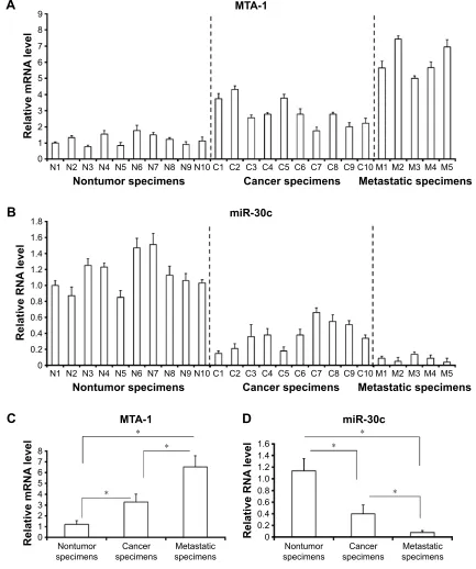
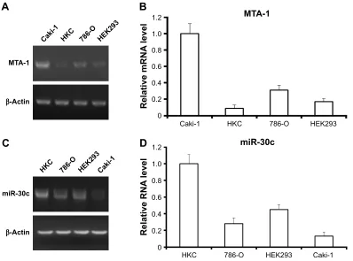
To examine the baseline expression of miR-30c and MTA-1 in ccRCC tissues, quantitative RT-PCR was performed. The expression of MTA-1 mRNA was evaluated in samples of ccRCC rather than in the adjacent nontumor specimens, and the highest level of MTA-1 was detected in metastatic speci-mens (Figure 1A and C). In parallel, the expression of miR-30c is inversely associated with MTA-1 (Figure 1B and D). Next, the endogenous expression of MTA-1/miR-30c was examined in ccRCC or nontumor kidney cell lines. Four different cell lines, including two nontumor kidney cell lines HKC and HEK293, as well as ccRCC cell line 786-O and a highly aggressive cell line Caki-1, were examined by qRT-PCR. As shown in Figure 2, the mRNA level of MTA-1 was markedly elevated in ccRCC cell lines com-pared to the HKC or HEK293 cell line (Figure 2A and B).
Table 1 The concentrations of antitumor drugs used in this work Compounds Concentration (μmol/L)
Paclitaxel 0.003 0.01 0.03 0.1 0.3 1.0 3.0
sorafenib 0.01 0.03 0.1 0.3 1.0 3.0 10.0
OncoTargets and Therapy downloaded from https://www.dovepress.com/ by 118.70.13.36 on 25-Aug-2020
Dovepress Yang et al
Figure 1 The endogenous expression of mir-30c is inversely associated with MTa-1 in clinical specimens.
Notes: endogenous mrna level of MTa-1 (A, C) or rna level of mir-30c (B, D) was detected by qrT-Pcr in 10 nontumor (n1–n10), 10 primary ccrcc (c1–c10),
and 5 metastatic ccrcc specimens (M1–M5). results were shown as mean ± sD from three experiments with similar results. *P,0.05 versus with nontumor specimens or
cancer specimens; *P,0.05 versus with primary ccrcc specimens or metastatic ccrcc specimens.
Abbreviations: ccrcc, clear cell renal cell carcinoma; mir, microrna; qrT-Pcr, quantitative real-time polymerase chain reaction; sD, standard deviation.
5HODWLYH51$
OHYHO
1RQWXPRUVSHFLPHQV &DQFHUVSHFLPHQV
PL5F
0HWDVWDWLFVSHFLPHQV 1 1 1 1 1 1 1 1 1 1 & & & & & & & & & &00 0 00
%
5HODWLYHP51$
OHYHO
1 1
1RQWXPRUVSHFLPHQV &DQFHUVSHFLPHQV
07$
0HWDVWDWLFVSHFLPHQV 1 1 1 1 1 1 1 1 & & & & & & & & & &00 00 0
$
07$
5HODWLYHP51$
OHYHO
1RQWXPRU
VSHFLPHQV VSHFLPHQV&DQFHU VSHFLPHQV0HWDVWDWLF
PL5F
5HODWLYH51$
OHYHO
1RQWXPRU
VSHFLPHQV VSHFLPHQV&DQFHU VSHFLPHQV0HWDVWDWLF
&
'
The highest expression of MTA-1 was identified in Caki-1 cell (Figure 2A and B). In addition, the expression of miR-30c was inversely associated with MTA-1 in cells (Figure 2C and D). These results indicated that MTA-1 would participate in ccRCC metastasis and that the low level of miR-30c would contribute to the aberrant expression of MTA-1.
mir-30c inhibits the ability of caki-1 cells
to proliferate and anchorage-independent
growth
To elucidate the effect of MTA-1 in Caki-1 cells proliferation, we generated miR-30c expressing lentivirus (LV) vectors. Caki-1 cells, which were infected with control (LV-control)
OncoTargets and Therapy downloaded from https://www.dovepress.com/ by 118.70.13.36 on 25-Aug-2020
Dovepress mir-30c suppresses MTa-1 to attenuate aggressive ccrcc proliferation
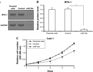
or miR-30c (LV-miR-30c), were harvested for MTT assays. As shown in Figure 3A and B, the expression of MTA-1, a target of miR-30c, was significantly reduced after LV-miR-30c infection. The proliferation of Caki-1 cells infected with miR-30c is much lower than cells infected empty vectors (LV-control) or the parental cells (Figure 3C). There was no significant difference between parental cells (negative control) and LV-control infected cells (Figure 3C).
Next, the soft agar methods were performed to test the anchorage-independent growth of Caki-1 cells. Overexpression of miR-30c significantly inhibited the anchorage-independent growth of Caki-1 cells (Figure 4). Taken together, these data showed that down-regulation of MTA-1 expression via miR-30c inhibits proliferation or anchorage-independent growth of Caki-1 cells.
mir-30c inhibits in vitro invasion
and migration of caki-1 cells
To further explore the roles of miR-30c in Caki-1 cells, we examined in vitro invasion and migration by trans-well methods. Infection of miR-30c decreased the ability of Caki-1 cells to invade as well as migrate (Figure 5A and B) com-pared with control. The inhibition rate for relative invasion or
migration cell number of Caki-1 was 84.20% (for invasion) or 79.41% (for migration), respectively. These results indicated that MTA-1 may be a key regulator of ccRCC metastasis and that decreasing MAT-1 expression via miR-30c significantly inhibits in vitro invasion or migration of Caki-1 cells.
mir-30c attenuates in vivo growth
of caki-1 cells in nude mice mode
Next, in vivo growth of Caki-1 cells was examined. As shown in Figure 6, infection of miR-30c decreased the in vivo growth of Caki-1 cells compared with control. These results further confirmed the effect of miR-30c on ccRCC cells proliferation.
mir-30c inhibits caki-1 cells proliferation
via MTa-1
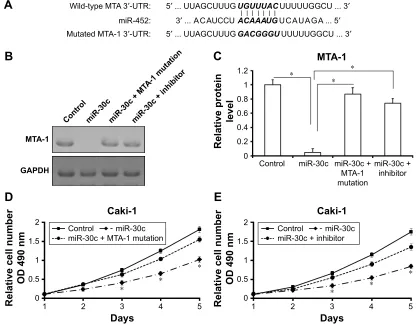
To further reveal the specificity of miR-30c on Caki-1 proliferation, the inhibitor of miR-30c or mutated-MTA-1 vector (MTA-1Mut) was also used (Figure 7). miR-30c reduced the expression of MTA-1 but not mutated MTA-1 (Figure 7B and C). Furthermore, co-transfection inhibitor of miR-30c or MTA-1 mutation almost blocked the effect of miR-30c in Caki-1 cells (Figure 7D and E). Taken together,
Figure 2 expression of mir-30c is inversely associated with MTa-1 in kidney or ccrcc cell lines.
Notes: The expression of MTa-1 (A, B) or mir-30c (C, D) in kidney nontumor cell lines hKc or heK293, and ccrcc cell lines caki-1 or 786-O was detected by
quantitative real-time polymerase chain reaction assays. Results are shown as representative figures or mean ± sD from three experiments with similar results.
Abbreviations: ccrcc, clear cell renal cell carcinoma; mir, microrna; sD, standard deviation.
$
%
'
&
&DNL
07$
β$FWLQ
+.& 2 +(.
07$
5HODWLYHP51$
OHYHO
&DNL +.& 2 +(.
&DNL
PL5F
β$FWLQ
+.& 2 +(.
PL5F
5HODWLYH51$
OHYHO
&DNL
+.& 2 +(.
OncoTargets and Therapy downloaded from https://www.dovepress.com/ by 118.70.13.36 on 25-Aug-2020
Dovepress Yang et al
these results suggested that miR-30c attenuates Caki-1 cells proliferation by targeting MTA-1.
mir-30c enhances the sensitivity
of caki-1 cells to antitumor agents
As shown in Figure 8, sorafenib and paclitaxel inhibited Caki-1 cells’ survival in a dose-dependent manner. The inhi-bition rates (IR) of sorafenib and paclitaxel on Caki-1 cells were significantly enhanced after miR-30c infection. The 50% inhibiting effect concentration values correspondingly decreased from 4.48±0.33 μmol/L to 0.87±0.074 μmol/L, or from 0.79±0.11 μmol/L to 0.082±0.01 μmol/L, respectively (Table 2). Transfection of miR-30c inhibitor or MTA-1 muta-tion almost blocked the activity of miR-30c.
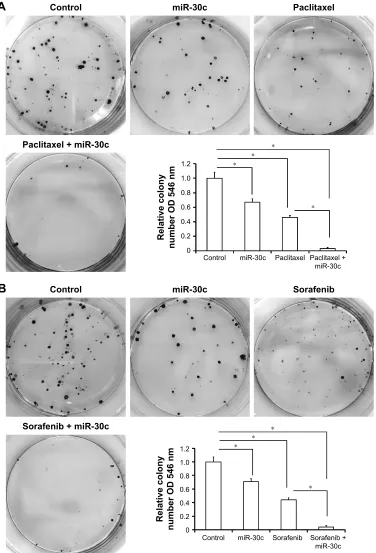
Then, the colony formation was examined (Figure 9). Infection of miR-30c significantly enhanced the inhibitory effect of paclitaxel (Figure 9A) and sorafenib (Figure 9B) on Caki-1 colony formation. Infection of miR-30c inhibitor or MTA-1 mutation almost blocked the activity of miR-30c on Caki-1 colony formation. These data indicated that MTA-1 would participate in the chemotherapy of ccRCC and that
down-regulation of MTA-1 via miR-30c enhances the sen-sitivity of Caki-1 cells to antitumor agents.
Discussion
In this work, our data identified that MTA-1 would be a key regulator of metastatic ccRCC proliferation of metastasis, and revealed the proliferation inhibiting roles of miR-30c in metastatic ccRCC by targeting MTA-1. This notion is supported by results that decreased expression of MTA-1 via miR-30c significantly attenuated the proliferation or anchorage-independent growth of Caki-1 cells. As highly aggressive ccRCC cell lines, infection of miR-30c disrupted the migration and invasion of Caki-1. Moreover, miR-30c also enhanced the sensitivity of Caki-1 cells to antitumor agents. Therefore, MTA-1 may be an interesting novel target for the treatment of highly aggressive ccRCC, and expression of miR-30c would be a potential strategy for ccRCC treatment.
The ccRCC is the most common malignance of adult kidney, and the overall survival of ccRCC is highly variable, ranging from 1 to 10 years.22 It has been confirmed
Figure 3 mir-30c represses MTa-1 to inhibit the proliferation of caki-1 cells.
Notes: (A) Western blot was used to examine the effect of miR-30c on MTA-1 expression. Results were shown as representative figure (A) or mean ± sD (B) from three
experiments with similar results. (C)The proliferation of parental caki-1 cells or caki-1 cells infected with control (lentivirus-control) or mir-30c (lentivirus-mir-30c) was determined
by MTT assays. *P,0.05 versus with parental caki-1 cells infected with control or mir-30c.
Abbreviations: mir, microrna; OD, optical density; sD, standard deviation.
%
$
07$
3DUHQWDOFHOOV &RQWURO PL5F
5HODWLYHSURWHLQOHYHO
07$
3DUHQWDO
FHOOV &RQWURO PL5F
*$3'+
&
5HODWLYHFHOOQXPEHU
2'QP
'D\V &DNL
3DUHQWDOFHOOV &RQWURO PL5F
OncoTargets and Therapy downloaded from https://www.dovepress.com/ by 118.70.13.36 on 25-Aug-2020
Dovepress mir-30c suppresses MTa-1 to attenuate aggressive ccrcc proliferation
Figure 4 mir-30c inhibits anchorage-independent growth of caki-1 cells.
Notes: The parental caki-1 cells (A), cali-1 cells infected with control (B) or mir-30c (C) were then measured by soft agar assays for anchorage-independent growth.
Results are shown as representative figures (A–C) or mean ± sD from three experiments with similar results(D). *P,0.05 versus with parental caki-1 cells infected with
control or mir-30c.
Abbreviations: mir, microrna; sD, standard deviation.
$
3DUHQWDOFHOOVP P
&RQWURO
%
P
PL5F
&
3DUHQWDOFHOOV &RQWURO PL5F
&DNL
&RORQ\QXPEHU
'
$
&RQWUROP
PL5F
P
&RQWURO
P P
PL5F
&RQWURO PL5F
&DNL
5HODWLYHPLJUDWLRQ
FHOOQXPEHU
&DNL
&RQWURO PL5F
5HODWLYHLQYDVLRQ FHOOQXPEHU
%
Figure 5 mir-30c inhibits in vitro invasion and migration of caki-1 cells.
Notes: caki-1 cells, infected with control or mir-30c, were then measured by trans-well assays for in vitro invasion (A) or migration (B). results are shown as representative
figures or mean ± sD from three experiments with similar results. *P,0.05 versus with caki-1 cells infected with control or mir-30c.
Abbreviations: mir, microrna; sD, standard deviation.
OncoTargets and Therapy downloaded from https://www.dovepress.com/ by 118.70.13.36 on 25-Aug-2020
Dovepress Yang et al
Figure 6 microrna (mir)-30c inhibits the in vivo growth of caki-1 cells in nude mice model.
Notes: caki-1 cells, which were infected with mir-20c or control, were injected into BalB/c nude mice. The effect of mir-30c on caki-1 in vivo growth is shown as
photograph (A), tumor volumes (B), or tumor weight (C). The tumor iD numbers (1–8) are shown in Figure A. *P,0.05 versus with mir-30c or control.
7XPRUZHLJKWJ
&RQWURO
PL5F
&
%
7XPRUYROXPH
PP
'D\V
&RQWURO PL5F
$
&RQWURO
PL5F
Figure 7 mir-30c would inhibit caki-1 cells proliferation via decreasing MTa-1 expression by targeting its 3′-UTr sequence.
Notes: (A) italicized font and bold font indicate the mir-30c targeted sites or mutated sequences of within MTa-1 3′-UTr. (B, C) caki-1 cells were infected with control,
mir-30, or co-transfected with mutated-MTa-1 or mir-30c inhibitor. The endogenous MTa-1 level was detected by Western blot (B). results are shown as representative
figures or mean ± sD (C) from three experiments with similar results. (D, E) The effect of mutated-MTa-1 or inhibitor on mir-30c’s inhibitory activity was determined.
*P,0.05 versus with mutated-MTa-1 or not; *P,0.05 versus with mir-30c inhibitor or not.
Abbreviations: mir, microrna; OD, optical density; sD, standard deviation.
$
:LOGW\SH07$′875 ′88$*&888*0XWDWHG07$′875 ′88$*&888* PL5 ′$&$8&&8
8*888$&88888**&8′
*$&***888888**&8′
$&$$$8*8&$8$*$′
%
&
07$
&RQWURO PL5FPL5F07
$PXWDWLRQ
PL5FLQKLELWRU
*$3'+
5HODWLYHSURWHLQ
OHYHO
07$
&RQWURO PL5F
PL5F 07$ PXWDWLRQ
PL5F LQKLELWRU
'
(
&DNL
'D\V
5HODWLYHFHOOQXPEHU
2'QP
&RQWURO PL5F PL5F07$PXWDWLRQ
&DNL
'D\V
5HODWLYHFHOOQXPEHU
2'QP
&RQWURO PL5F PL5FLQKLELWRU
OncoTargets and Therapy downloaded from https://www.dovepress.com/ by 118.70.13.36 on 25-Aug-2020
Dovepress mir-30c suppresses MTa-1 to attenuate aggressive ccrcc proliferation
that patients with advanced stage or metastatic ccRCC may have a poor prognosis owing to insensitive chemotherapy or radiotherapy.22 There is therefore an urgent need to identify
the detailed mechanisms of or novel options for ccRCC treatment. Our previous work showed that KLF4 expres-sion was lower in ccRCC tissues and that patients with weakly or negatively stained KLF4 had larger tumor size and worse prognosis.15 Another work also revealed
aber-rant expression of Tubulin cofactor A (TBCA) in ccRCC, and revealed its function as a novel positive regulator of ccRCC progression, invasion, and metastasis. Exploration of the mechanisms suggested that TBCA could function via modulating cytoskeleton integration and influencing cell cycle progress.3 Although these data enhanced our
knowledge about ccRCC, more work is needed to declare mechanisms for insensitive chemotherapy or radiotherapy during ccRCC treatment.
MTA-1 is one of the key regulators of human cancer metastasis. It would promote invasion, metastasis, and angiogenesis and associate poor clinical outcome tumor by up-regulating the expression of fibronectin, MMP2, or MMP9.5,23 In addition, MTA-1 would also be a positive
regu-lator of cancer cells proliferation.5,24 MTA-1 can also enhance
growth of nasopharyngeal carcinoma cells by promoting G1/S-phase transition.24 Yao et al5 reviewed the related
research works and indicated that MTA-1 would modulate the cell proliferation and G1/S-phase transition via Cyclin D1 and P21.5 In this work, up-regulation of MTA-1 can be detected
both in metastatic ccRCC cell lines and in specimens. The expression of miR-30c, which targets to MTA-1, is inversely associated with MTA-1. Infection of miR-30c inhibits Caki-1 cells proliferation, anchorage-independent growth, in vitro invasion, and migration by repressing MTA-1. These results revealed the functions of MTA-1 in highly aggressive ccRCC, and expression of miR-30c via lentivirus vectors would be a potential approach in ccRCC treatment. In addition, Kaur et al6 showed that MTA-1 can promote the EMT process by
repressing E-cadherin transcription.6 It also promotes
tum-origenesis and metastasis by up-regulating TGF-β signaling activity.6 Human cancer cells may develop recurrence or
resistance to therapies via the EMT process. It has been shown that MTA-1 participates in antitumor drugs resistance
$
3DFOLWD[HO
&RQWURO PL5F
,&
YDOXH0
&RQWURO
PL5F 6RUDIHQLE
,&
YDOXH0
5HODWLYHVXUYLYDO FHOOQXPEHU
0
0
5HODWLYHVXUYLYDO FHOOQXPEHU
%
&
'
3DFOLWD[HOFRQWURO 3DFOLWD[HOPL5F
6RUDIHQLEFRQWURO 6RUDIHQLEPL5F
Figure 8 mir-30c enhances the sensitivity of caki-1 cells to anticancer drugs.
Notes: (A, B) caki-1 cells, which were infected with control or mir-30c, were treated with the indicated concentrations of paclitaxel (A) or sorafenib (B) for 72 h. next,
cells were harvested for MTT assay. (C, D) The ic50 values of antitumor agents are shown. *P,0.05 versus with caki-1 cells infected with control or mir-30c.
Abbreviations: ic50, 50% inhibiting effect concentration; mir, microrna.
Table 2 mir-30c enhances the sensitivity of anticancer drugs Compounds Control miR-30c miR-30c +
mutated MTA-1
miR-30c + inhibitor
IC50 value (μmol/L)
Paclitaxel 0.79±0.11 0.082±0.01 0.57±0.24 0.44±0.31 sorafenib 4.48±0.33 0.87±0.074 4.01±0.25 3.79±0.57 Abbreviations: ic50, 50% inhibiting effect concentration; mir, microrna.
OncoTargets and Therapy downloaded from https://www.dovepress.com/ by 118.70.13.36 on 25-Aug-2020
Dovepress Yang et al
Figure 9 mir-30c up-regulates the inhibitory activity of paclitaxel and sorafenib on caki-1 cell colony formation.
Notes: (A, B) caki-1 cells, which were infected with control or mir-30c, were treated with the ic50 concentration of paclitaxel (0.8 μmol/l) or sorafenib (4.5 μmol/l), and
were harvested for colony formation analysis. The results are shown as photographs or mean ± sD of three experiments with similar results. *P,0.05 versus with caki-1
cells infected with control or mir-30c.
Abbreviations: mir, microrna; sD, standard deviation.
$
%
&RQWURO
PL5F 3DFOLWD[HO 3DFOLWD[HO PL5F
5HODWLYHFRORQ\
QXPEHU2'QP
&RQWURO PL5F 3DFOLWD[HO
3DFOLWD[HOPL5F
&RQWURO
PL5F 6RUDIHQLE 6RUDIHQLE PL5F
5HODWLYHFRORQ\
QXPEHU2'QP
&RQWURO PL5F 6RUDIHQLE
6RUDIHQLEPL5F
of breast cancer.7 A high level of MTA1 suggests
develop-ment of resistance to tamoxifen.8 Therefore, identification of
the effects of MTA-1/miR-30c on ccRCC EMT and related signaling pathways, eg, TGFβ/Smads, will be future work.
Further, a large cohort study to reveal the expression of mTA-1/miR-30c in ccRCC is also needed.
It has been well known that ccRCC, especially highly aggressive or metastatic ccRCC, is resistant to conventional
OncoTargets and Therapy downloaded from https://www.dovepress.com/ by 118.70.13.36 on 25-Aug-2020
Dovepress mir-30c suppresses MTa-1 to attenuate aggressive ccrcc proliferation
chemotherapy.25 Although small molecular kinase inhibitors,
eg, sunitinib and sorafenib, are foremost choices for patients suffering from advanced or metastatic ccRCCs, these treat-ments rarely produce complete responses, and curative effects are not observed.25 Thus, further work is needed to address the
appropriate biomarkers and therapeutic targets of ccRCC. Some important genes, eg, MDR-1, VHL, or Notch-1, would par-ticipate in regulation of ineffective chemotherapy.26–29 Gururaj
et al8 reported that MTA-1 would mediate the resistance of ERα
negative breast cancer cells to antitumor agents. In the present study, we found that down-regulation of MTA-1 expression via miR-30c significantly enhanced the sensitivity of Caki-1 cells to antitumor drugs. Moreover, there are still some other pathways involved in chemotherapeutic or radiotherapeutic resistance, eg, Notch-1 or MDR-1 (multidrug resistance 1). Notch-1 signaling pathway mediates cancer cell-fate decision via pro-survival, antiapoptosis, or EMT process.16 MDR-1 and
some other resistance protein, eg, BCRP, are transcripted by some nuclear receptors, eg, PXR or CAR.19 Thus, it is
neces-sary to identify the effect of MTA-1 on Notch-1 or interaction between MTA-1 and PXR or CAR, in future.
Conclusion
In this work, we reported that MTA-1 may be an important regulator of ccRCC metastasis or chemotherapy resistance. Infection of miR-30c inhibited the proliferation and enhanced the sensitivity of highly aggressive ccRCC Caki-1 cells to anticancer agents by repressing MTA-1 expression. In light of this, we showed that expression of miR-30c could participate in the epigenetic downmodulation of MTA-1 and would guide further development of novel therapies to benefit ccRCC patients.
Acknowledgments
The authors thank Dr Peng Zhang at the State Key Laboratory of Kidney Diseases Department of Urology, PLA General Hospital, Beijing, People’s Republic of China, for his help-ful advice. This study was supported by The Science and Technology Development Project of Changshu (CS201417).
Author contributions
HLY, ELS, GRS and THZ carried out experiments. ELS and TWJ drafted the manuscript. HS, LPN, BW and ZYL partici-pated in experiments. JPQ performed the statistical analysis. TWJ designed the study. All authors made substantial con-tributions to the design and conception; acquisition, analysis or interpretation of data; took part in revising the manuscript; gave final approval of the version to be published; and agree to be accountable for all aspects of the work in ensuring that
questions related to the accuracy or integrity of any part of the work are appropriately investigated and resolved.
Disclosure
The authors report no conflicts of interest in this work.
References
1. Zhang N, Wu Y, Gong J, et al. Germline genetic variations in PDZD2 and ITPR2 genes are associated with clear cell renal cell carcinoma in Chinese population. Oncotarget. Epub January 14, 2016.
2. Nuerrula Y, Rexiati M, Liu Q, Wang YJ. Differential expression and clinical significance of serum protein among patients with clear-cell renal cell carcinoma. Cancer Biomark. 2015;15:485–491.
3. Zhang P, Ma X, Song E, et al. Tubulin cofactor A functions as a novel positive regulator of ccRCC progression, invasion and metastasis.
Int J Cancer. 2013;133:2801–2811.
4. Escudier B, Eisen T, Stadler WM, et al; TARGET Study Group. Sorafenib in advanced clear-cell renal-cell carcinoma. N Engl J Med. 2007;356:125–134.
5. Yao Y, Feng S, Xiao M, Li Y, Yang L, Gong J. MTA1 promotes prolif-eration and invasion in human gastric cancer cells. Onco Targets Ther. 2015;8:1785–1794.
6. Kaur E, Gupta S, Dutt S. Clinical implications of MTA proteins in human cancer. Cancer Metastasis Rev. 2014;33:1017–1024. 7. Tuncay-Cagatay S, Cimen I, Savas B, Banerjee S. MTA-1 expression
is associated with metastasis and epithelial to mesenchymal transition in colorectal cancer cells. Tumour Biol. 2013;34:1189–1204. 8. Gururaj AE, Holm C, Landberg G, Kumar R. Breast cancer-amplified
sequence 3, a target of metastasis-associated protein 1, contributes to tamox-ifen resistance in premenopausal patients with breast cancer. Cell Cycle. 2006;5:1407–1410.
9. Qin H, Sha J, Jiang C, et al. miR-122 inhibits metastasis and epithelial-mesenchymal transition of non-small-cell lung cancer cells. Onco Targets Ther. 2015;8:3175–3184.
10. Ma D, Jia H, Qin M, et al. MiR-122 Induces radiosensitization in non-small cell lung cancer cell line. Int J Mol Sci. 2015;16:22137–22150. 11. Zhang Y, Han L, Pang J, Wang Y, Feng F, Jiang Q. Expression of
microRNA-452 via adenoviral vector inhibits non-small cell lung cancer cells proliferation and metastasis. Tumour Biol. 2016;37:8259–8270. 12. Chen Y, Feng F, Gao X, et al. MiRNA153 reduces effects of
chemo-therapeutic agents or small molecular kinase inhibitor in HCC cells.
Curr Cancer Drug Targets. 2015;15:176–187.
13. Kong X, Xu X, Yan Y, et al. Estrogen regulates the tumour suppressor MiRNA-30c and its target gene, MTA-1, in endometrial cancer. PLoS One. 2014;9:e90810.
14. Heinzelmann J, Henning B, Sanjmyatav J, et al. Specific miRNA sig-natures are associated with metastasis and poor prognosis in clear cell renal cell carcinoma. World J Urol. 2011;29:367–373.
15. Song E, Ma X, Li H, et al. Attenuation of krüppel-like factor 4 facilitates carcinogenesis by inducing g1/s phase arrest in clear cell renal cell carcinoma. PLoS One. 2013;8:e67758.
16. Jia H, Yang Q, Wang T, et al. Rhamnetin induces sensitization of hepa-tocellular carcinoma cells to a small molecular kinase inhibitor or che-motherapeutic agents. Biochim Biophys Acta. 2016;1860:1417–1430. 17. Leonov G, Shah K, Yee D, Timmis J, Sharp TV, Lagos D. Suppression
of AGO2 by miR-132 as a determinant of miRNA-mediated silencing in human primary endothelial cells. Int J Biochem Cell Biol. 2015; 69:75–784.
18. Ma H, Yao Y, Wang C, et al. Transcription factor activity of estrogen receptor α activation upon nonylphenol or bisphenol A treatment enhances the in vitro proliferation, invasion, and migration of neuro-blastoma cells. Onco Targets Ther. 2016;9:3451–3463.
19. Zhao J, Bai Z, Feng, F, et al. Cross-talk between EPAS-1/HIF-2α and PXR signaling pathway regulates multi-drug resistance of stomach cancer cell. Int J Biochem Cell Biol. 2016;72:73–88.
OncoTargets and Therapy downloaded from https://www.dovepress.com/ by 118.70.13.36 on 25-Aug-2020
OncoTargets and Therapy
Publish your work in this journal
Submit your manuscript here: http://www.dovepress.com/oncotargets-and-therapy-journal OncoTargets and Therapy is an international, peer-reviewed, open access journal focusing on the pathological basis of all cancers, potential targets for therapy and treatment protocols employed to improve the management of cancer patients. The journal also focuses on the impact of management programs and new therapeutic agents and protocols on
patient perspectives such as quality of life, adherence and satisfaction. The manuscript management system is completely online and includes a very quick and fair peer-review system, which is all easy to use. Visit http://www.dovepress.com/testimonials.php to read real quotes from published authors.
Dovepress
Dove
press
Yang et al
20. Feng F, Lu YY, Zhang F, et al. Long interspersed nuclear element ORF-1 protein promotes proliferation and resistance to chemotherapy in hepa-tocellular carcinoma. World J Gastroenterol. 2013;19:1068–1078. 21. Yang FQ, Zhang HM, Chen SJ, Yan Y, Zheng JH. MiR-506 is
down-regulated in clear cell renal cell carcinoma and inhibits cell growth and metastasis via targeting FLOT1. PLoS One. 2015;10:e0120258. 22. Cano-González A, López-Rivas A. Opposing roles of TGF-β and EGF
in the regulation of TRAIL-induced apoptosis in human breast epithelial cells. Biochim Biophys Acta. 2016;1863:2104–2114.
23. Song Q, Zhang H, Wang M, et al. MTA1 promotes nasopharyngeal carcinoma growth in vitro and in vivo. J Exp Clin Cancer Res. 2013; 32:54.
24. Shaikh T, Handorf EA, Murphy CT, et al. Contemporary trends in the utilization of radiotherapy in patients with renal cell carcinoma.
Urology. 2015;86:1165–1173.
25. Zhou L, Liu XD, Sun M, et al. Targeting MET and AXL overcomes resistance to sunitinib therapy in renal cell carcinoma. Oncogene. 2016; 35:2687–2697.
26. Walsh N, Larkin A, Kennedy S, et al. Expression of multidrug resistance markers ABCB1 (MDR-1/P-gp) and ABCC1 (MRP-1) in renal cell carcinoma. BMC Urol. 2009;9:6.
27. Cautain B, de-Pedro N, Schulz C, et al. Identification of the Lipodepsi-peptide MDN-0066, a novel inhibitor of VHL/HIF pathway produced by a new pseudomonas species. PLoS One. 2015;10:e0125221. 28. Sjölund J, Johansson M, Manna S, et al. Suppression of renal cell
car-cinoma growth by inhibition of Notch signaling in vitro and in vivo.
J Clin Invest. 2008;118:217–228.
29. Santoni M, Rizzo M, Burattini L, Berardi R, Carteni G, Cascinu S. Novel agents, combinations and sequences for the treatment of advanced renal cell carcinoma: when is the revolution coming? Curr Cancer Drug Targets. 2013;13:313–325.
OncoTargets and Therapy downloaded from https://www.dovepress.com/ by 118.70.13.36 on 25-Aug-2020