Correlation between the shape of the lateral meniscus
and risk of extrusion based on MRI of knee joint
Joshi B.* and Chaudhary R.
Department of Radiology, TU Teaching Hospital (TUTH), Maharajgunj, Kathmandu, Nepal.
Accepted 19 April, 2019
ABSTRACT
Meniscus extrusion can be considered as a serious and relatively frequent clinical problem and can be also considered as a risk factor for osteoarthritis progression and knee pain symptom. Previous studies have shown a strong association between degenerative radial meniscal tears and extrusion. The aim of this study was to correlate the shape of lateral meniscus and the risk of its extrusion based on MRI examination of the knee joint. A prospective study was done in 75 patients of age group between 18 years and 50 years of age who were referred for MRI of knee joint in a tertiary hospital. The MRI was performed on a Magnetom Amira 1.5 Tesla MRI scanner following TUTH MRI routine protocol of Knee joint. All the cases who met the inclusion criteria were included in the study. After the procedure, images were evaluated. The measurements for the parameters used for this study were done in T1 Coronal image at the level of inter-condylar eminence and Proton density(PD) fat saturated sagittal image at the mid bow tie level where the morphology of the lateral meniscus were clearly delineated. Then the recorded data for each patient was correlated with the extrusion of the lateral meniscus along with body mass index (BMI) of the patient. The study showed a strong positive linear correlation of BMI and lateral meniscus-bone angle [LMBA] with meniscus extrusion, where (r=0.94; p=0.000) and (r=0.857; p=0.000) respectively. However, lateral meniscus-cartilage angle [LMCA] and lateral meniscus-cartilage height [LMCH] showed a moderate positive linear correlation with meniscus extrusion (r=0.564; p=0.000) and (r=0.305; p=0.26) respectively. The height of the anterior and posterior horns of the lateral meniscus showed the mean dimension of about 6.13(SD±0.62) mm and 6.30 (SD±0.58) mm respectively in our study. Similarly, the mean antero-posterior diameter of the anterior and posterior horns of the lateral meniscus were found to be 11.3(SD±1.06) mm and 11.4(SD±1.02) mm, respectively. Finally, the mean meniscus extrusion of the lateral meniscus observed beyond the tibial margin were about 2.57 (SD±0.93) mm whereas, the maximum meniscus extrusion was found to be 4.7 mm. In conclusion, increased BMI and LMBA showed significant increased risk of lateral meniscus extrusion beyond the tibial margin. However, LMCA and LMCH showed statistically moderate relation with the risk of lateral meniscus extrusion.
Keywords: Extrusion, knee joint, meniscus, MRI.
*Corresponding author.E-mail: [email protected].
INTRODUCTION
MRI is a non-invasive imaging modality that has increased its demand for the detail imaging of soft tissue anatomy as well as pathology. Different structures of the knee joint such as meniscus, ligaments, articular cartilages and the joint spaces are more prone to delineate clearly in MRI than other imaging modalities. Due to the high contrast resolution of MRI as compared
to other modalities, it has become the choice of interest for soft tissue imaging regarding the knee joint (Fox et al., 2014).
The menisci are crescent-shaped, intra-articular fibro-cartilaginous disks located on the medial and lateral condyles of the tibia and they also play a vital role in force distribution across the knee joint. Due to the
2017). The lateral meniscus is displaced more than the medial meniscus during compression because of its semilunar anatomy as the load is transmitted away from the center of the femoral condyles (Fox et al., 2014). Hence, meniscus extrusion can be considered as a serious and relatively frequent clinical problem and a risk factor for osteoarthritis progression. Previous studies have shown a strong association between degenerative radial meniscal tears and extrusion (Szarmach et al., 2016).
The objectives of the study were to measure the lateral meniscus-cartilage height, lateral meniscus-cartilage angle, lateral meniscus-bone angle, and the antero-posterior [AP] diameter and height of anterior and posterior horns of lateral meniscus; to determine the extrusion of lateral meniscus and to correlate meniscus extrusion with BMI, lateral meniscus-cartilage height, lateral bone angle and lateral meniscus-cartilage angle respectively.
METHODOLOGY
This prospective study was conducted in a tertiary center in Kathmandu, Nepal during the period from September 2018 to February 2019. The cases were performed using Siemens Magnetom Amira 1.5 T MRI Scanner. The study was conducted on patients age ranging 18 to 50 years knee joint pain. Patient with BMI upto 25 kg/m2 and without a history of meniscal injury or diagnosed knee joint space disease were included in this study. Both male and female patient were included.
Patients less than 18 years and over 50 years old were excluded from this study. Patients with metal implants, patients with history of any kind of knee injury or disease and patients with postural abnormalities such as valgus or varus deformity were excluded in this study.
Written consent from participants and ethical approval was received from Institutional Review Board. Non-probability sampling technique was used in selecting the patients referred for MRI of the Knee joint in Radiology Department. All the cases who met the inclusion criteria and consenting to the study were included in the study. Weight and height of the respective patient were taken before the MRI of knee joint. Measurements for the lateral meniscus-cartilage height (LMCH), lateral meniscus-cartilage angle (LMCA) and lateral meniscus-bone angle (LMBA) were taken at the central slices at the level of the inter-condylar eminence where there was optimal delineation of the meniscus body. Meniscus Extrusion (ME) beyond the tibial margin was determined on T1 coronal image, at level of the mid-point of the lateral femoral condyle. The height and AP diameter of the anterior horn and the
superior and inferior margin (maximum meniscal height) of the meniscus (Figure 2).
Lateral Meniscus-Bone Angle (LMBA): Angle between tibial plateau and superior margin of the meniscus (maximum meniscal height) (Figure 3).
Meniscus Anterior Horn Height (MAHH): Maximum distance from the superior surface of the anterior horn to the inferior surface of the anterior horn
Meniscus Anterior Horn Antero-Postero Diameter (MAHAPD):
Distance from the base of the anterior horn to the apex of the anterior horn.
Meniscus Posterior Horn Height (MPHH): Maximum distance from the superior surface of the posterior horn to the inferior surface of the posterior horn.
Meniscus Posterior Horn Antero-Postero Diameter (MPHAPD):
Distance from the base of the posterior horn to the apex of the posterior horn at the mid bow tie level.
Meniscus Extrusion (ME): Distance from the most peripheral aspect of the meniscus to the border of the tibial plateau (Figure 4).
Figure 1. Measurement of LMCH.
RESULTS
The total number of MRI Knee joint cases evaluated in this study were 75 [[38 (50.67%) male patients and 37 (49.33%) female].
Figure 2. Measurement of LMCA.
Figure 3. Measurement of LMBA.
Figure 4. Measurement of meniscus extrusion which is the distance from the most peripheral aspect of the meniscus to the border of the tibial plateau as shown above.
Figure 5. Bar diagram showing age group distribution of patients.
The mean BMI was found to be 22.92 (SD ± 1.79) kg/m2, maximum 25.22 kg/m2 and minimum 18.69 kg/m2. The mean lateral meniscus-cartilage height was 6.45 (SD ± 0.53) mm. The lateral meniscus-cartilage height was 5 mm maximum and 20 mm minimum (Figure 6).
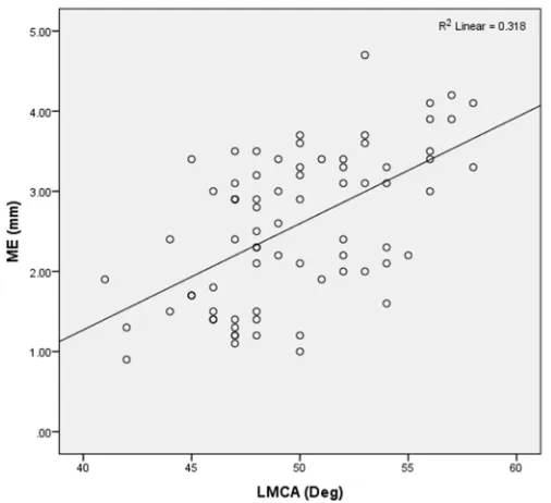
The mean lateral meniscus-cartilage angle was 49.7° (SD±3.96 with maximum 58° and minimum 41°. Mean angle between the lateral meniscus and the tibial plateau (LMBA) was 41.09° (SD±3.74). The lateral meniscus-bone angle was 49° maximum and 33° minimum (Figure 7).
The height of the anterior and posterior horns were 6.13 (SD ± 0.62) mm and 6.30 (SD ± 0.58) mm respectively. The maximum height of anterior and posterior horns were 7.6 and 7.7 mm, respectively. The mean antero-posterior diameter of the anterior and posterior horns were 11.3 (SD ± 1.06) mm and 11.4 (SD ± 1.02) mm, respectively (Figure 8).
There was a strong positive linear correlation between BMI and LMBA (r = 0.799) and meniscus extrusion(r = 0.904) with (p = 0.000) (Table 2). BMI showed a moderate positive linear correlation with LMCH (r = 0.39; p = 0.01) and LMCA (r = 0.467; p = 0.000). There was a weak positive linear correlation between LMCH (r = 0.257; p = 0.26) and moderate positive linear correlation between LMBA (r=0.305; p=0.08). LMCH showed a moderate correlation with meniscus extrusion (r = 0.305; p = 0.008).
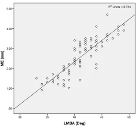
A strong positive correlation was between LMCA and LMBA (r = 0.707; p = 0.000) and between LMBA and meniscus extrusion (r = 0.857; p = 0.000). The mean meniscus extrusion was about 2.57 (SD ± 0.93) mm.
DISCUSSION
MPHAPD (mm) 9.70 15.10 11.37 1.02
ME (mm) 0.90 4.70 2.57 0.93
Figure 6. Scatter plot showing strong positive linear correlation between BMI and ME.
the meniscus extends beyond margin of the tibial plateau (Wang et al., 2010).The extrusion of the lateral meniscus is a multifactorial process involving not only its shape, but also due to the relationship of the lateral meniscus with position and structure of the tibia.
The results of Szarmach et al. (2016) showed a strong positive correlation between LMCA and LMBA (r=0.789, p<0.001). LMCA had a moderate positive linear correlation with meniscus extrusion and the mean lateral meniscus-cartilage angle. However, they measured the slope angle (arc-tangent of the quotient of the lateral meniscus-cartilage height to its width) instead of lateral meniscus extrusion. Their study demonstrated a strong positive correlation between lateral meniscus bone angle and slope angle of the lateral meniscus which can induce meniscus extrusion. They mentioned the cut off value of
Figure 7. Scatter plot showing moderate positive correlation between LMCA and ME and value of r2.
lateral meniscus-bone angle of 42°. The values of slope angle and lateral meniscus-bone angle if increased by one degree, the risk of meniscus extrusion increased by a factor of 1.157 and 1.078 respectively. In our study, the lateral meniscus extrusion increased as the lateral meniscus-bone angle increased above 42°.
The study of Szarmach et al. (2016) demonstrated that an increase in the meniscus-cartilage height by 1 mm significantly elevated the risk of lateral meniscus extrusion. The results of Erbagci et al. (2004)showed that the measurements of the height and of the anterior horn of the lateral meniscus were found to be 4.33 and 8.88 mm, respectively. Their study demonstrated the mean dimensions of the height and AP diameter of the posterior horn measured of about 5.36 and 9.70 mm, respectively.
Figure 8. Scatter plot showing strong positive linear correlation between LMBA and ME.
Table 2. Pearson correlation coefficient values.
Variables BMI LMCH LMCA LMBA ME
BMI 1 0.39 0.467 0.799 0.904
LMCH 0.39 1 0.257 0.305 0.305
LMCA 0.467 0.257 1 0.707 0.564
LMBA 0.799 0.305 0.707 1 0.857
ME 0.904 0.305 0.564 0.857 1
extrusion as minor when the meniscus was extruded 3 mm and major when the meniscus was extruded above 3 mm. Choi et al. (2010) considered 3 mm as the physiological limit of meniscal subluxation and beyond this limit, the meniscus was considered to be extruded.
One of the limitations of our study is small samples size. The morphology and relative position of the meniscus between non-bearing and weight-bearing and may mimic some differences in the measurements.
Another limitation is the use of coronal and sagittal slices can display partial volume effects of some parts of the lateral meniscus. 3-Dimensional segmentation process software was not available in our image processing system so the complete morphology of the anterior horn and the posterior horn of the lateral meniscus could not be determined. An advanced statistical software and a weight bearing position of knee joint are preferred to determine the accurate degree lateral meniscus extrusion.
CONCLUSION
The study suggested a positive correlation between the shape of the meniscus and the risk of its displacement in those patients who were referred for MRI of the knee joint with knee joint pain. The increase in the LMBA increased the risk of meniscal extrusion in patients with higher BMI. However, LMCH and LMCA had moderate effect on meniscal extrusion.
REFERENCES
Choi C, Choi Y, Lee J, Choi C, 2010. Magnetic resonance imaging evidence of meniscal extrusion in medial meniscus posterior root tear. Arthroscopy: The Journal of Arthroscopic & Related Surgery, 26(12): 1602-1606.
Costa C, Morrison W, Carrino J, 2004. Medial meniscus extrusion on knee MRI: Is extent associated with severity of degeneration or type of tear. American Journal of Roentgenology, 183: 17-23.
subchondral bone in osteoarthritic knees. Rheumatology, 49(5): 997-1004.