ADAPTATIVE IMAGE
WATERMARKING SCHEME BASED ON
NEURAL NETWORK
SAMEH OUESLATI
University of Sciences of Tunis, Department of Physics, Laboratory of Signal Processing, 1060, Tunisia
ADNENE CHERIF
University of Sciences of Tunis, Department of Physics, Laboratory of Signal Processing, 1060, Tunisia
BASSEL SOLAIMANE
Higher National School of Telecommunication of Bretagne Technopole, Brest Iroise, 29285, France
Abstract:
Digital image watermarking has been proposed as a method to enhance medical data security, confidentiality and integrity. Medical image watermarking requires extreme care when embedding additional data, given their importance to clinical diagnosis, treatment, and research. In this paper, a novel image watermarking approach based on the human visual system (HVS) model and neural network technique is proposed. The watermark was inserted into the middle frequency coefficients of the cover image’s blocked DCT based transform domain. In order to make the watermark stronger and less susceptible to different types of attacks, it is essential to find the maximum amount of interested watermark before the watermark becomes visible. In this paper, neural networks are used to implement an automated system of creating maximum-strength watermarks. The experimental results show that such method can survive of common image processing operations and has good adaptability for automated watermark embedding.
Keywords: watermarking security; discrete cosine transform; neural network; human visual system (HVS).
1. Introduction
As digital technology pervades our society, a vast amount of medical images now exists in electronic format for easy storage and maintenance. The convenience of data access and distribution also poses a great challenge on privacy protection for patients’ information. Constant efforts have been made to provide security solutions [14], [15], [16], [17] to ensure: Confidentiality, Reliability and Availability.
Recently, watermarking has been proposed for medical information protection. Even though most of the work on watermarking has concerned medical images in order to verify image integrity or improve confidentiality watermarking also provides a new way to share data. Medical image watermarking requires extreme care when embedding additional data within the medical images because the additional information must not affect the image quality. And because modifying gray levels of a medical image may interfere with its interpretation and consequently with the diagnosis.
too many neurons. Later Zhang [11] improved the algorithm to some extent again. Wang [12] proposed a blind watermarking algorithm based on neural network. Chang [13] proposed a watermarking algorithm based on a full counter-propagation neural network (FCNN), which has a good performance, but the watermark is embedded in the synapses of FCNN instead of the cover image.
Neural networks are used to improve the watermark extraction. However, neural networks can also be used to decide the watermark strength. Zhang [5] proposed a RBF neural network to achieve maximum-strength watermark according to the frequency component of the cover image. Liu [6] also proposed a watermarking scheme based on RBF neural network. A good watermarking scheme should be robust enough to defend against attacks while being invisible such that the dissimilarity among the watermarked image and the host image should not be differentiated by the human eyes. Alternatively, the removal of the watermark embedded in the host image should not be easy and the quality of the host image should not be damaged.
In terms of the domain in which the watermark is embedded, watermarking techniques can be classified into two categories: spatial-domain and frequency-domain techniques. Spatial-domain schemes embed the watermark by directly modifying the pixel values of the original image [1–4]. On the other hand, transform-domain methods embed the watermark by modulating the coefficients in a transform transform-domain such as Discrete Cosine Transform (DCT), Discrete Fourier Transform (DFT), and Discrete Wavelet Transform (DWT) [2–3].
In this work, one would like to insert the watermark with maximum strength before it becomes visible to the human visual system (HVS). As a result the way the strength of the added watermark is chosen is of highest importance. This paper attempts to define an adaptive watermarking scheme based on Multi-Layer Feed-forward (MLF) neural networks. We use it to model human visual system (HVS) to determine the strength of watermark bit that should be embedded into the middle-frequency coefficients of block DCT domain of the host image.
The rest of this paper is organized as follows: Section 2 provides a detailed a description of the HVS model and the neural technique used to create maximum watermark. Section 3 describes our algorithm for embedding and extraction of the watermark. Section 4 contains simulation results and discussions about the robustness of the proposed approach against common attacks and the comparison of its performance over other watermarking techniques and finally Section 5 concludes the paper.
2. Proposed Method
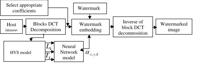
Our watermarking system comprises a watermark embedding and a detection process. Host image is first decomposed into non-overlapping 8x8 blocks, and the DCT process is performed for each block. Coefficients are then selected for watermark insertion. Human visual system (HVS) is adopted to further ensure the watermark invisibility. Then, the luminance sensitivity, frequency sensitivity, texture sensitivity and entropy sensitivity are computed and used to as the inputs of the NNS. In this paper, neural networks are used to automatically control and create the maximum image-adaptive strength watermark. After embedding process, an inverse transformation is applied. To obtain a watermarked image. Below the watermark embedding process is shown in Figure1.
2.1. Watermarking based on the Human Visual System Model
Robustness, perceptual transparency and capacity are the three basic requirements of digital watermarking techniques. However, there is a disagreement among these requirements. Integrating HVS into watermarking
process can assist in satisfying these conflicting requirements. HVS essentially results in the fact that each pixel
Fig. 1. Embedding process based on the Human Visual System and Neural Network model
Inverse of block DCT decomposition Blocks DCT
Decomposition
HVS model
Neural Network
model
Watermark embedding
k
L
k y x, ,
Select appropriatecoefficients Watermark
k
T
Host image
image quality. The literature provides several HVS model based watermarking techniques [14, 15, 20, 21, and 22]. But until now there is no lossless watermarking method proposed has utilized the properties of HVS. Three basic properties of human vision: frequency sensitivity, luminance sensitivity and masking effects are employed by the majority HVS models in image processing. The sensitivity of human eye to various spatial frequencies is determined by the frequency sensitivity. The effect of the detectability threshold of noise on a constant background is calculated by luminance sensitivity. In accordance with the change of background luminance, the frequency sensitivity is corrected. The effect of decreasing visibility of one signal in the presence of another signal called masker is known as masking [22]. We focus here on the sensitivity of brightness, frequency, texture and entropy. The HVS model used in this work has been suggested in [26, 27]. This model is also used in many insertion algorithm and detection of the watermark.
Luminance sensitivity (
L
k)Brightness masking proves the effects of detectable noise thresholds on a constant background. The brighter the background is, the larger the embedded signal could be [22]. The luminance sensitivity is estimated by the following formula: ) ( , DC k DC k V V
L (1)
Where VDC,kis the
DC
coefficientof the DCT of thek
thblock, VDCis the mean value of all VDC,kcoefficientsof a specific image, and
is set to 0.649 to control the degree of luminance sensitivity. Frequency sensitivity (F
k)If we divide the image into 8x8 blocks and DCT is applied to each block, there will be 8x8 matrix of DCT coefficients for each block. This matrix is divided into three areas of high frequency (H), low frequency (L), and medium frequency (M). The 2D DCT matrix's top left comer represents lowest frequency coefficient while the bottom right comer is the highest frequency. The Energy content of image is placed in low frequency DCT coefficients. It has been proved that watermark embedding in low frequency, places the water-mark perceptibly in the image [28]. On the other hand embedding the watermark in high frequency coefficients causes the watermark to be removed from the image after the image compression, since compression process causes the DCT coefficients to be removed in high frequencies. So the medium frequencies are used to embed the watermark.
Texture sensitivity (
T
k)An image can be divided into smooth, textured, and edge block. Extensive HVS research shows that the sensitivity of information distortion to human eyes decreases down from edge block, smooth block, and texture block respectively [21, 19, and 23]. Dissimilar sensitivity degree means different significance of these three blocks [18, 25].The texture sensitivity can be estimated by quantizing the DCT coefficients of an image using the JPEG quantization table. The result is then rounded to nearest integers. The number of non-zero coefficients is then computed. This method can be calculated by the following formula:
( , ) ) , ( ( 1, Q x y
y x V cond T k N y x
k (2)
Where
(
x
,
y
)
represents the location in thek
thblock. Andcond
(
R
)
takes the rounded value ofR
and returns ‘1’ if the value is not equal to zero, ‘0’ otherwise. Entropy sensitivity (
E
k)The entropy and variance value of image blocks represent the amount of the information, and they play an important role in sorting image blocks. The entropy value of the smooth block is smaller than that of the edge and the textured block, and the variance of textured block is smaller than that of the edge block. The entropy sensitivity is calculated by the following formula:
7 0 , ( , ) 1 log ). , ( y x k k k y x p y x pE (3)
Where
7 0 , ) , ( ) , ( ) , ( y x k k k y x X y x X y x2.2. Application of Neural Network in Digital Image Watermarking
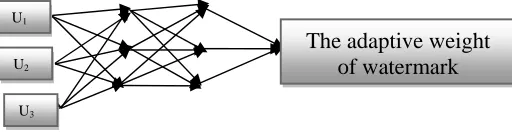
Artificial neural network is important embranchment of artificial intelligence field, and it has certain advantage on the aspect of simulating biology neural computation [29], [30]. It has self-learning, self-organizing, association of ideas, blur extend abilities, etc. and has great comparability with human visual system. Therefore, the selected NN in this paper is a feed-forward NN. There are three layers including an input layer, hidden layers and an output layer. The desired outputs of NN are the maximum watermarking strength. Figure 2 shows the architecture of this network. The first layer is to present the input variables of network. Each HVS feature vector has the elements to be input variables in this study. The four feature components form a feature vector
u
(
u
1,
u
2,
u
3,
u
4)
. Theu
1,u
2,u
3andu
4represent the sensitivity value of luminance, frequency,texture, and entropy of respectively. Each u represents one of the input components in NNS algorithm and each pixel generates a corresponding feature vector u. These inputs are then fed to a hidden layer using tan sigmoid activation functions (output in the range -1 to 1); this is then fed to the output layer. During training samples, information is propagated back through the network and used to update connection weights. It repeats learning many times for every example in the training set until it has minimized the output errors. This neural network, when combined with the available rules in perceptual watermarking of an image and ability to query the network, does provide an automatic system for generating near maximum power invisible watermarked images. This system does not fully replace humans, its they are still needed to generate the training data and should check the results periodically to ensure the neural network is working at peak performance.
3. Watermarking Scheme
3.1. Watermarking Embedding
The first step of our watermarking system is decomposed into non-overlapping 8x8 blocks, and the DCT is computed for each block. We browse the set of blocks, and only one coefficient is chosen to be coded. The DCT coefficients where the watermark bits will be encoded are chosen from the medium frequency band of the
transformed blocks in order to provide additional resistance to lossy compression while avoiding significant modifications or distortions to the cover image [30]. Instead of choosing arbitrarily the coefficients locations, we can increase the robustness to compression by basing our choice on the recommended JPEG table [29]. In fact if two locations are chosen as they present identical quantization values, any scaling of the first coefficient will scale the second by the same factor preserving their relative size [31]. On the other hand to augment the survival chances of the embedded watermarks against a large set of attacks and reduces the probability of detection errors [32]. The study consists on encoding the watermark progressively on the DCT coefficients beginning by using one coefficient to the 22nd coefficient of the medium frequency band, to determine the optimal number of coefficients that can be used which gives a maximum of robustness against a minimum of distortion (PSNR). The next step consists in choosing the size of the watermark (PP),Pis variable according to the number of coefficient, i.e: if we have to select one coefficient in each DCT block then
P
is equal to 32, it increases with the number of selected coefficients. Everything depends on the information to insert; the watermarks in our proposed scheme are patient data such as Patient’s Name, Birth and Gender, Mother's Birth Name, Country and Region of Residence, Ethnic Group, Patient’s Religious Preference and so on. Then, the mark will be linearized vector. We thus obtain a binary sequence of (PP) composed by 0 or 1 element. W [0,1,1,0,1,1,....]The watermark matrix is transformed into a binary sequence. 1 coefficient from the medium frequency band of the transformed blocks is used. Block-based reverse DCT transform after modifying, the block-basedDCT
I transform inverse is implemented and gets the watermarked imageIW. To reinforce the robustness of the image against the various distortions which it can undergo, we use a force of insertion while keeping compromise robustness invisibility. This force depends on the characteristics of the zones of insertion and must be lower than a visual threshold of perceptibility. Neural network applied to digital watermark embedded
Fig. 2. Architecture of NN with textural features.
The adaptive weight
of watermark
U1
U2
endured by middle frequency coefficients in every one of (88)image block DCT coefficients. The embedding process is formulated as follows.
) 1 ( ,, ,, , , ' ,
,yk xyk xyk xyk
x X W
X (5)
Where Xx,y,kdenotes the DCT coefficient of the position
(
x
,
y
)
of theth
K block, and
x,y,kis the adaptive weight of watermark Wx,y,k in the position (x,y) of theth
K blocks.
3.2.Watermarking Extraction
The watermark can be extracted by performing the inverse process of embedding the watermark. The first step of the process is to transform the watermarked image and original image into DCT domain respectively. The embedded coefficients are subtracted and divided by the weights, to extract the corrupted watermark. The weights
x,y,kare then computed from the original image by using the same processes as that used in the watermark embedding process. The watermark detection process can he described as:) . /( ) ( ,, , , * , , , , * k y x k y x k y x k y x
i X X X
W (6)
Where the
X
*x,y,kis the DCT coefficient of thecorrupted image in the position(
x
,
y
)
of theK
th blocks, andk y x, ,
is the adaptive weight of watermark and theW
i*is the corrupted watermark which was formed by combining with all the watermark elements.4. The Experimental Results
In order to evaluate the performance of watermarking technique, and especially for the imperceptible capability, a quantitative index, Peek Signal-to-Noise Ratio (PSNR), is employed to evaluate the difference between an original image
I
and a watermarked imageI
*. For the robust capability, the normalized correlation (NC) measures the difference between the original watermark W(i,j)and the extracted watermark *(, )j i
W . These two indices are, respectively, defined by formula (7) and (9) Where MSE stands for the average mean square error between the original and the watermarked image. The watermarking scheme has been tested on the medical images with size of 512x512 pixels. The tested images are estimated by the human visual system that described in Section 3, and the extracted features are clustered by the NNS algorithm. The experimental results of JPEG compression, cropping, filtering, and noise for the watermarked images are described in this section. Figure 3 (a) Original medical image1; (b) Watermarked medical image1 (PSNR=40.34) (NC =1); (c) Original medical image2; (d) Watermarked medical image2 (PSNR=39.81) (NC =1); There is no difference between the original image and corresponding watermarked image in view of visual quality. A larger PSNR indicates that the watermarked image more closely resembles the original image, meaning that the watermarking method makes the watermark more imperceptible.
) 255 ( log 10 ) ( log 10 2 10 2 max 10 MSE PSNR MSE X
PSNR (7)
WhereXmax: The maximum luminance.
Where MSE is the mean-square error between the original image and the distorted one. MSE is defined as:
NM I I MSE N i M j ij ij
1 1*
²
(8)
1
0 1
0
2 1
0 1
0
*
1 2
1 2
)] , ( [
) , ( ) , (
M
i M
j M
i M
j C
j i W
j i W j i W
N (9)
Here, for an M1M2Watermark image, W(i,j)and *(, )
j i
W denote the (i,j)pixel’s values in the original image
watermark and that in the extracted watermark, respectively.
4.1. Robust against JPEG Compression
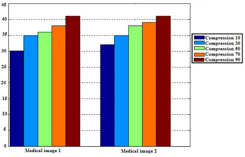
Robustness against JPEG compression is of crucial importance. We always need to apply JPEG compression to the medical images for archive or transmission. To assess the watermark performance from this point of view, we iteratively applied JPEG compression to the watermarked image, each time decreasing the quality factor from 90%, 70%, 50%, 30% et 10%. The tested image is compressed using JPEG in different quality factors. The lower quality factor value corresponds to the greater compression. When the JPEG compression quality factor (QF) is larger than 90%, the watermark can be extracted. When the QF is 30%, the extracted watermark image can be identified. Figure 5(a) shows the watermarked image under JPEG compression attack with quality factor 30%. Figure 5(b) shows the response to detector is higher than the response of false watermarks. Results we obtained are summarized in Figure 4, which shows the NC values of the extracted logo image after JPEG
compression attacks. As can be seen, the proposed algorithm can resist against JPEG compression attack with quality factor as low as 10. We can see our method shows better performance to be against JPEG compressions. Moreover, the JPEG compression scheme reduces the high frequency components of an image, whereas our method embeds the watermark into the DCT coefficients of the frequency mid-band. Whereas whereas in our work, using the HVS model with neural network system, the watermark can be adjusted for each different image that provides a maximum and suitable watermark subject to the imperceptibility constraint. Indeed, the watermark scheme proposed by Cox and al has a weakness that the HVS model has not been taken into account. For the sake of the imperceptibility constraint of a watermark, Cox and al’s scheme must embed the low strength watermark to avoid degrading the image quality. Unfortunately, it reduces the robustness of the watermark. The detector responses of our method are still stronger than Cox and al's method. In our experiments, the proposed watermarking scheme has a better PSNR as shown in Figure 6 and has stronger detection responses than Cox and al's method. Thus, our method is more imperceptible and robust than Cox and al's method.
(a) (b)
(c) (d)
Fig. 3. (a) Original medical image1, (b) watermarked medical image1 (PSNR= 40.34), (c) Original medical
Fig. 4. Results of JPEG compression attack experiment.
Fig. 6. Mean values of PSNR between the original images and watermarked images attacked by different rate of compression.
0 100 200 300 400 500 600 700 800 -0.2
0 0.2 0.4 0.6 0.8 1 1.2
Indexe de la signature
Co
rr
é
la
ti
o
n
Corrélation entre la signature et le dictionnaire
Fig. 5. (a) JPEG compression (with quality factor 30%) of the watermarked image (b) Watermark detector response of attacked by JPEG compression (with quality factor 30).
10 20 30 40 50 60 70 80 90 0.65
0.7 0.75 0.8 0.85 0.9 0.95 1
JPEG compression attacks
JPEG compression quality factor (QF)
PSN
R
4.1 Robust against other attacks
In order to prove the robustness of the proposed watermarking technique, we have carried out a range of attacks on watermarked images in our experiments. Figure 7 shows the results of different image attacks such as noise addition, filtering, and cropping. Table 1 displays the correlation coefficient between the original watermark and the extracted watermark from the attacked watermarked images. The experimental results demonstrate that the correlation coefficient’s value is above 0.5. The robustness of the proposed scheme is evident from the experimental evaluation. The proposed method has better performance, especially suffering from JPEG compression. At the same time we also find that the proposed method can not fight median filtering. The reason is that the neighbor pixel relations are destroyed when the image is processed by median filtering.
Table 1. Nc of extracted watermark under various attacks.
Acknowledgments. The authors express gratitude to Dr. AZAIEZ Mustapha, clinic from the Manar II of Tunisia, for providing the medical images used in this paper.
Attacks Parameters
NC of the extracted watermark Watermarked medical
image1
Watermarked medical image2
Noise addition
Gaussian (0.03) 0.9352 0.9178
Salt and pepper (0.03) 0.9980 0.9943
Speckle (0.03) 0.9674 0.9710
Filtering
Median filtering (windows)
3x3 0.7225 0.7019
5x5 0.6743 0.6501
Mean filtering (Windows)
3x3 0.8283 0.8747
5x5 0.7421 0.6985
Cropping Cropping 25% 0.9023 0.9548
(a) (b) (c ) (d)
(e) (f) (g) (h)
5 Conclusion
A new method to obtain the invisibility and the robustness in DCT domain watermarking has been described. HVS characteristics are taken into consideration so as to make the watermark invisible during the process of watermark embedding and artificial neural network is applied and for enhancing the performance of conventional watermarking techniques. Simulation results demonstrated that the proposed method significantly improve the perceptual quality of a watermarked image while preserving the robustness against various attacks. Our current research provides a promising technique for watermarking images as perspective we can extend research in the multiresolution field and many improvements still remain to be implemented.
References
[1] Chang, C.C.; Chen, T.S.; Chung, Z. (2002): A steganographic method based upon JPEG and quantization table modification. Inf. Sci. vol. 141, pp.123–38.
[2] Cox, I.J.; Kilian, J.; Leighton, F.T. (1997): Shamoon, T.: Secure spread spectrum watermarking for multimedia. IEEE Trans. Image Process. Vol.6, pp.1673–87.
[3] Liu, J.L.; Lou, D.C.; Chang, M.C., Tso, K. (2006): A robust watermarking scheme using self-reference image. Comput Standards Interfaces, vol.28, pp.356–67.
[4] Celik, M.U., Sharma, G., Tekalp, A.M., Saber, E. (2005): Lossless generalised-LSB data embedding. IEEE Trans Image Process. Vol.14, pp.253–66.
[5] Zhi-Ming, Z.; Rong-Yan, L.; Lei, W. (2003): Adaptive Watermark Scheme with RBF Neural Networks. In Proc. International Conf Neural Networks and Signal Processing, vol. 2, pp. 1517-1520.
[6] Quan, L.; Jiang, X. (2006): Design and Realization of a Meaningful Digital Watermarking Algorithm Based on RBF Neural Network. Proceedings of the Sixth World Congress on Intelligent Control and Automation, WCICA 2006. vol. 1, pp. 2878-2881.
[7] Yu, P.; Tsai, H.; Lin, J. (2001): Digital watermarking based on neural networks for color images. Signal Processing, vol. 81, no. 3, pp. 663-671.
[8] Kutter, M.; Jordan, F.; Bossen, F. (1998): Digital watermarking of color images using amplitude modulation. Journal of Electronic Imaging, vol.7, no. 2, pp. 326–322.
[9] Fan, Z.; Hongbin, Z. (2005): Applications of a neural network to watermarking capacity of digital image. Neurocomputing, 67, pp.345-349.
[10] Zhang, F.; Hongbin Z. (2004): Applications of neural network to watermarking capacity. Proceedings of IEEE International Symposium on Communications and Information Technology, vol. 1, pp. 340 -343.
[11] Xinhong, Z.; Fan, Z. (2005): A Blind Watermarking Algorithm Based on Neural Network. Proceedings of International Conference on Neural Networks and Brain, vol. 2, pp. 1073-1076.
[12] Zhenfei, W.; Nengchao, W.; Baochang, S. (2006): A Novel Blind Watermarking Scheme Based on Neural Network in Wavelet Domain. Proceedings of the 2006 Sixth World Congress on Intelligent Control and Automation, WCICA, vol. 1, pp. 3024 – 3027. [13] Chuan-Yu C.; Sheng-Jyun, S.: The application of a full counterpropagation neural network to image while preserving high diagnostic
quality.
[14] HEMA, “DICOM: Digital imaging and communication in medicine,” [Online], Available: http://medical.nema.org/
[15] Cao, F.; Huang, H.; Zhou, X. (2003): Medical image security in a HIPAA mandated PACS environment. Computerized Medical Imaging and Graphics, vol. 27, pp. 185-196.
[16] Owens, J.; Tachakra, S.; Banitsas, K.; Istepanian, R. (2001): Securing a medical wireless LAN system. Proc. 23rd Annual Conf. IEEE Engineering in EMBS, Istanbul, Turkey.
[17] Coatrieux, G.; Henry, M.; Sankur, H.; Rolland, Y.; Collorec, R. (2000): Relevance of watermarking in medical imaging,” Proc. IEEE EMBS Conf. on Inform. Tech. Appl. in Biomedicine, Arlington.
[18] Miladi, B.; Sayadi, M.; Fnaiech, F. (2010): Textures synthesis methods. International Conference on Electrical Systems and Automatic control, Tunisia.
[19] Davoine, F.; Pateux, S. (2004): Tatouage de documents audiovisuels numériques. Livre édition Hermès Lavoisier, traité IC2 Série Traitement du signal et de l’image.
[20] Patrick, B., François, C.: Natural Watermarking: a secure spread spectrum technique for WOA Information Hiding, pp.1-14, 4437. [21] Patrick, B. ; Cayre, F.; Mathon, B. (2007): Techniques sures de tatouage pour l’image. Compression et Représentation des Signaux
Audiovisuels, Montpellier, France.
[22] Mathon, B.; Patrick, B; Cayre, F. (2007): Practical performance analysis of secure modulations for WOA spread-spectrum based image watermarking. Multimedia and Security Workshop, Dallas, Texas, USA.
[23] Vincent, M. (2006): Contribution des filtres LPTV et des techniques d’interpolation au tatouage numérique. PhD Thesis presented in Toulouse.
[24] Rey, C. (2003) : Tatouage d’image: Gain en robustesse etintégrité des images. PhD Thesis, University of Avignon and Pays de Vaucluse.
[25] Geetha S.; Siva, S.; Kamaraj, N. (2008): Close Color Pair Signature Ensemble Adaptive Threshold based Steganalysis for LSB Embedding in Digital Images. Transactions on Data Privacy, pp. 140 – 161.
[26] Watson, A.: DCT (1993): A technique for visual optimization of DCT quantization matrices for individual images. Society for Information Display Digest of Technical Papers, pp. 946-949.
[27] Jayant, S.; Johnson, J.; Safranek, R. (1993): Signal compression based on models of human perception”. Proceedings of IEEE, Volume 81, No. 10, pp. 1385-1422.
[28] Quan, L.; Xuemei, J. (2005): Design and Realization of a Meaningful Digital Watermarking Algorithm Based on RBF neural Network. Neural Networks and Brain, ICNN & Bapos; 05. International Conference on Volume 1, Issue, 13-15, pp.214 – 218.
[29] Barni, M.; Bartolini, F.; Cappellini, V.; Piva, A. (1998): A DCT domain system for robust image watermarking. Signal Proces, vol. 66, no. 3, 1998, pp. 357-372.
[30] Bors, A.; Pitas, I. (1999): Image watermarking using DCT domain constraints. Proceedings ICIP, Vol. 3, Lausanne, pp. 231–234. [31] Hartung, F.; Kutter, M. (1999): Multimedia watermarking techniques. Proc. IEEE 87, pp. 1079–1107.