University of Pennsylvania
ScholarlyCommons
Publicly Accessible Penn Dissertations
1-1-2013
Characterization and Targeting of Thromboxane
Receptor Dimerization: A Gateway to Novel
Therapeutic Developments
Alexander Jack FreyUniversity of Pennsylvania, [email protected]
Follow this and additional works at:http://repository.upenn.edu/edissertations Part of thePharmacology Commons
This paper is posted at ScholarlyCommons.http://repository.upenn.edu/edissertations/861
For more information, please [email protected].
Recommended Citation
Frey, Alexander Jack, "Characterization and Targeting of Thromboxane Receptor Dimerization: A Gateway to Novel Therapeutic Developments" (2013).Publicly Accessible Penn Dissertations. 861.
Characterization and Targeting of Thromboxane Receptor Dimerization:
A Gateway to Novel Therapeutic Developments
Abstract
Thromboxane A2(TXA2) contributes to cardiovascular disease (CVD) by activating platelets and vascular
smooth muscle cell constriction and proliferation. Despite their preclinical efficacy, pharmacological antagonists of the TXA2 receptor (the TP), a G protein-coupled receptor (GPCR), have not been clinically successful, raising interest in novel approaches to modifying TP function. We sought to examine molecular mechanisms underlying auto-upregulation of the TP in response to agonist activation. We first determined a lack of agonist-induced TP mRNA modulation, focusing our attention on post-translational TP regulation. GPCR dimerization contributes to post-translational regulation of receptor expression and function, therefore we characterized how TP forms dimers with itself (homodimerization) or other related receptors
(heterodimerization) and defined the relative affinities. To determine how disruption of TP dimerization impacts its regulation and function, we targeted a GxxxGxxxL helical interaction motif, reportedly involved in transmembrane protein-protein interactions between other membrane proteins and GPCRs, that is located in the human TP's (α isoform) 5th transmembrane domain. We determined that disruption of this motif suppressed TP agonist-induced Gq signaling and TPα homodimerization, but not its cell surface expression, ligand affinity or Gq association. Heterodimerization of TPα with the functionally opposing receptor for prostacyclin (the IP) shifts TPα to signal via the IP-Gs cascade contributing to prostacyclin's restraint of TXA2 function. Interestingly, and in contrast to the TPα homodimer, disruption of the TPα-TM5 GxxxGxxxL motif did not modify either TPα-IP heterodimerization or its Gs-cAMP signaling. Our study indicates that distinct regions of the TPα receptor direct its homo- and hetero- dimerization and normal homodimerization appears necessary for efficient TPα-Gq activation. Targeting the TPα-TM5 GxxxGxxxL domain may allow development of biased TPα- homodimer antagonists that avoid suppression of TPα-IP heterodimer's predicted beneficial "IP-like" effects. Such novel therapeutics may prove superior in CVD compared to non-selective suppression of all TP functions with TXA2 biosynthesis inhibitors or traditional TP antagonists.
Degree Type
Dissertation
Degree Name
Doctor of Philosophy (PhD)
Graduate Group
Pharmacology
First Advisor
Emer M. Smyth
Keywords
dimerization, GGL motif, GPCR, thromboxane, thromboxane receptor
Subject Categories
Pharmacology
CHARACTERIZATION AND TARGETING OF THROMBOXANE RECEPTOR
DIMERIZATION: A GATEWAY TO NOVEL THERAPEUTIC DEVELOPMENTS
Alexander Frey
A DISSERTATION
in
Pharmacology
Presented to the Faculties of the University of Pennsylvania
in
Partial Fulfillment of the Requirements for the
Degree of Doctor of Philosophy
2013
Supervisor of Dissertation
__________________________________________________
Emer M. Smyth, Associate Research Professor of Pharmacology
Graduate Group Chairperson
___________________________________________________
Julie Blendy, Professor of Pharmacology
Dissertation Committee
David R. Manning, Professor of Pharmacology, Committee Chair
Lawrence F. Brass, Professor of Medicine
Tilo Grosser, Research Assistant Professor of Pharmacology
CHARACTERIZATION AND TARGETING OF THROMBOXANE RECEPTOR DIMERIZATION: A
GATEWAY TO NOVEL THERAPEUTIC DEVELOPMENTS
COPYRIGHT
2013
Alexander Jack Frey
This work is licensed under the Creative Commons Attribution- NonCommercial-ShareAlike 3.0 License
To view a copy of this license, visit
iii
This work is dedicated to every person; friend, family, colleague, and acquaintance, who has touched my life and guided me to this place I find myself today. I would not be who I am, and this
iv
ACKNOWLEDGMENTS
Thanks go out to Emer Smyth for being an amazing advisor and mentor even beyond what expectations I had upon arriving here at Penn. For my having been your first graduate student, it seems that it came naturally to you how to strike the ideal balance between knowing when to answer my questions and when to tell me to go find it myself, and how to keep encouraging me through it all – even the really tough parts.
Thanks go out to all the members of the Smyth Lab, past and present. Mazell for being so welcoming to a chemist joining a microbiology lab; Nune and Salam for not only helping me when I needed it, but being great role models of experienced scientists; Ed for being a great friend and labmate and walking along this
graduate school road alongside me; and Vicky for always being there to help keep the lab moving but, more importantly, to always be so ready with a laugh and a smile. And thanks to the numerous members of the FitzGerald lab, for providing so much support over the years, which I couldn’t hope to expound upon in such a small space here. And thanks to Garrett FitzGerald as well, for your guidance and contributions to my work.
Thanks go out to my thesis committee, for finding the right balance between guidance and challenge that allowed me to stay on track while still giving me room to grow as a scientist.
Thanks go out to the faculty of the Pharmacology Graduate Group, from whom I’ve learned so much during my years here at Penn, especially Vlad Muzykantov and Julie Blendy for their efforts as chairpersons of the graduate, and Dave Manning and Randy Pittman for all the work they’ve done to ensure that early-year students have the guidance they need to succeed.
Thanks go out to the pharmacology graduate students, especially the rest of the entering class of 2007. Gabe, Mike, Trisha, Rob, Graham, Bobby, and Obe, the times we shared as both friends and students have been invaluable to me.
Thanks go out to my family, for the time and effort they put into raising me to be the best that I could be, and the support that I’ve garnered from the love you show me. I know that without the freedom you granted me to grow and find my own way in life I couldn’t have come to this amazing point.
v
ABSTRACT
CHARACTERIZATION AND TARGETING OF THROMBOXANE RECEPTOR DIMERIZATION: A
GATEWAY TO NOVEL THERAPEUTIC DEVELOPMENTS
Alexander J. Frey
Emer M. Smyth
Thromboxane A2 (TXA2) contributes to cardiovascular disease (CVD) by
activating platelets and vascular smooth muscle cell constriction and
proliferation. Despite their preclinical efficacy, pharmacological antagonists of the
TXA2 receptor (the TP), a G protein-coupled receptor (GPCR), have not been
clinically successful, raising interest in novel approaches to modifying TP
function. We sought to examine molecular mechanisms underlying
auto-upregulation of the TP in response to agonist activation. We first determined a
lack of agonist-induced TP mRNA modulation, focusing our attention on
post-translational TP regulation. GPCR dimerization contributes to post-post-translational
regulation of receptor expression and function, therefore we characterized how
TP forms dimers with itself (homodimerization) or other related receptors
(heterodimerization) and defined the relative affinities. To determine how
disruption of TP dimerization impacts its regulation and function, we targeted a
GxxxGxxxL helical interaction motif, reportedly involved in transmembrane
protein-protein interactions between other membrane proteins and GPCRs, that
vi
determined that disruption of this motif suppressed TP agonist-induced Gq
signaling and TP homodimerization, but not its cell surface expression, ligand
affinity or Gq association. Heterodimerization of TP with the functionally
opposing receptor for prostacyclin (the IP) shifts TP to signal via the IP-Gs
cascade contributing to prostacyclin’s restraint of TXA2 function. Interestingly,
and in contrast to the TP homodimer, disruption of the TP-TM5 GxxxGxxxL
motif did not modify either TP-IP heterodimerization or its Gs-cAMP signaling.
Our study indicates that distinct regions of the TP receptor direct its homo- and
hetero- dimerization and normal homodimerization appears necessary for
efficient TP-Gq activation. Targeting the TP-TM5 GxxxGxxxL domain may
allow development of biased TP- homodimer antagonists that avoid
suppression of TP-IP heterodimer’s predicted beneficial “IP-like” effects. Such
novel therapeutics may prove superior in CVD compared to non-selective
suppression of all TP functions with TXA2 biosynthesis inhibitors or traditional TP
vii
TABLE OF CONTENTS
ACKNOWLEDGMENTS ... IV
ABSTRACT ... V
LIST OF TABLES ... IX
LIST OF FIGURES ... X
CHAPTER 1: INTRODUCTION ... 1
Thromboxane A2 biosynthesis ... 1
Physiological and pathophysiological actions of thromboxane A2 ... 1
Actions in the vasculature ... 1
Actions in the nervous system ... 2
Other actions ... 3
The thromboxane receptor ... 3
Structure ... 3
Signaling ... 6
Regulation: Desensitization ... 8
Regulation: Intracellular trafficking ... 10
Interplay between thromboxane and prostacyclin and their receptors ... 11
G protein-Coupled Receptor Dimerization ... 11
Dimerization motifs as mediators of GPCR pairing. ... 14
Current Therapeutics Targeting TXS and TP... 15
Project aims ... 17
CHAPTER 2: MATERIALS AND METHODS... 20
Constructs ... 20
Cell culture ... 22
Transient transfection of cell lines ... 24
Bioluminescence Resonance Energy Transfer (BRET) assay: original protocol. ... 25
BRET assay optimization ... 30
Cell surface expression of the TP ... 38
Measurement of second messenger generation ... 39
Radioligand binding and displacement ... 40
viii
Treatment of cells with the CHAMP peptide ... 41
Quantitative-PCR ... 42
Receptor modeling ... 42
CHAPTER 3: CHARACTERIZATION OF THROMBOXANE RECEPTOR REGULATION. ... 44
TPα auto-upregulation is not driven by increases in mRNA levels ... 44
A modified version of the BRET assay provides greatly increased sensitivity ... 47
Dimerization of the TP occurs with TP, IP, and DP1, but not CCR5 ... 48
Dimerization does not contribute to auto-upregulation of the TP following agonist activation. ... 52
CHAPTER 4: TARGETING THE SPECIFIC GGL MOTIF OF THE TP AS A MEANS OF RECEPTOR REGULATION ... 57
Identification and mutation of a GxxxGxxxL motif in the 5th transmembrane of the TP ... 57
Disruption of the TM5 GGL motif suppressed TP function ... 58
Disruption of the TM5 GGL motif did not reduce TP cell surface expression ... 60
Ligand affinity and is not modified by mutation of the TM5 GxxxGxxxL motif ... 62
Association of TP with Gq is not modified by mutation of the TM5 GxxxGxxxL motif ... 64
Disruption of the TM5 GxxxGxxxL motif modifies TP homodimerization ... 67
TM5 GGL domain disruption does not modify TP-IP heterodimerization or function ... 72
A TM GxxxGxxxL motif is found in numerous class A GPCRs ... 74
A peptide targeted against a TM GxxxGxxxL motif modifies TP signaling ... 76
Introduction of a peptide derived from the TP TM1 modifies receptor signaling ... 80
CHAPTER 5: DISCUSSION ... 83
A role for homodimerization in TP regulation ... 83
Heterodimeric partners of the TP and modification of signal transduction ... 87
Exploration of the role of the GxxxGxxxL motif in TP dimerization and function ... 89
Promise of the GGL as a target for future therapeutics ... 93
ix
LIST OF TABLES
Table 1: Sequences of primers used in the generation of mutants employed in
this work ... 21
Table 2: Prevalence of the GxxxGxxxL motif in GPCR transmembrane domains
x
LIST OF FIGURES
Figure 1: Snake plot of the human TPα ... 4
Figure 2: Summary of the reported major and minor signaling pathways of TP activation ... 7
Figure 3: Location and positioning of the GxxxGxxxL motif within the TP ... 16
Figure 4: Design of the TP TM1 interacting peptide ... 23
Figure 5: Illustrated summary of Bioluminescent Resonance Energy Transformation (BRET) methodology ... 26
Figure 6: Indicative responses from BRET assays ... 28
Figure 7: Signal loss inherent in the original BRET methodology ... 31
Figure 8: Decay in BRET and component emissions over time ... 32
Figure 9: Decay of aqueous coelenterazine at 27 °C ... 34
Figure 10: Decay in BRET and component emission measurements after initial modifications to BRET protocol ... 36
Figure 11: Decay in BRET and component emission measurements after final modifications to BRET protocol ... 37
Figure 12: Levels of TP and IP mRNA after a 2-hour IBOP treatment ... 45
Figure 13: Activation of the TP does not alter TP transcription ... 46
Figure 14: Homodimerization of TPWT by BRET ... 50
Figure 15: Heterodimerization of TPWT by BRET ... 51
Figure 16: Impact of agonist treatment on TPα homodimerization ... 53
Figure 17: Effect of ligand treatment on maximal BRET values in TPWT homodimerization ... 55
Figure 18: Inositol Phosphate Signaling through WT and mutant TP ... 59
Figure 19: Cellular localization of TPWT or TPL205, L209, Y213 mutant ... 61
Figure 20: Surface Expression of Wild Type and Mutant TP ... 63
Figure 21: Displacement of 3H-SQ 29,548 by Various Ligands ... 65
Figure 22: Binding of 3H-SQ 29,548 to whole cells... 66
xi
Figure 24: Homo- and hetero- dimerization of TPWT and TPL205,L209,Y213 by BRET ... 70
Figure 25: Quantification of homo- and hetero- dimerization of TPWT and
TPL205,L209,Y213 by BRET ... 71
Figure 26: Competition of TPL205,L209,Y213, TPL50,L54, and TPWT for binding with
TPWT by BRET ... 73
Figure 27: Cyclic AMP Signaling through TPWT or TPL205,L209,Y213 heterodimerized
with the IP ... 75 Figure 28: The β-CHAMP peptide ... 78 Figure 29: Inositol Phosphate Signaling through TP in presence of the β-CHAMP ... 79
1
Chapter 1: INTRODUCTION
Thromboxane A2 biosynthesis
Thromboxane (TxA2) is generated by thromboxane synthase (TS) metabolism of
prostaglandin H2, the immediate product of cyclooxygenase (COX) action on
arachidonic acid (1–3). It has a half-life of about 30s prior to non-enzymatic
degradation into the inactive thromboxane B2 metabolite (4). Platelet COX-1, the
only COX isoform expressed in mature platelets, is the dominant source of TxA2
synthesis under normal conditions (5). Other cells, including macrophages and
monocytes, contribute to TxA2 generation via both COX-1 and COX-2 with the
latter isozyme being particularly relevant during inflammation (2, 6). COX and TS
also act on 5,8,11,14,17-eicosapentaenoic acid (EPA) to form TxA3, a less potent
relative of TxA2 believed to play a role in the cardioprotective effects of n-3
polyunsaturated fatty acids consumption, most frequently found in fish oils.
Physiological and pathophysiological actions of thromboxane A2
Actions in the vasculature
TxA2 acts as a local autocrine or paracrine mediator to mediate a range of
physiological and pathophysiological responses that include platelet activation,
vasoconstriction, and smooth muscle cell proliferation (3, 7–11). TxA2 acts in a
paracrine manner activating adjacent platelets to generate more TxA2 and
2
processes are of particular relevance to cardiovascular disease (CVD) in which
TXA2 generation is markedly elevated and expression of its receptor, the TP, is
increased (14–16). In humans inhibition of platelet COX-1 with low-dose aspirin
is widely used for prevention of heart attack and stroke (17–20), while in mouse
models of atherogenesis and injury-induced vascular proliferation or remodeling,
disease severity was blunted by antagonism or deletion of the TP (8, 21, 22).
Interestingly, in hyperlipidemic mice TP antagonist was more effective in
reducing atherogenesis that COX inhibition (23). This may reflect antagonism of
COX-independent TP ligands, such as the isoprostanes, free-radical derived
metabolites of arachidonic acid that can activate the TP in vivo (24). These and
other studies have placed significant emphasis on the TP as a therapeutic target
in CVD(8, 12, 23). Despite their potential, however, pharmacological antagonists
of the TP have been clinically disappointing compared to low-dose aspirin, in
large part because none replicate aspirin’s irreversible inhibitory effect on
platelets (12, 25–27).
Actions in the nervous system
Within the central nervous system, TxA2 acts to promote proliferation and survival
of oligodendricytes (28) as well as increased secretion of interleukin 6 (29) and
peripheral adrenal catecholamine (30). It is also involved in astrogliosis in
astrocytes and astrocytoma cells (31) as well as the dipsogenic response to
angiotensin II (32). In the peripheral nervous system, TxA2 can elicit pulmonary
3
35) and has been implicated in transduction of allergic itching responses in a
murine model (36).
Other actions
In the kidney, TxA2 acts on mesangial cells to cause cell contraction and
proliferation (37, 38), modulate cellular ion fluxes (39), and has been implicated
in the progression of nephritis and nephrotic disease (40, 41). Within the immune
system, TxA2 stimulates apoptosis and DNA fragmentation of CD4+/CD8+ cells
(42) and modulates acquired immunity through various effects upon native T
cells (43). Additionally, studies report a role for TxA2 in inflammation in the lung
(44), heart (45), and liver (46), and in the pathophysiological development of
asthma, rhinitis, and atopic dermatitis (47–49). During carcinogenesis, TxA2 may
contribute to angiotensin II-induced neovascularization (50) and to metastasis
(11).
The thromboxane receptor
Structure
The TP is a class A cell-surface G protein-coupled receptor (GPCR) exhibiting
the typical seven-transmembrane domain structure characteristic of this class of
receptors (Figure 1). In humans, but not in other species, there are two splice
variants, the TPα and TPβ, which differ structurally only in their C termini. As
ligand binding domains for the receptor are considered to be located on
4
5
isoforms are thought to have identical ligand binding sites, an assumption that is
supported experimentally (51).
Transcription of the and ß TP variants is modulated by different upstream
promoters (52, 53) and the isoforms both maintain differences in some of their
post-translational modifications, interacting proteins, and agonist-induced
regulation (54–59). In transfected CHO cells, for example, stimulation of cells
expressing TPα led to an increase in cAMP levels, while similar activation of
TPβ-transfected cells cause a decrease in cAMP, suggesting a preference for
coupling with Gs and Gi, respectively; however, both receptor isoforms responded
similarly to agonist in terms of inositol phosphate generation (56). In another
study, activation of either TPα or TPβ led to ERK1/2 phosphorylation. For
TPα-expressing cells, this action was inhibited by H89, an inhibitor of protein kinase A,
whereas for TPβ-expressing cells it was abrogated through overexpression of
p115-RGS, which has an inhibitory action towards G12/13 (55). These and other
examples suggest that the unique C-termini of each isoform may provide
differential preferences of G protein association.
Despite these differences, studies have not established significant physiological
or pathophysiological divergence between the two TP isoforms (12). As Smyth
notes, however, the fact that only humans express both TP isoforms should be a
principal consideration in the analysis of in vivo studies of the receptor in model organisms that lack TPβ (12). There is evidence for an anti-angiogenic role for
6
shown that TxA2 is a positive regulator of blood vessel growth, particularly in
tumors, in murine models using SQ 29,548 to inhibit TP activation (11, 63).
Signaling
The TP is expressed in a wide variety of tissues and cells including platelets,
smooth muscle cells, endothelial cells, lungs, kidneys, heart, thymus, and spleen
(64–66). A number of tissues appear to express both splice variants (67, 68)
although TPα is the only isoform expressed in platelets (69). This thesis work
focused on the TP α isoform. Thus, unless otherwise noted, references made in
this thesis to “TP” refer to TPα. Research from our group and others has defined
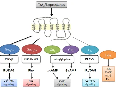
the TP’s functional and regulatory pathways (51, 54, 69–72) (Figure 2). Signaling
via the TP can be transduced through multiple G protein pathways, including Gq,
G11, G12/13, G15, G16, Gi, Gs and Gh (51), though some of these associations have
only been reported in isolated studies.
The two signaling pathways that appear most relevant to the biological actions of
TP are Gq and G12/13 (73), which stimulate the phospholipase-C pathway of
inositol phosphate/intracellular calcium elevation and RhoA activation,
respectively (74). TP-mediated signaling via Gq causes activation of
phospholiase C (75) and, through phosphoinositide hydrolysis, generation of
1,4,5-trisphosphate (IP3) and diacylglycerol (DAG), thereby mobilizing
intracellular Ca2+ and activating protein kinase C (PKC)(76–78). Signal
7
Figure 2: Summary of the reported major and minor signaling pathways of TP activation.
8
Rho signaling cascade, modulating such responses as regulation of the actin
cytoskeleton, cytokinesis, cell motility, contraction, cell proliferation, apoptosis,
thymic cellularity, Na+/H+ exchanger and myosin light chain kinase (51).
These systems both appear to contribute to platelet function - G12/13-mediated
stimulation of RhoA signaling induces myosin light chain phosphorylation leading
to platelet shape change, with subsequent activation of Gq-PLCβ signaling
causing aggregation (80). In mice, platelets lacking Gq or G13 are completely
unresponsive to TxA2, showing that Gq and G13 are required for platelet activation
(74). It is also interesting to note that low concentrations of the TP agonist
U46619 are sufficient to cause platelet shape change, while high concentrations
are necessary to induce aggregation (81).
In addition to signaling through Gα subunits, studies have reported
Gβγ-mediated activation of phosphatidylinositol 3-kinase (PI3K), phospholipase C-β2
and p44/42 mitogen-activated protein kinase (p44/42 MAPK)/extracellular
signal-regulated kinase 1/2 (ERK1/2), though the precise role in TP function and biology
has not been clearly defined (73, 82).
Regulation: Desensitization
The TP undergoes both homologous (following its own activation) and
heterologous (following activation of another receptor) desensitization (58, 83–
86) via phosphorylation of residues in the C-terminus. Here distinctions between
9
isoform, Ser329 is a phosphorylation site for protein kinase A (PKA) activated by
cAMP, allowing heterologous desensitization by Gs-coupled receptors like the
prostacyclin receptor (86). Ser331 is a target for protein kinase G
(PKG)/cGMP-mediated desensitization, and Thr337 is a site for protein kinase C (PKC)
phosphorylation (51).
The TPß, on the other hand, undergoes phosphorylation at Thr299 and Ser145 by
PKC (87), and at Ser357 (in tandem with Ser239) by G protein-coupled receptor
kinase (predominantly GRK2; lesser effects seen with GRK3, GRK5, or GRK6)
(88). GRK-mediated phosphorylation leads to recruitment of β-arrestin and
subsequent decoupling of G proteins from the receptor followed by internalization
(89). Arrestin-mediated internalization has been implicated in regulation of the
TPß contributing to lower basal surface expression levels of the receptor (54),
and its more ready internalization, compared to TP, following activation (88).
Of noteworthy relevance to the discussion of TP is an autoupregulation system
downstream of TP activation. Work by Wilson et al. uncovered a reactive oxygen
species (ROS)-dependent mechanism through which stimulation of the TP leads
to activation of NADPH oxidase, in turn leading to increased TP protein stability
in early biogenesis and, ultimately, increased receptor expression at the cell
surface (70). Though the mechanism underlying this pathway is as yet ill-defined,
exogenous ROS can also increase in TP protein stability and expression, which
may be particularly relevant in cardiovascular disease where ROS levels are
10
Regulation: Intracellular trafficking
As previously noted, TPα has been seen to be generally expressed at higher
levels at the cell surface compared to TPβ, at least in the transfected cell models
found in the literature, likely because the latter binds to protosomal subunit α7
and proteasome activator PA28γ through the unique TPβ the C-terminal domain,
leading to TPβ degradation by PA28γ-dependent protease activity (90). TPβ
endocytosis also occurs in a Rac-1-dependent manner through interaction with
Nm23-H2 (91), a process that requires interaction with the actin cytoskeleton
(92). TPβ that has been thus internalized may be recycled to the cell surface
through an interaction with Rab11 and the its GTPase-positive recycling
endosome (57, 93). Studies disagree on whether or not TPα is internalized
suggesting that internalization of TPα may cell- or context- specific (88, 94).
Successful trafficking to the TP to the cell surface appears to be strictly
dependent on glycosylation of Asn4 and Asn16 at the N-terminus, a process
common to both isoforms. Treatment of TPα and TPβ with tunicamycin (a
specific inhibitor of N-linked glycosylation) significantly reduced the binding of SQ
29,548, a TP antagonist, in both isolated cell membranes (95) and whole stably
receptor-expressing cells (96). Further, targeted mutation of either of these sites
resulted in a reduction by half of the Bmax for SQ 29,548 binding, while mutation
of both lead to near-complete retention of the receptor within the endoplasmic
11
Interplay between thromboxane and prostacyclin and their receptors
The actions of TxA2 are generally opposed by prostacyclin (PGI2), another
short-lived prostanoid that is generated from arachidonic acid predominantly through
COX-2 and prostacyclin synthase in the vascular endothelium (97). Acting
though the prostacyclin receptor (IP), PGI2 inhibits platelet activation, reduces
vascular proliferation, and causes vasodilation (98). The IP is coupled to Gs,
thus activation leads to increased intracellular cyclic AMP generation. Mice
lacking the IP display heightened thrombotic responses (99) and accelerated
development of atherosclerosis (8). A critical function of the IP is to restrain TP
function – mice that lack the IP show increased platelet and proliferative
responses to vascular injury in vivo with the opposite phenotype in TP deficient
mice and normalization of both phenotypes in double receptor knock out animals
(21). With their opposing roles, the interplay between TxA2 and PGI2 is an
important component of cardiovascular function and disease and is particularly
relevant in later parts of this work.
G protein-Coupled Receptor Dimerization
Substantial evidence has emerged in the field of GPCR research that these
seven-transmembrane proteins do not function only as monomeric receptors in
physiological systems, but rather as dimeric, and possibly oligomeric, units (100–
103). Receptor dimerization appears necessary for normal physiological
12
become a novel target for therapeutic research, with the goal of modifying
dimerization through the use of small molecules. Both homodimer and
heterodimer formation has been noted for a variety of GPCRs. For the GABAB
receptor, dimerization has been described as “obligatory”, with the pairing
necessary for proper biogenesis and receptor function (106, 107). In comparison,
dimer formation does not appear essential for other receptors, like the dopamine
receptor, but does modulate the receptor’s signaling response to agonist (108,
109). In addition, “‘non-obligatory’ GPCR heterodimer” formation has been noted
for a number of GPCRs (100), including the TP (see below) with significant
changes in downstream signaling of a given receptor when it forms a
heterodimer as compared to its homodimer (110, 111).
Dimerization may contribute early in GPRC biosynthesis (112) at certain
quality-control checkpoints (100). Homodimerization can occur early in the biosyntetic
pathway, most likely in the endoplasmic reticulum (112, 113), and is a
prerequisite for receptor trafficking to the cell surface for a number of other
GPCRs including the GABAB receptor (107), α1D- and α1B-adrenoceptors (114),
and β2-adrenergic receptor (115). Mutations that cause GCPR retention in the
ER/Golgi can act in a dominant-negative manner to block cell surface expression
of wild-type receptor (116, 117), as the mutants remain able to dimerize, yet
cannot pass through quality control checkpoints to exit the ER and continue
through biogenesis to be transported to the cell surface. These studies further
13
pathway, and have raised interest in developing ways to disrupt dimer formation
as a novel approach to modifying receptor function. For example, a small
molecule that prevents dimerization would also prevent cell surface expression,
an alternative approach to antagonism to reduce receptor function. If the target
receptor were one involved in disease pathophysiology, such as the TP in CVD,
then disease could be prevented or ameliorated.
Across the GPCR superfamily, there is substantial evidence for receptor
dimerization (118, 119) and a significant contribution therein to receptor
trafficking, ligand recognition, signaling and regulation (100, 102, 115, 120). As
mentioned above, previous work in our lab reported that the TP forms dimeric
receptor complexes (94, 121–123). In addition to homodimerization, TPα can
heterodimerize with TPβ, leading to enhanced isoprostane responsiveness (122).
Further, as part of the work outlined in this thesis, we observed equal propensity
for TPα to heterodimerize with the receptor for PGI2, the IP (123). As mentioned
above, the PGI2, a predominantly COX-2-derived mediator, acts via the IP to
activate the Gs-adenylyl cyclase signaling pathway causing vasodilation and
inhibition of platelet activation (98). In mice, the restraint placed by the PGI2-IP
system on TxA2-TP function limits the proliferative and platelet response to
vascular injury (21) demonstrating the in vivo relevance of this interplay. Further,
the elevated cardiovascular hazard in patients treated with COX-2 inhibitors can
be explained by selective suppression of COX-2-derived PGI2 without alteration
14
the TP with the IP contributes to the PGI2-TXA2 interplay - dimerization with the
IP dramatically shifts TP function from a lipid raft-excluded Gq-coupled receptor
to a raft-associated Gs-coupled receptor that yields a robust Gs-cAMP response,
concomitant with reduced Gq-inositol phosphate signaling, to TP agonists (121,
123). In effect therefore, the IP can bias the response of TXA2-TP toward a PGI2
-IP “like” signal predicted as beneficial in CVD. Loss of this shift in TP function in
individuals heterozygous for a signaling deficient IP mutant, IPR212C, may
contribute to their accelerated CVD (125).
Dimerization motifs as mediators of GPCR pairing
The importance of transmembrane (TM) helical interactions to protein structure
and function is evident across multiple diverse integral membrane protein
families (126, 127). Consequently, there is significant interest targeting TM
domains to modulate the function of membrane-spanning proteins, including
GPCRs (107, 118, 120, 128–130). Various studies, include those resolving
GPCR crystal structures (131), have shown that dimerization interfaces are
predominantly found in the TM domains (132–134).
Among all TM domains, a GxxxG motif, in which two glycines are separated by
any three other residues, is strongly over-represented (135), highly conserved
across species (136, 137) and can direct homologous or heterologous helical
interactions (126, 127, 138). Neighboring residues, especially the large aliphatic
helix-15
helix interactions (136, 137). In a number of proteins (139–142), including at least
two GPCRs (104, 115), placement of a leucine three residues after the second
glycine, to create a GxxxGxxxL motif, directs protein-protein interaction and
function. We identified a GxxxGxxxL motif within the 5th transmembrane of the
TPα (Figure 3) and, as part of this thesis research, examined its relevance for TP
function.
Current Therapeutics Targeting TXS and TP
Therapeutics that interfere with TP signaling generally act in one of three ways:
inhibition of COX-1/2 conversion of arachidonic acid to PGH2, inhibition of TXS
conversion of PGH2 to TxA2, or antagonism of the TP itself. Low-dose aspirin
which inhibits platelet COX-1-derived TxA2 is used widely to protection against
heart attack and stroke. Although true aspirin resistance is likely extremely rare
(143), heterogeneity in the beneficial response and weak benefit in some
pathophysiologies (e.g., diabetes, peripheral artery disease (144, 145), as well as
possible gender differences (146)) underscore the need for greater mechanistic
understanding to advance new therapeutic TXA2-TP approaches in CVD.
Over the past decade, attempts to use therapeutics that targeted TxA2 synthase
or the TP itself have generally met with limited to no clinical success. Most
recently, the selective TP receptor antagonist terutroban showed comparable,
but not superior, efficacy as low-dose aspirin in preventing recurrent ischemic
16
G205
G209
L213
G205
G209
L213
Figure 3: Location and positioning of the GxxxGxxxL motif within the TP.
Homology modeling (SWISS-MODEL) of the human TPα based on a 2.8Å
17
ifetroban (148), seratrodast (used for treatment of asthma) (149), and sulotroban
(150).
A number of other drugs act as tandem TP antagonists/TxA2 synthesis inhibitors.
Dipyridamole has been shown to be effective in adjunctive therapy with aspirin
for secondary prevention following stroke (151). Picotamide is a platelet
aggregation inhibitor that has shown antiplatelet efficacy in some studies (152).
Ridogrel has been tested for use in helping to treat Crohn’s disease and
ulcerative colitis, but without promising results (153, 154).
Furegrelate is a potent inhibitor of thromboxane synthase with little effect on
other enzymes essential for arachidonate metabolism, and has been investigated
for use in treatment of pulmonary arterial hypertension with promising early
results in model animals (155). Ozagrel also acts to inhibit synthesis, and has
been seen to reduce neurological impairment suffered with stroke, though
without improving long-term morbidity or other endpoints (156).
Project aims
While previous work by Wilson established the presence of the ROS-dependent
auto-upregulation paradigm, the underlying mechanisms of TP regulation within
the cell remained ill-defined. As such, one aim of my thesis work sought to
explore the mechanism(s) for the increase in cell surface TP expression in
response to activation. One possibility was that stimulation of the receptor was
18
However, quantitative real-time PCR analysis did not strongly support this
hypothesis.
In light of emerging evidence for dimerization as a mode of GPCR regulation, I
also sought to confirm TP homodimerization, which was apparent in
co-immunoprecipitation studies previously performed in the laboratory. Not only was
I able to confirm TP homodimerization, I also characterized the relative efficiency
of TP interaction with other prostanoid receptors, including the IP and the DP1
receptor for prostaglandin D2.
The findings from dimerization studies in this first aim, taken together with
research in the literature describing the significance of the GxxxGxxxL helical
interaction motif, a motif we identified within the TPs 5th TM, led to the
development of the second aim. The second aim sought to determine if the
GxxxGxxxL motif contributed to TP dimerization and, if so, whether or not
targeting of this motif would be a novel therapeutic approach to suppress TP
function. We determined that the TP TM5 GxxxGxxxL motif does contribute to TP
dimer formation. Further, using mutant forms of the TP, we demonstrated that
this motif was selectively involved in TP homodimer formation and signaling but
not TP-IP heterodimerization or signaling by the TP-IP heterodimer. These
studies provided the first proof-of-principle that the homo- and hetero-dimeric
functions of the TP and TP-IP can be discriminated to suppress the
CVD-deleterious TP function and preserve the CVD-beneficial TP-IP function.
19
a similar transmembrane domain in the II integrin (128), provided an exciting
initial indication of the potential for targeting this region to develop a biased
therapeutic directed against the deleterious side of TP signaling. Full analysis of
20
CHAPTER 2: Materials and Methods
Constructs
Hemagglutinin- (HA) tagged human IP and TPα cloned into the mammalian
expression vector pcDNA3 (Invitrogen, CA) were previously created in the lab
(123). QuikChange site-directed mutagenesis (Stragagene, CA) was used to
replace G205 and G209 with leucines, a small-to-large replacement that disrupts
helix-helix interaction (139, 141, 142). We replaced L213 with a tyrosine based on
the studies of the GxxxGxxxL motif in the ß2-adrenergic receptor(115). The
resulting mutant was termed TPL205,L209,Y213. Similar mutations were made at the
partial GxxxG motif at the beginning of the first intracellular loop to create the
TPL51,L55 mutant. See Table 1 for the list of primers designed for mutagenesis.
HA-tagged IP, TPα, and TPL205,L209,Y213 were fused at their C-termini to either
Renilla luciferase (rLuc) or yellow fluorescent protein (YFP), via restriction
enzyme cutting and reassembly after purification of DNA segments (157). Briefly,
the stop codon was removed by PCR and each stop-less construct cloned into
pRLuc-N3(h) (Perkin-Elmer, MA) and pEYFP-N1 (Clontech, CA) plasmids in
frame with the fusion protein start site. All sequences were verified by DNA
sequencing.
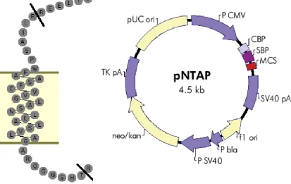
The TP first transmembrane domain (TM1) interacting peptide was created
through PCR amplification of the TP sequences between residues R23 and T59,
21
Table 1:Sequences of primers used in the generation of mutants employed in this work.
Note that some primers must be used following the introduction of a prior mutation due to overlap in the sequences.
Target
Mutation Primer Sequence
G205xxxG209xxxL213 → G205xxxG209xxxY213
Sense 5'- CTG TCC TTC TAC CTG AAC ACG GTC -3'
Antisense 5'- GAC AGG AAG ATG GAC TTG TGC CAG -3'
G205xxxG209xxxY213 → G205xxxL209xxxY213
Sense 5'- CC ATG CTG GGC GGC CTC TCG GTC TTG CTG TCC TTC -3'
Antisense 5'- GAA GGA CAG CAA GAC CGA GAG GCC GCC CAG CAT GG -3'
G205xxxL209xxxY213 → L205xxxL209xxxY213
Sense 5'- GG CTG CTC TTC TCC ATG CTG GGC CTC CTC TCG GTC -3'
Antisense 5'- GAC CGA GAG GAG GCC CAG CAT GGA GAA GAG CAG CC -3'
G51xxxG55 → G51xxxL55
Sense 5'- CGC GCG GCA GTT GGG TTC GCA CAC GCG CTC -3'
Antisense 5'- GAG CGC GTG TGC GAA CCC AAC TGC CGC GCG -3'
G51xxxL55 → L51xxxL55
Sense 5'- CTG AGC GTG CTG GCG CTC GCG CGG CAT TG -3'
Antisense 5'- CAA TGC CGC GCG AGC GCC AGC ACG CTC AG -3'
22
restriction enzyme digestion and religation, into the pNTAP vector obtained as
part of the InterPlay N-terminal Mammalian TAP System (Stratagene, LaJolla,
CA) to provide necessary the constitutive cytomegalovirus promoter and
SV40/poly-adenosine tail for stability (Figure 4).
Cell culture
HEK 293 and Meg-01 cell lines were from the American Type Tissue Culture
Collection (ATTC; Rockville, MD). HEK 293 cells were maintained following
established and published protocol in the lab (48); HEK cells were grown in
DMEM High Glucose medium (Invitrogen) containing 10% fetal bovine serum,
1% penicillin-strepomycin, 2mM L-glutamine, and 25mM HEPES buffer. Cells
were grown in 75 cm2 surface area flasks and passaged in a 1:4 ratio upon
reaching 80-90% confluency by allowing cells to lift in 37°C Hank’s Balanced Salt
Solution containing 0.02% EDTA prior to collection and redistribution into new
plates.
Meg-01 cells were grown in RPMI-1640 (Invitrogen) containing 10% fetal bovine
serum and 1% penicillin-strepomycin. Cells were grown in 20 mL of medium in
75 cm2 surface area flasks. Passaging was performed according to ATTC
literature that accompanied the cells, every 2-3 days by scraping the bottom of
the flask with a disposable cell scraper and addition of 5 mL of this cell
23
24
Human aortic smooth muscle cells (HuAoSMCs, Biowhittaker Inc., Walkersville,
MD) were cultured in smooth muscle cell basal medium supplemented with fetal
bovine serum (5%), human recombinant epidermal growth factor (hEGF; 0.5
ng/ml), insulin (5 g/ml), human recombinant fibroblast growth factor (hFGF; 2
ng/ml) plus gentamicin (50 g/ml), and amphotericin-B (50 ng/ml) (all supplies via
Lonza, Allendale, NJ). HuAoSMCs of passages 5–9 were used in experiments.
Transient transfection of cell lines
Transient transfections of HEK 293 cells were initially performed using FuGENE
6 (Roche Applied Science, IN) following manufacturer’s instructions (123). After
discontinuation of FuGENE 6 production, transfections were performed using
X-tremeGENE 9 (Roche Applied Science, IN), following manufacturer’s
instructions. This replacement reagent was created by Roche as an improved
form of FuGENE 6 with reduced cytotoxicity and the need for less reagent per
transfection. The serum-free medium used in transfections was Opti-MEM
medium (Invitrogen). DNA transfected ranged widely, based on the needs of the
given experiments, but always following the proscribed DNA:transfection
reagent:Opti-MEM medium ratio provided in the literature from the manufacturer.
Exact amounts of DNA transfected varied, based on the assay to be performed
and quantity of cells needed, and are noted in subsequent sections below.
Transient transfections of Meg-01s were performed by nucleofection using an
25
manufacturer’s instructions, introducing a total of 3 μg of DNA to the cells as
instructed. The range of DNA quantities used in HEK 293 transfection was not
possible given the constraints of the nucleofection system. Experimental design
with transfected Meg-01 cells was adjusted based on this constraint, dictating
how many duplicate measurements or treatment groups were possible in a given
replicate.
In all cases, DNA levels were equalized in all transfections using empty pcDNA3
vector. Assays were performed 48 hours after transfection.
Bioluminescence Resonance Energy Transfer (BRET) assay: original
protocol.
Dimerization of rLuc and YFP fused receptors was examined by measuring
bioluminescence resonance energy transfer (BRET) from an energy donor (rLuc
fused) receptor to an energy acceptor (YFP-fused) receptor following addition of
substrate for rLuc (coelenterazine h; Molecular Probes, Life Technologies, NY)
(Figure 5). Coelenterazine h was supplied solid, 250 µg, and stored at -20°C. A
stock vial, in which the compound was resuspended to a 2.5 mM solution in
200-proof ethanol, was maintained for further dilution into a working solution (50 μM)
for the assay.
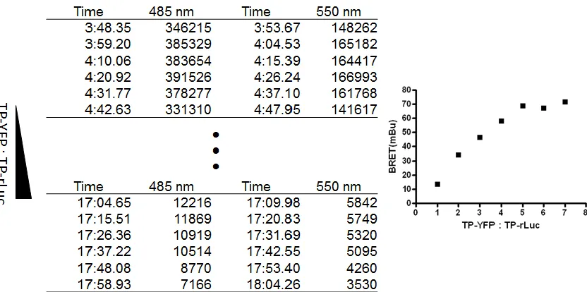
In BRET saturation experiments, cells were transfected with a fixed amount of
rLuc-receptor (0.25µg) together with increasing amounts of YFP receptor
26
27
dimerization resulted in a characteristic saturation curve (Figure 6A) that allowed
for calculation of the BRET50 - the level of acceptor receptor YFP-rLuc tagged
receptor (expressed as fold over basal total YFP, excited with an external light
source; see details below) at which half of the maximal BRET signal was
detected.
The BRET50 served as a quantitative measurement of affinity for dimerization,
with a lower BRET50 indicating a higher affinity for dimerization. This allows for
comparison of affinities between receptor pairings. Changes in the maximal
BRET values may reflect absolute levels of dimer formed. However, the absolute
BRET value also can be influenced by the distance and orientation of the donor
and acceptor molecules, which are variable based on the molecular arrangement
of a particular dimer, rather than simply the number of dimers formed. Similarly,
binding of a ligand to either protomer can change the three-dimensional structure
of a receptor, potentially changing the distance between the rLuc donor and YFP
acceptor molecules and, thus, the absolute BRET values. This possibility is
discussed further in the relevant section of the results. Thus, the BRET50 is
particularly useful as a readout of dimerization affinity that is independent of
changes in donor-acceptor distance.
In BRET competition assays, increasing amounts of a competitor receptor
(without a donor or acceptor moiety) were co-transfected together with a fixed
ratio (1:7) of receptor-rLuc + receptor-YFP (and hence a fixed BRET value). A
28
29
indicates that the competing receptor can interact with either (or both) protomer,
allowing an initial characterization of dimerization affinity (Figure 6C). This assay
is useful for initial screen of receptor pairings of interest, as it does not require
the time-consuming work of stop codon removal generation of donor and
acceptor fused proteins.
Initially, BRET measurements were performed following protocols published by
Bouvier, et al. (121). Forty eight hours after transient transfection, cells were
harvested (phenol red-free Hank’s Balanced Salt Solution containing 0.02%
EDTA), washed twice with phosphate-buffered saline and resuspended in DPBS
containing 5.56 mM glucose (Invitrogen, 14287), then redistributed in two 96-well
plates (first: black, clear-bottomed; second: white, opaque-bottomed; 100,000
cells/well) and maintained at 37°C. Total YFP (Ex485nm, Em555nm) was first
collected using a luminescence multi-plate reader (VICTOR3, Perkin Elmer) with
the black, clear-bottomed plate and acceptor expression calculated as fold over
basal. Following this, coelenterazine h (Invitrogen, stock resuspended to 2.50
mM in ethanol for working solution) was diluted to 5μM in phosphate buffered
saline containing Ca2+ and Mg2+. A fresh solution was made each time, added to
all cells in the white plate, and emission from the donor (485nm) and acceptor
(555nm) were gathered sequentially from each well across the entire plate. Milli
BRET units (mBU) were calculated as the ratio of Em555 over Em485 nm
corrected for cells expressing the rLuc receptor alone, and arbitrarily multiplied
30
BRET assay optimization
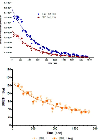
After several months of using this standard approach, concerns surfaced as to
the stability of the BRET signal over the time taken to process a single plate (i.e.
from the time of substrate addition to the time of the last BRET reading, typically
20 minutes). A simple experiment was designed to examine signal stability: a
single population of HEK 293 cells were transiently transfected with a set ratio
(1:7) of rLuc- and YFP-tagged TP and the established protocol used to
establish whether YFP and BRET readings were consistent across both plates.
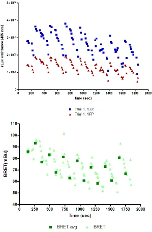
Total YFP values were stable across the black plate; however, the BRET signal
was strikingly unstable (Figure 7, Figure 8), with a significant loss of signal. This
loss of signal across a homogenous plate raised concerns for interpretation of
the BRET assay going forward.
A variety of different experimental adjustments were made in attempts to
minimize the loss of BRET signal observed with the original assay protocol: [1]
the protocol was amended to take a reading of one sample set (6 replicates) from
the black total YFP plate followed by one reading from the white BRET plate,
alternating so as to more closely align the time of measurement of the two values
for a given sample. [2] 2-hydroxypropyl-β-cyclodextrin, a ring-shaped compound
that increases solubility and stability of compounds in water (158), was added to
31
32
Figure 8: Decay in BRET and component emissions over time. HEK 293 cells were transfected with rLuc- TPWT (donor, 0.25µg) + YFP- TPWT (acceptor, 0.75µg). Two 96-well plates were prepared containing identical samples in each well (except for the control rLuc alone cells), and assayed following the original BRET protocol: coelenterazine was diluted to 50 μM in PBS containing Ca2+
33
taken to perform the readings. Neither adjustment improved the signal stability
across a homogenous set of plates.
A search of data sheets and technical literature for coelenterazine h as a
substrate for Renilla luciferase revealed (159) that coelenterazine undergoes a
predictable deterioration upon introduction to aqueous solution with a 40-50%
loss of functional capacity over the first 20 minutes. After this period has passed,
however, the solution maintains a mostly linear functional response over the next
four hours (Figure 9). Additionally, this decay appears to be caused in part by the
presence of calcium cations in solution. Based on this information, the BRET
assay was modified to include [3] a 20-minute waiting period at room
temperature after addition of the coelenterazine into Ca2+-free phosphate
buffered saline before addition to the wells.
Because of the time taken from the first addition of colenterazine to the final
reading of the 96th well (20 min), loss of signal due simply to substrate catabolism
by rLuc was a further issue. One approach to addressing this issue was imaging
of the whole plate at one time through use of an IVIS imaging system. Luciferase
and YFP activity was captured for the plate as a whole, and binning (manually
overlaying a 96-square grid onto the image produced by the IVIS so as to allow
quantitation of activity by well) was employed to measure the YFP and BRET
signals given off by samples. This proved significantly less sensitive that the
Victor multiwell plate reader. However, a significant observation was made in the
34
35
itself emitted significant amounts of background light emission during the
measurement. Thus, despite the established protocols calling for the use of two
separate plates for the BRET assay – the clear bottomed black plate for total
YFP and the opaque white plate for BRET, it appeared that it was preferential to
avoid the white plate (hence reducing background) and instead to [4] use a single
clear-bottomed black plate to measure first the YFP signal and then, after
addition of coelenterazine h (rested in Ca2+-free buffer for 20 mins), the BRET
signal. This change to the method had the added benefit of taking paired YFP
and BRET measurements from the exact same sample of cells, as opposed to
two different distributions of the same cell mixed in two parallel plates, further
improving precision (Figure 10).
To avoid loss of signal due to enzymatic substrate catabolism, [5] coelenterazine
was added to one sample (six replicate wells) and the BRET signal was
measured before moving to the next sample. This approach greatly reduced the
time that the substrate sat in the well with the enzyme before the BRET reading.
Altogether, these 5 changes to the assay resulted in a marked improvement in
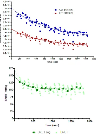
signal stability across a plate (Figure 11) as well as the following improved
protocol that is now standard in the laboratory:
Coelenterazine h (Invitrogen, stock resuspended to 2.5 mM in 200-proof ethanol
for working solution) was diluted to 5μM in Ca2+
- and Mg2+-free phosphate
buffered saline and allowed to rest at room temperature for 20 minutes. Cells
36
37
Figure 11: Decay in BRET and component emission measurements after final modifications to BRET protocol. HEK 293 cells were transfected with rLuc-TPWT (donor, 0.25µg) + YFP- TPWT (acceptor, 0.75µg) assayed following the a further modified BRET protocol: coelenterazine was diluted to 50 μM in PBS without Ca2+
38
Balanced Salt Solution containing 0.02% EDTA), washed twice with
phosphate-buffered saline and resuspended in DPBS containing 5.56 mM glucose
(Invitrogen, 14287), then redistributed into a black, clear-bottomed 96-well plate
(100,000 cells/well) and maintained at 37°C. Total YFP (Ex485nm, Em555nm)
was first collected using a luminescence multi-plate reader (VICTOR3, Perkin
Elmer) and calculated as fold over basal (no YFP-fused receptor present).
Following this reading, coelenterazine h was added a set of six replicate wells
and donor (485nm) and acceptor (555nm) emissions gathered sequentially for
each sample. Colenterazine addition and BRET readings in sets of 6 replicate
wells were repeated until all data was collected. Milli BRET units (mBU) were
calculated as the ratio of Em555 over Em485 nm corrected for cells expressing
the rLuc receptor alone, and multiplied by 1000.
Cell surface expression of the TP
HEK 293 and Meg-01 cells were transfected with HA-tagged TPWT or
HA-TPL205,L209,Y213. Cells were harvested into ice-cold FACS buffer (DPBS containing
1% BSA and 0.1% sodium azide). Cell suspensions were stained with anti-HA
mouse IgG1 (Monoclonal 16B12) conjugated to Alexa Fluor® 488 (Invitrogen,
CA) for 30 minutes prior to washing. Median fluorescence intensity (MFI) was
collected using a BD FACSCalibur flow cytometer (Becton Dickinson, Franklin
Lakes, NJ, maintained by UPenn flow cytometry core). Cells were first gated to
39
scatter measurements, then gated on the FL1 filter (488nm excitation, 530/30nm
filter emission) to obtain MFI values, which were corrected by subtraction of
collected MFI values of non-transfected, antibody-stained HEK 203 of Meg-01
cells.
Measurement of second messenger generation
Measurement of intracellular inositol monophosphate (InosP) or cyclic AMP
(cAMP) was performed using the IP-One Tb kit (Cisbio Bioassays, MA) or
LANCE cAMP 384 kit (PerkinElmer, MA), respectively, according to the
manufacturer’s instructions. Both kits are based around the same principle of
fluorescence resonance energy transfer (FRET) between two fluorophores. In the
IP-One InosP assay, cells are treated with a mixture of [a] monoclonal anti-InosP
antibody (Ab) tagged with crypate (a fluorophore) and [b] InosP tagged with the
dye “d2”, in addition to [c] LiCl, which inhibits inositol-phosphate phosphatase.
The d2-InosP forms a complex with the crypate-Ab that allows for FRET from the
crypate to the d2 upon excitation of the former. Unlabeled InosP, produced by
the cell, competes for binding to the crypate-Ab. The more InosP is produced by
the cell, the less FRET-capable complex is created, with the ratio of light emitted
by the crypate to light emitted by the d2 dye as the quantitative readout of cellular
InosP production after comparison to a standard curve of InosP concentrations.
This same principle is employed by the LANCE cAMP assay, with
40
replacing the d2-tagged InosP. IBMX (3-isobutyl-1-methylxanthine) is employed
to inhibit cAMP degradation through phosphodiesterase inhibition. Cells were
stimulated with or without the TP agonist U46619 (Cayman Chemicals, MI) over
a range of concentrations, as noted in the respective results section, for one
hour.
Radioligand binding and displacement
HEK 293 cells, transfected with HA-tagged TPWT or TPL205,L209,Y213 in poly-L
lysine-coated 12 well plates, were washed with radioligand binding buffer (HBSS
with 2% BSA). For saturation binding 3H-SQ 29,548 (PerkinElmer, Waltham, MA
or American Radiolabeled Chemicals, St. Louis, MO) was distributed to cells at
concentrations ranging from 25 μM to 250 μM. After 60 minutes at 37°C, cells
were washed with ice-cold binding buffer to remove unbound ligand, lysed with 1
M NaOH for 30 minutes at 37°C and radioactivity measured by scintillation
counting.
For displacement analysis, 3H-SQ 29,548 was held constant at 0.25 nM and
competing ligands were added 5 minutes prior to the radioligand. Competing
ligands I-BOP and cold SQ 29,548 were applied as treatments ranging from 5 nm
to 500 nm, while U46619 and the isoprostane E2III (iPE2III) concentrations ranged
from 25 nm to 2500 nm, due to lower affinity for the TP. In either experiment,
non-specific binding was accounted for by the addition of a 100-fold excess of
41
binding buffer to remove unbound ligand, lysed with 1 M NaOH for 30 minutes at
37°C and radioactivity measured by scintillation counting.
Immunoprecipitation and immunoblotting
HA-tagged TPWT or TPL205,L209,Y213 were immunoprecipitated from transfected
HEK 293 cells using Pierce HA Tag immunoprecipitation/Co-immunoprecipitation
Kit (cat# 23610,Thermo Scientific, Waltham, MA), according the manufacturer’s
instructions. This kit uses anti-HA antibody conjugated to agarose beads for
immunoprecipitation of HA-tagged proteins. Eluted proteins were resolved via
NuPAGE electrophoresis (Invitrogen, CA) under reducing conditions. HA-tagged
TPWT or TPL205,L209,Y213 were visualized with anti-TP (Cayman Chemicals, MI;
1:100) while immunoprecipitated Gqα was visualized with anti-Gq/11α (Millipore,
CA; 1:1000). Antigen-antibody complexes were revealed using horseradish
peroxidase conjugated anti-rabbit IgG (Jackson ImmunoResearch, PA; 1:10,000)
and visualized by enhanced chemiluminesence (ECL Plus, GE
Healthcare/Amersham, NJ). Quantification of proteins was by densitometry.
Treatment of cells with the CHAMP peptide
A computed helical anti-membrane protein (CHAMP) peptide (128, 142) was
supplied by the lab of Dr. Joel Bennett (University of Pennsylvania School of
Medicine) as 1 mM CHAMP in DMSO. As directed by members of the Bennet
42
second messenger generation assays. To avoid vehicle effects, the total final
concentration of DMSO in media did not exceed 0.1%.
Quantitative-PCR
RNA isolated from human aortic smooth muscle cells grown in culture was
quantified (NanoDrop Spectrophotometer) and reverse transcribed into cDNA
(MultiScribe Reverse Transcriptase, Applied Biosystems) according to
manufacturer’s instructions. Quantitative real-time polymerase chain reaction
(Q-PCR) was carried out using inventoried primer/probe gene expression assays
with TaqMan Universal PCR Master Mix (Applied Biosystems) for the human
thromboxane receptor gene (TBXA2R, cat# 4331182). Q-PCR products were
monitored using the ViiaTM 7 Real-Time PCR System (Applied Biosystems) and
data was analyzed using the 2-ΔΔCt method of relative quantification (RQ) using
18S for normalization (160).
Receptor modeling
Working with the Hwa Laboratory (Yale Medical School), the human hTP
sequence was aligned with solved crystal structures, bovine rhodopsin (OPSD,
UniProt identifier P02699) and the human ß2-adrenergic receptor (ADRB2,
UniProt identifier P0755) in ClustalW [http://www.clustal.org]. Both the PAM250
and BLOSUM evolution matrix modeling algorithms (161) indicated closer
43
with ADRB2 (33.48). Each bundle of seven transmembrane -helices was
therefore based on a 2.8Å crystallographic bovine rhodopsin template (1HZX)
(162) using the internet-based protein-modeling server, SWISS-MODEL
[http://swissmodel.expasy.org] (GlaxoSmithKline, Geneva, Switzerland), and
energy minimized using the Gromos96 force field in DeepView
[http://spdbv.vital-it.ch]. Extracellular and cytoplasmic loop regions were manually constructed, built
according to JPred consensus, and energy-minimized using the NAMD molecular
dynamics simulator (163).
Fluorescence microscopy
HEK 293 cells were grown to 80-90% confluency in 60 mm dishes, then lifted in 1
mL 0.25% trypsin, then added to 8 mL HEK growth medium. Cells were
transfected in 0.4 mL aliquots by 0.25 μg of either myc-TPWT or
myc-TPL205,L209,Y213, with each aliquot being dispensed into one well of an 8-well
poly-D-lysine-coated slide (Becton Dickinson, Franklin Lakes, NJ) and allowed to
grown for 48 hours. Cells were then fixed with 4% paraformaldehyde for 10
minutes at room temperature, followed by permeabilization with 0.2% Triton
X-100 in PBS. Staining was performed with 1:800 diulted anti-myc AlexFluor 555
conjugate (Millipore, Billerica, Massachusetts) for one hour with gentle shaking.
Cells were then mounted with Vectashield + DAPI (VectorLabs, Burlingame, CA)
and cover slips were sealed with clear nail polish. Imaging was performed on a
44
CHAPTER 3: Characterization of Thromboxane Receptor Regulation.
TPα auto-upregulation is not driven by increases in mRNA levels
The first possibility explored as a mechanistic explanation for auto-upregulation
of the TP following agonist activation (70) was increased receptor biogenesis
resulting from elevated levels of mRNA. If one downstream effect of TP activation
was to increase TP mRNA levels this could contribute to the increase in TP
protein and cell surface TP levels observed. To examine whether TP activation
leads to increases in mRNA levels TP-transfected HEK 293 cells or human
aortic smooth muscle cells (HuAoSMCs), which endogenously express TP, were
treated with either 100 nM IBOP, 1 μM SQ 29,548 (a TP antagonist), or a
combination of both for 2 hours. Cells were harvested and TP mRNA quantified
by real-time PCR. IP mRNA was also measured as a negative control since IP is
not stimulated by IBOP. Quantitative comparisons made to mRNA levels at
pre-treatment showed no significant change in mRNA levels for cells with any
treatment (Figure 12). Thus, upregulation of TP transcription in response to
short-term agonist treatment did not appear to contribute to auto-upregulation.
Additional studies were performed to query whether a longer-term agonist
treatment could lead to changes in mRNA levels that might explain that
auto-upregulation paradigm. In both transiently transfected HEK 293 cells as well as
natively expressing HuAoSMCs, 6- or 12-hour treatment with 100 nM IBOP lead
to no significant change in TPα mRNA levels (Figure 13A, B). Nor did we see any
45
(A)
(B)
46
(A)
(B)
(C)
Figure 13: Activation of the TP does not alter TP transcription.
47
course of up to 24 hours (Figure 13C). Thus, agonist activation in both and the
long and short terms did not cause any noticeable changes in mRNA levels that
could explain the auto up-regulation seen for the TPα.
A modified version of the BRET assay provides greatly increased
sensitivity
Preliminary co-immunoprecipitation studies carried out previously in the lab
suggested that, similar to other GPCRs (100–103, 107, 114, 115), the TP formed
homodimers within the cell. To address the question of dimerization in live cells
(as compared to the cell homogenates used in co-immunoprecipitation assays),
we used the bioluminescence resonance energy transfer (BRET) assay, which
measures transfer of energy from a donor (rLuc-fused receptor) to acceptor
(YFP-fused receptor) as their physical interaction (for details see Chapter 2,
Methods, Pages 26-30 and Figure 6). As outlined in Chapter 2 (Pages 31-39),
while establishing the BRET assay, we noticed a large decline in the raw values
for the emission readings from the beginning to end of reading the BRET plates.
We considered whether this decline in signal was a real reflection of changing
dimerization events or indicative of potential problems with the design of the
BRET assay itself. To examine this, one large, homogenous batch of cells were
transfected with a set amount of TP-rLuc and TP-YFP. These cells were
harvested, then resuspended in DPBS with glucose and sodium pyruvate, then