University of Pennsylvania
ScholarlyCommons
Publicly Accessible Penn Dissertations
Summer 8-14-2009
Structure and Mechanics of Proteins from Single
Molecules to Cells
Andre E. Brown
University of Pennsylvania, [email protected]
Follow this and additional works at:http://repository.upenn.edu/edissertations Part of theBiological and Chemical Physics Commons
This paper is posted at ScholarlyCommons.http://repository.upenn.edu/edissertations/9
For more information, please [email protected].
Recommended Citation
Brown, Andre E., "Structure and Mechanics of Proteins from Single Molecules to Cells" (2009).Publicly Accessible Penn Dissertations. 9.
Structure and Mechanics of Proteins from Single Molecules to Cells
Abstract
Physical factors drive evolution and play important roles in motility and attachment as well as in
differentiation. As animal cells adhere to survive, they generate force and “feel” various mechanical features of their surroundings and respond to externally applied forces. This mechanosensitivity requires a substrate for cells to adhere to and a mechanism for cells to apply force, followed by a cellular response to the mechanical properties of the substrate. We have taken an outside-in approach to characterize several aspects of cellular mechanosensitivity. First, we used single molecule force spectroscopy to measure how fibrinogen, an extracellular matrix protein that forms the scaffold of blood clots, responds to applied force and found that it rapidly unfolds in 23 nm steps at forces around 100 pN. Second, we used tensile testing to measure the force-extension behavior of fibrin gels and found that they behave almost linearly to strains of over 100%, have extensibilities of 170 ± 15 %, and undergo a large volume decrease that corresponds to a large and negative peak in compressibility at low strain, which indicates a structural transition. Using electron microscopy and X-ray scattering we concluded that these properties are likely due to coiled-coil unfolding, as observed at the single molecule level in fibrinogen. Moving inside cells, we used total internal reflection fluorescence and atomic force microscopy to image self-assembled myosin filaments. These filaments of motor proteins that are responsible for cell and muscle contractility were found to be asymmetric, with an average of 32% more force generating heads on one half than the other. This could imply a force imbalance, so that rather than being simply contractile, myosin filaments may also be motile in cells.
Degree Type
Dissertation
Degree Name
Doctor of Philosophy (PhD)
Graduate Group
Physics & Astronomy
First Advisor
Dennis E. Discher
Keywords
biophysics, fibrin, myosin, atomic force microscopy, fluorescence microscopy, mechanosensation
Subject Categories
Biological and Chemical Physics
ii
To Klara
iii
Acknowledgments
First I would like to thank my advisor Dennis Discher who has taught me to be persistent
and not give up when confronting a challenging problem. I hope his penchant for
penetrating analysis and his ability to perceive the most interesting seam of gold in a
mountain of data has rubbed off at least in part. I would also like to thank John Weisel
for being both a close collaborator and a friend. John’s inextinguishable enthusiasm for
science and life is a constant inspiration. Rustem Litvinov is a wonderful scientist and
working closely with him has been a great pleasure. This dissertation would be much
thinner if not for his diligent work. Special thanks are due Phil Nelson. Exposure to his
textbook as an undergraduate was the impetus for my application to Penn in the first
place and he has helped make sure that coming here was one of the best decisions I have
made. One of the other parts of my experience here that has made it so worthwhile is the
openness of the faculty to discussion with lowly graduate students. This has lead to a
productive collaboration with Prashant Purohit, who never hesitates to discuss whatever
bit of data I bring to his office. For the same sentiment, I thank Paul Heiney for not only
meeting with me whenever I had a question but also making visits to the lab to explain
(and fix) the X-ray apparatus on a regular basis. I also thank Paul Janmey, Yale
Goldman, and Tom Lubensky for their insights and suggestions. Finally, I have had the
privilege of sharing offices with Kun-Chun Lee in the early days, Florian Rehfeldt for
most of my time in the Discher lab, and most recently with Amnon Buxboim and Irena
iv ABSTRACT
STRUCTURE AND MECHANICS OF PROTEINS FROM
SINGLE MOLECULES TO CELLS
André E. Brown
Dennis E. Discher
Physical factors drive evolution and play important roles in motility and attachment as
well as in differentiation. As animal cells adhere to survive, they generate force and
“feel” various mechanical features of their surroundings and respond to externally
applied forces. This mechanosensitivity requires a substrate for cells to adhere to and a
mechanism for cells to apply force, followed by a cellular response to the mechanical
properties of the substrate. We have taken an outside-in approach to characterize several
aspects of cellular mechanosensitivity. First, we used single molecule force spectroscopy
to measure how fibrinogen, an extracellular matrix protein that forms the scaffold of
blood clots, responds to applied force and found that it rapidly unfolds in 23 nm steps at
forces around 100 pN. Second, we used tensile testing to measure the force-extension
behavior of fibrin gels and found that they behave almost linearly to strains of over
100%, have extensibilities of 170 ± 15 %, and undergo a large volume decrease that
corresponds to a large and negative peak in compressibility at low strain, which indicates
a structural transition. Using electron microscopy and X-ray scattering we concluded that
v
level in fibrinogen. Moving inside cells, we used total internal reflection fluorescence
and atomic force microscopy to image self-assembled myosin filaments. These filaments
of motor proteins that are responsible for cell and muscle contractility were found to be
asymmetric, with an average of 32% more force generating heads on one half than the
other. This could imply a force imbalance, so that rather than being simply contractile,
vi
Table of Contents
INTRODUCTION………..………..1
CHAPTER 1: Review of Unfolding in Cellular Mechanosensing..………..4
Summary……….4
Introduction……….4
Proteins Unfold under Force………...7
Unfolding in the Extracellular Matrix Influences Assembly………11
Force Modulates Cell-Matrix Interactions………14
The cytoskeleton responds to and actively applies force………..17
Towards a Characterization of the Cellular ‘Unfoldome’………22
Conclusion………26
CHAPTER 2: Single Molecule Mechanics of Fibrinogen………...…………28
Summary………...….……...28
Introduction………...28
Methods……….29
Results and Discussion………..33
Conclusions………...36
CHAPTER 3: Multiscale Mechanics of Fibrin Clots.….……….37
Summary………...……37
Introduction………....…...37
Results and Discussion………...…..38
Conclusion………...…….48
Supplement………...……50
CHAPTER 4: Asymmetry of Myosin Filaments……..………84
Summary………...84
Introduction………...85
Materials and Methods..………86
Results and Discussion………..89
Conclusion………...107
Supplement………..108
vii
List of Figures
CHAPTER 1
FIGURE 1: Transcription on elastic substrates………...6
FIGURE 2: Protein unfolding in cell substrate interactions………8
FIGURE 3: Nano tools for protein folding and funnels………10
FIGURE 4: Cys-shotgun labeling………..25
CHAPTER 2 FIGURE 1: Fibrinogen unfolding schematic……….30
FIGURE 2: Electron microscopy of fibrinogen oligomers…….……..……….32
FIGURE 3: Force extension curves of fibrinogen and histograms...……….34
CHAPTER 3 FIGURE 1: Multiple scales of fibrin mechanics...……….39
FIGURE 2: Mechanics of fibrin clots...……….40
FIGURE 3: Structural changes in stretched fibrin clot….……….43
FIGURE 4: Small angle X-ray scattering of stretched fibrin clots…...……….44
FIGURE 5: Longitudinal and lateral strain measurement……….………55
FIGURE 6: Image-pull-image AFM of fibrin…...………56
FIGURE 7: Schematic of water loss in stretched fibers………57
FIGURE 8: Force relaxation in a hydrated clot……….………58
FIGURE 9: Volume change of polyacrylamide………59
FIGURE 10: 8-chain model schematic………..………61
FIGURE 11: Confocal reconstruction of fibrin network………...………..……..62
FIGURE 12: 8-chain model applied to collagen………...……….68
FIGURE 13: 8-chain model applied to fibrin….………...…...……....….……73
FIGURE 14: Change in network volume………..76
FIGURE 15: 3-chain model applied to fibrin………77
FIGURE 16: Isotropic network model applied to fibrin……….……...………79
FIGURE 17: Isotropic network model with buckling……….………...82
CHAPTER 4 FIGURE 1: TIRF vs. AFM resolution..……….91
FIGURE 2: Intensity and height profiles of myosin filament………..……….…….93
FIGURE 3: Intensity-height scaling………….……….96
TABLE 1: Myosin per filament……….………98
FIGURE 4: Fractional asymmetry of myosin filaments………..101
FIGURE 5: Pyramidal lattice model……….………...103
FIGURE 6: Schematic of an asymmetric filament………..106
FIGURE 7: Geometry for scaling correction………...109
1
Introduction
Biological materials have evolved to serve a wide variety of functions ranging from
structural (bone and exoskeleton) to optical (eyes) to thermal (fur). Cells themselves are
also increasingly being studied as complex, active materials for their interesting optical
and especially mechanical properties. Furthermore, many cell types are also able to sense
and respond to the mechanical properties of their surroundings. However, the processes
by which cells sense these properties are far from exhaustively characterized, let alone
completely understood. Current research is aimed at understanding every aspect of
cellular mechanosensing from the mechanical properties of the extracellular matrix that
cells adhere to in vivo, to how cells apply forces to their surroundings, and ultimately
how the reaction forces from the substrate affect cell behaviors. In this dissertation I will
touch on aspects at each of these levels.
The first chapter reviews work on cell mechanosensation with an emphasis on
protein unfolding as a possible force-sensitive process. Because of the variety of systems
where unfolding is thought to play a role, this chapter also ties the remaining chapters
together thematically.
Chapters 2 and 3 describe investigations into the mechanical properties of fibrin
and its precursor fibrinogen. Fibrin is the principal protein component of blood clots so
in addition to its mechanical role in stopping blood flow at sites of injury it is also an
extracellular matrix protein involved in wound healing. We first used single molecule
2
force and found that their coiled-coil regions unfold in 23 nm steps. Using this
information we then performed a series of experiments on macroscopic clots to
understand how the unfolding observed in single molecules is manifested in the
mechanics at larger scales. Interestingly, unfolding seems to endow clots with their large
extensibility despite their relatively rigid microstructure and leads to a large volume loss
upon stretching. This volume loss corresponds to a negative compressibility that is likely
driven by bundling of hydrophobic residues that are exposed upon unfolding.
Finally, Chapter 4 moves inside of cells to consider the structure of the force
generating components of the cellular cytoskeleton. Results were collected using a
combined total internal reflection fluorescence and atomic force microscope to study
self-assembled myosin filaments. These are in vitro mimics of the contractile apparatus that powers muscle and also drives contraction in non-muscle cells. When self-assembled,
these filaments are found to be asymmetric, meaning that there are more force generating
units on one side than the other. This suggests that these structures may not be purely
contractile but also motile. These results agree qualitatively with the predictions of a
simple equilibrium lattice model of filament assembly.
All of these studies have been highly collaborative and so they are the combined
work of multiple co-authors. They are based in whole or in part on the following
manuscripts:
1. AB and Dennis E. Discher (2009) Current Biology. To appear.
3
3. AB, Rustem I. Litvinov, Dennis E. Discher, Prashant Purohit, John W. Weisel
(2009) Science. To appear.
4. AB, Alina Hategan, Daniel Safer, Yale E. Goldman, Dennis E. Discher (2009)
4
Chapter 1
Summary
Conformational Changes and Signaling in Cell and Matrix Physics
Physical factors drive evolution and play important roles in motility and attachment as well as in differentiation. As animal cells adhere to survive, they generate force and "feel" various mechanical features of their surroundings – with mechanosensation mechanisms based in part on force-induced conformational changes. Single molecule methods for in vitro nano-manipulation together with new in situ proteomic approaches that exploit mass spectrometry are helping to identify and characterize the molecules and mechanics of structural transitions within cells and matrices. Given the diversity of cell and molecular responses, networks of biomolecules with conformations and interactions sculpted by force seem more likely than singular mechanosensors. Elaboration of the proteins that unfold and change structure in the extracellular matrix and in cells is needed, particularly the force-driven kinetics, to understand the systems biology of signaling in development, differentiation, and disease.
Introduction
Evolutionary and developmental pressures such as outpacing pursuants, pumping
5
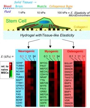
indirectly – signals to the tissue-integrated cells. Since the beginnings of cell biology, various micro-tools have been developed to push or pull on cells and determine effective responses in terms of elastic, viscous, and yield parameters [1], though often without much molecular insight. Brownian and non-Brownian motions of particles within cells have also been tracked to analytically distinguish passive, thermally-scalable properties of cells from active, energy-driven characteristics [2]. The latter responses often involve signaling and exhibit molecular specificity: when a cell-matrix adhesion is stretched by an external force, for example, select proteins are post-translationally modified and actively enriched at the site of applied force [3]. When cells test the mechanics of their microenvironment through adhesive engagement and ATP driven contraction of actin-myosin ‘stress’ fibers [4], gene expression can change over hours to days, with recent examples including matrix elasticity directed lineage specification of stem cells (Fig. 1) [5] and malignant transformations of breast epithelial cells on stiff substrates [6].
Regardless of process, however, the underlying molecular mechanics need to be clarified. A number of signaling pathways have been described that transduce mechanical signals into biochemical responses which could then lead to complex cell behaviors [7, 8]. Interactions within or between molecules that are directly modulated by force or by some other mechanical property of the microenvironment are likely to be critical in any
6
Figure 1: Cells can ‘feel’ the physical properties of their microenvironment. In one recent example with mesenchymal stem cells,
7
Of the various mechanisms that have been proposed for mechanosensing [7, 8, 10, 11], here we focus on tension-induced changes in protein structure (Fig. 2A). Forced unfolding can be localized to a part of a domain or loop, or it can involve complete domains; force can also re-orient domains or straighten unstructured regions such as hinges between domains. We review the experimental methods that have been used to probe transitions and also review how these observations fit into a cellular context. From outside to inside a cell, we first consider extracellular matrix, which defines a mechanical substrate for cells (Fig. 2B) and which interacts with cell surface receptors such as
integrins to couple matrix to cell cytoskeleton (Fig. 2C). We then focus on the
cytoskeleton itself, including its contractile myosin components with which a cell exerts pulling forces and probes its surroundings (Fig. 2D). More physical tools and
biochemical insights are certainly needed, but it seems fair to claim that ‘molecular mechanobiology’ is not just emerging but perhaps burgeoning.
Proteins Unfold under Force
8
Figure 2: (A) When force is applied to proteins their native structure can be perturbed. This can involve a conformational change in
which only quaternary structure is perturbed or unfolding in which secondary or tertiary structure are disrupted locally or over an
entire domain. Lower panel: Adherent cells attach to the extracellular matrix (ECM), spread, and apply contractile forces using
acto-myosin stress fibers. The image in the lower left shows the detergent extracted cytoskeleton of a mesenchymal stem cell adhering to
its substrate. This can in principle activate several force sensitive processes that may be mediated by protein unfolding. (B) ECM
proteins such as fibronectin are extended, exposing cryptic binding sites that promote fiber assembly as well as cell adhesion (inset
adapted from [23]). (C) Focal adhesions sense applied force and respond by recruiting additional proteins to shore up the
cell-substrate interaction. A possible contributing mechanism is the force-induced exposure of vinculin binding sites in the talin rod
domain, shown here to unfold under force in molecular dynamics simulations (adapted from [55]). (D) The cytoskeleton is actively
contractile and these forces impact not only the ECM and focal adhesions, but also the cytoskeleton itself. The inset shows
simulations of the forced unfolding of an actin binding protein called filamin (adapted from [76]). The schematic (adapted with
permission from original by Hyungsuk Lee, Jorge M. Ferrer, and Matthew J. Lang) to the left shows how filamin unfolding and actin
9
residing in the Earth’s gravitational field. Forced unfolding of single proteins was first shown about a decade ago [13, 14]. A ‘grip and break’ approach is perhaps the most intuitive way to think about disrupting any structure, but force is a vector (with magnitude and direction) that is difficult to apply to proteins tumbling in solution. Immobilization is required, as occurs with structural proteins within a cell or matrix, and there is also a need for nanoscale techniques to apply a force and monitor the effects of force at the molecular level.
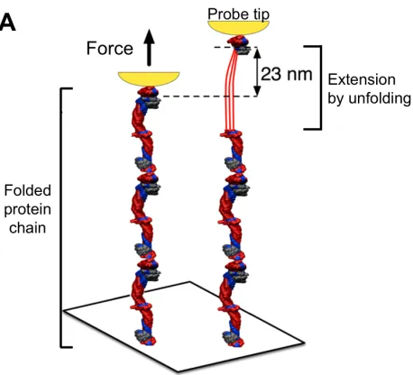
The most commonly used tools today for single molecule force spectroscopy are atomic force microscopes (AFM) (Fig. 3A) and optical tweezers as reviewed elsewhere [15]. Based on forced unfolding measurements of many proteins, forces in the range of f = 10 pN to 200 pN will unfold most proteins within a second or less. At lower forces, proteins unfold more slowly [16, 17], which one can understand from the simplest possible expression, widely attributed to Bell [16], for the rate k of reaction that is accelerated by force above a basal rate ko:
k = ko exp( f d / kBT ) Eq.1
The distance d is specific to a protein state, and kBT (= 4 pN nm) is the standard
10
Figure 3: Nano-tools for protein folding and funnels. (A) Proteins can be extended and unfolded by force using a probe (shown in
yellow) either in AFM or optical tweezers (adapted from [33]). (B) The energy of an extended protein conformation is typically higher
than the folded native state and this is often conceptualized as an energy landscape in the form of a folding funnel where chain entropy
dominates with the many unfolded states and energetic interactions pull the protein into one or a few well-defined structures. With
11
With full unfolding of a typical repetitive domain within a multi-domain structural protein, extensions of 5-10 fold correspond to distensions of 20-40 nm, which is large compared to the dimensions of most other cytosolic proteins. While extension can serve as a strain-release mechanism that allows some biological scaffolds to distend without dissociating, for a growing list of proteins, unfolding seems to have additional functional consequences, such as exposure of previously hidden assembly or binding sites.
Unfolding in the Extracellular Matrix Influences Assembly
Adherent cell types are viable only when they are attached to a solid substrate, which in vivo is the extracellular matrix (ECM)—a crosslinked hydrogel complex of protein and polysaccharide that is produced by either the cell it surrounds or specialized cells such as fibroblasts. ECM proteins are the most abundant proteins in animals and play critical mechanical roles for tissues by providing them with substantial tensile strength and, in the case of bone—through calcification, compressive strength. The role of ECM in regulating cell behavior, and in particular how it can be modulated by force, is highlighted here.
12
was due to a force-induced unfolding was plausible given that fibronectin unfolding under tension had already been predicted theoretically [21]. This early prediction was shortly followed up by molecular dynamics simulations [22, 23] and single molecule experiments on fibronectin domains from the related ECM protein tenascin [24]. These ‘force spectroscopy’ experiments also showed that the domains could refold in seconds after removal of force. Further evidence for some type of conformational change with the matrix under tension was provided by Foerster Resonance Energy Transfer (FRET) changes between fluorescent dyes attached to Cys and Lys residues in fibronectin [25, 26]. Although such Cys labeling tends to block disulfide formation that is essential to matrix crosslinking within the oxidative, extracellular space, the FRET results are quantitative and compelling.
The results for fibronectin suggest that unfolding is activated by cell tension in the ECM, contributing to assembly, but fibronectin interacts with many other proteins
13
Fibrin, fibronectin, and collagens (types 1 and 3) are linked physically through binding—perhaps with tension dependent mechanisms—and they are linked functionally during wound healing. An initial fibrin clot is replaced by ECM components that include fibronectin and then more permanent (and stiffer [31]) crosslinked collagen [32].
Fibrinogen binds several proteins in addition to fibronectin, and some of these
interactions might also be regulated by force. The coiled-coils of fibrinogen have been shown to unfold under force (see Chapter 2 for more details) [33, 34]. More recent studies of fibrin gel stretching (Chapter 3) further indicate at small strains (<50%) that: (i) a volumetric phase transition occurs with a large negative compressibility that represents loss of water and association of unfolded protein, and (ii) a reversible order-disorder transition occurs as seen by small angle X-ray scattering [35]. Despite such macroscopic evidence of coiled-coil unfolding, it is not yet clear that binding sites found in fibrinogen’s globular end domains undergo conformational changes under force. Mechanically labile coiled-coils might protect the globular domains from unfolding and thus play the reverse role of unfolding in fibronectin. Another possibility is that force regulates the conformation of the C-terminal end of the !-chain in fibrinogen, a natively
unstructured region that contains an AGDV sequence involved in platelet adhesion [36, 37].
14
makes unfolding seem less likely, simulations have shown a variety of failure modes at high forces [39]. This could be relevant after traumatic injury, even if not under normal circumstances from cell-generated tension, but an active role of cells in any aspect of ECM remodeling should not be underestimated. Growth factors—so essential in development and differentiation—provide a valuable recent lesson in that many have long been known to bind ECM, but more recent experiments show that release of at least the canonical Transforming Growth Factor-" (TGF-") is directly modulated by the
elasticity of the matrix and by tension generated by cells [40].
Force Modulates Cell-Matrix Interactions
Cells have evolved many ways of modulating their physical interaction with each other and with the surrounding ECM. Most cells possess integrin receptors that bind to specific ECM peptides and mediate stable adhesion [28]. Forced unfolding with exposure (or protection) of these sequences is not the only way for mechanics to influence cell behavior; integrins and perhaps other focal adhesion proteins are also sensitive to force through changes in protein structure (Fig. 2C). Methods for
interrogating forces from single adhesions on living cells [41] continue to be developed [42] and should provide more insight into the mechanics of adhesion that accompany these conformational changes, as they have for other processes that regulate adhesion, including mechanics [43].
15
suggested by recent work with fluid shear forces imposed on adherent cells [46]. With increasing stress, the average bond force per integrin increases, but this increase depended on exposure of a synergy site in fibronectin. This scenario is consistent with the formation of “catch-bonds” that exhibit an increased binding strength when put under mechanical load. This behavior has been directly observed at the single molecule level in other proteins [47-49], and similarly well-controlled single molecule experiments with integrins are to be anticipated.
In addition to regulating adhesion strength, integrin clustering acts as a signal involved in the formation of focal adhesions, which are currently estimated to contain around 160 component proteins [50]. When cells are stretched with a glass microneedle, they respond by locally recruiting focal adhesion proteins to shore up their attachment to substrate [3]. In this sense, focal adhesions are mechanosensitive complexes, however, changes in focal adhesion size and even the recruitment of specific proteins or the activation of particular kinases do not by themselves explain how mechanical cues are sensed since it is not yet clear how these processes could be modulated by force. Protein unfolding is one possibility.
16
of talin can be unfolded under moderate forces of ~10-100 pN in single molecule experiments [55]. Importantly, based on single molecule fluorescence during magnetic trapping and extension of proteins, vinculin is more likely to bind the talin rod domain when the rod domain is under tension. The number of binding events increased in constructs with tandem talin rod domains, while the low number of binding events to a-actinin was independent of the force, helping to rule out some possible experimental artifacts. These results are suggestive, but there are still several critical questions that remain unanswered. For example, what is the nature of the talin/vinculin binding? Simulations have provided insight [56], but experiments are lacking; rod domains with mutations in the binding sequence could help address whether the force-activated binding is similar to that observed in a talin construct that has been mutated to adopt the active conformation [57]. More importantly, a central open question is whether this
mechanism is in fact operative in focal adhesions in cells. This question is now
becoming addressable with new methods for the in situ labeling of exposed protein sites [9] as discussed in more detail below.
17
protein is incorporated structurally into a focal adhesion where it experiences an applied force, it could play a mechanosensitive role in which the affinity of one or both of its binding partners is altered when it completely or partially unfolds. There is recent evidence that this situation occurs for the mechanical activation of p130cas [58]. An important future task, perhaps best suited to higher throughput efforts, will be to
determine which of the many candidate interactions in the adhesome are indeed directly modified by force. Interaction forces or ‘strengths’ will not provide immediate answers, however, because entropy dictates that more frequent structures or pathways will tend to be affected more often than the unitary Boltzmann factor of Eq.1 would predict. In other words, a more appropriate weight or probability P in comparing processes a and b that respectively exist Na or Nb times is:
Pa!b = [Na koa exp( fa da / kBT )] / [Nb kob exp( fb db / kBT )] Eq.2
where f is the force applied to the protein a distance d along the reaction coordinate. Analogies to the width of a protein folding funnel (Fig. 3B), which represents the number of chain conformations, could perhaps be usefully formalized to multi-domain proteins in multi-protein network modules.
The cytoskeleton responds to and actively applies force
18
and are likely involved in mechanosensing (Fig. 2D). The cytoskeleton also possesses a unique feature not present in the ECM or most focal adhesion components: it contains active assemblies that consume energy and are therefore capable of doing mechanical work [59]. This means that the cytoskeleton not only responds to force but also applies force to its surroundings [60]. This closes the mechanical loop described above and allows a cell to alter its surroundings by the forced-remodeling of proteins like fibronectin and also to probe the mechanics of its substrate [4].
One of the central components of the cytoskeleton is actin. Actin monomers polymerize to form filaments that are an important structural component of cells. For example, gels made from purified filamentous actin and actin crosslinkers recapitulate aspects of cellular mechanics [61]. Actin polymerization consumes ATP and, because it is directional, it can apply forces to the cell periphery that drive cell motility [62]. In order to control cell morphology and to help regulate this complex actin dynamics, a host of actin binding proteins regulate its assembly and interact with other pathways [63]. An interesting example that has received significant attention is filamin.
19
expression has been shown to be required for active cell stiffening in response to substrate mechanics but not for passive stiffening in response to an external force [70]. For Dictyostelium discoideum filamin (ddFLN), this seems to be its main function as it is only known to bind to one other protein in addition to actin [71]. In contrast to ddFLN, human filamin has a more elaborate structure including hinge regions with important mechanical consequences and 24 instead of 4 Ig-like repeats. More than 20 binding partners have been identified for human filamin [72, 73]. These include proteins with a range of functions, including signaling. Particularly relevant here, filamin has also been shown to compete with talin in binding the cytoplasmic domain of integrin [74]. Thus there is a direct link between filamin and proteins known to be critical in
mechanosensing. Given this connection with focal adhesions and the cytoskeleton as well as filamin’s structural similarity with Ig domains to proteins like fibronectin, it is natural to ask how filamin behaves under force and whether this behavior has functional consequences.
The mechanics of filamin have been measured at the single molecule level using AFM, and the Ig domains were found to unfold in an abrupt all-or-nothing manner at forces of order 100 pN [75], consistent with molecular dynamics simulations [76]. An important finding from work on ddFLN is that the force required to break the
20
More recently, the mechanics of filamin have been measured in an elegant native-like system in which single filamin molecules are connected on one end to a fixed actin filament and on the other to a filament connected to a bead [78]. When the bead is trapped using optical tweezers and the sample stage is moved, the force required to either break the filamin-actin bond or to unfold one or more of the domains of filamin can be measured. One reason that this experiment is significant is that it gets directly to the issue of competition between alternative stress-release mechanisms. In this geometry at the rates studied, filamin was found to unbind most of the time, but unfolding was also observed. Similarity in the force required for unfolding and unbinding could have
functional consequences in regulating cytoskeletal remodeling under force and highlights the importance of thinking about timing, rates, and kinetics. Furthermore, it is not known whether any of the proteins that bind the filamin repeats affect their stability or
conversely whether applied stress can affect the binding affinity of the filamin binders. This question becomes especially interesting in light of recent work that suggests that filamin can serve to trap a transcription factor (i.e. PEBP2/CBF) in the cytoplasm [79], thereby regulating its action in the nucleus. If these results are confirmed, force-induced shifts in transcription factor binding could provide an elegant mechanism for mechanics to alter gene-expression, which is known to occur in (filamin-expressing) mesenchymal stem cells in response to substrate stiffness [5].
21
signaling complex that ultimately regulates a transcription factor well-known to be regulated by the cytoskeleton, namely serum response factor (SRF). Molecular dynamics calculations [81] and single molecule AFM provide insight into a possible mechanism, suggesting that the kinase domain unfolds at lower force than individual immunoglobulin domains of titin [82] – although Eq.2 would suggest some of titin’s ~200 Ig domains are likely to unfold if the kinase domain unfolds. Mutations in the titin kinase domain interfere with the signaling pathway and lead to a hereditary muscle disease in humans [80], which implicates force-controlled unfolding of this domain and perhaps also some of titin’s Ig domains.
Another class of actin-binding proteins that have been relatively well studied also contain an actin-binding domain followed by a series of compact repeats, but instead of Ig-like domains of "-strands, they consist predominantly of #-helical bundles. The spectrin
superfamily of proteins with their triple-helical bundle domains is prototypical: #
-actinins are the shortest and ubiquitous isoforms, while spectrins—originally isolated from red blood cell membranes—are generally found at all cell membranes and are well known to be essential for membrane stability. Additional members of the family include dystrophin, which links the contractile apparatus of striated muscle to ECM, and
nesprins, which link the nuclear envelope to the actin cytoskeleton. Dystrophin
22
proteins tested thusfar have been found to be remarkably labile, unfolding at forces around 15-30 pN even at the high loading rates typical in AFM experiments. Compared to "-sheet Ig-like domains, the force required to separate the helices of the spectrin bundles are relatively small and the helices themselves unfold easily because the hydrogen bonds holding them together are arranged in series in the direction of the applied force. In contrast, the bonds holding "-strands together are arranged in parallel and must be simultaneously sheared apart when the domains are pulled at their N- and C-termini [88]. Since proteins soften at higher temperatures [89, 90], spectrin domains will unfold at even lower forces in vivo. In fact, an exhaustive study of the thermal stability of the domains of spectrin in humans found that several domains have melting
temperatures " 37oC [91]; most single molecule experiments have been done at room temperature and will miss such effects. Strong effects of temperature (beyond kBT) and
the stability of multiple serial versus parallel bonds are important to consider in future experiments with single molecules as well as in extrapolations or direct study of cellular structures.
Towards a Characterization of the Cellular ‘Unfoldome’
23
small number of cases that have been examined in some detail so far, it is clear that there is no singular protein transition or master mechanical switch at the heart of cellular mechanical responses. Instead, we are likely at the early stages of discovering what will prove to be a wide variety of force sensitive proteins and reactions. In terms of signaling networks, we have just begun to identify the machanosensitive nodes embedded in the cell’s broader system of signaling pathways. A more complete characterization of the proteins that are unfolded by external forces applied to cells or by their own myosin-driven contractility will help guide efforts to understand the molecular basis of
mechanotransduction. Just as the study of focal adhesions will progress by understanding the interactions in the adhesome, the study of mechanotransduction more generally will benefit from a more complete characterization of the cellular ‘unfoldome,’ the set of proteins that can be unfolded as part of their physiological function. Although the focus of this review is on cell mechanics and transduction mechanisms, the unfoldome concept can be extended more broadly to other processes that induce changes in protein
conformation or quaternary structure including heat shock and other pleiotropic perturbations.
24
protein to be investigated seems unlikely to provide significant coverage of the unfoldome in the near future. Methods are therefore required to search for a wider variety of proteins whose conformations are force sensitive.
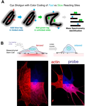
Advances in mass spectrometry based proteomics have set the stage to develop a method to screen the conformational state of a large number of proteins in living cells [9]. The experimental scheme uses cysteine accessibility as a probe (Fig. 4A): as a
moderately hydrophobic amino acid, cysteines are often buried or partially buried within protein tertiary and/or quaternary structure and are thus not accessible to small molecules in the solvent. If a cysteine-reactive fluorescent dye is introduced into cells, only those cysteines that are surface-exposed will be labeled. If changing conditions alter the conformational state of a protein with a buried cysteine, a difference in dye labeling will be detected. Thus, the spatial distribution of unfolding can be monitored to some extent in cells using standard fluorescence microscopy. More importantly, mass spectrometry of tryptic fragments from cell lysates can identify not only the proteins that show a change in conformation, but even the precise cysteines that are exposed so that force sensitive domains can be mapped within proteins.
When applied to the simplest possible cell, a red blood cell, this Cys shotgun approach identified a half dozen spectrin repeats that unfolded in response to
physiological shear stress. Many sites in spectrin showed no change, and other
25
Figure 4: (A)Cysteines are either buried within a protein fold or interaction site, or else they are exposed on the protein surface. The
latter surface sites are rapidly labeled with one color before perturbation while the buried sites are then exposed and colored
differently. This provides a ratiometric signal of unfolding and improves signal-to-noise ratio for exposed sites. (B) Addition of a
membrane-permeable dye to adherent cells (in this case mesenchymal stem cells) labels cysteines depending on cell state, and
relaxation of myosin (here with blebbistatin) leads to significant differences in protein labeling. Proteins that remain folded or
assembled upon relaxation show a site-specific decrease in cysteine accessibility that is pinpointed using mass spectrometry (adapted
26
This can be accomplished using a non-muscle myosin II inhibitor called blebbistatin. Cys shotgun labeling of mesenchymal stems cells attached to collagen-coated substrates in the presence or absence of blebbistatin gave a number of differential “hits” that included non-muscle myosin IIa (consistent with drug treatment preventing a conformational change in force generation), filamin, and the intermediate filament protein vimentin. The results were largely the same with embryonic cardiomyocytes that were labeled while beating spontaneously on a soft heart-like matrix or while static on a non-physiologically rigid matrix [92].
These initial studies serve as a proof-of-principle for Cys shotgun labeling of the unfoldome in cells and demonstrate the possibility of using such a proteomic-scale approach for the identification of protein conformational changes associated with a wide range of microenvironments or drug treatments. It will be important to explore
mechanically perturbed proteins in other adherent cell types using blebbistatin and to examine cell responses to changes in applied strain. Cys shotgun investigations will also suggest promising targets and domains for more detailed study by other approaches. In combination with phosphorylation networks widely studied now by mass spectrometry, the approach is poised to clarify the breadth of interplay between molecular mechanics and signaling networks.
Conclusion
27
seem to have functional consequences ranging from the regulation of matrix assembly and cell adhesion to cytoskeleton dynamics. Proximal effects of mechanics also have ramifications for more complex cell processes that require changes in gene expression because these changes in protein conformation couple into other signaling pathways that can have a broad range of effects downstream. Despite some successes in identifying proteins involved in mechanosensing and an increasing understanding of their mechanism of action, it seems likely that there are many more to be discovered and that this search will be aided by methods that can broadly pinpoint relevant conformational changes amidst the complex background of mechanically silent cytosolic and cytoskeletal proteins—recognizing that there is no single cellular mechanosensor. Ultimately, a diverse network of proteins will likely respond in specific ways to different mechanical cues such as matrix stiffness, applied force, and fluid flow. As hinted at by the species differences in filamin, a variety of sensing mechanisms might reflect the importance of mechanical homeostasis in the evolution of larger multicellular organisms. New perspectives on human diseases—beyond heart disease [93], muscular dystrophy, and
28
Chapter 2
Summary
Forced Unfolding of Coiled-Coils in Fibrinogen by Single-Molecule AFM
Fibrinogen is a blood plasma protein that, after activation by thrombin, assembles into
fibrin fibers that form the elastic network of blood clots. We used atomic force
microscopy (AFM) to study the forced unfolding of engineered linear oligomers of
fibrinogen, and we show that forced extension of the oligomers produces sawtooth
patterns with a peak-to-peak length consistent with the independent unfolding of the
coiled-coils in a cooperative two-state manner. In contrast with force plateaus seen for
myosin coiled-coils that suggested rapid refolding of myosin, Monte Carlo simulations of
fibrinogen unfolding confirm that fibrinogen refolding is negligible on experimental time
scales. We suggest that the distinct behavior of fibrinogen is due to its topologically
complex coiled-coils and an interaction between fibrinogen’s !C-domains and its central
region.
Introduction
A blood clot needs to have the right degree of stiffness and plasticity for hemostasis and
yet very stiff clots are not easily lysed and are associated with thrombosis and
thromboembolism, but the origin of these mechanical properties is unknown [1]. The
elasticity of self-assembled networks of fibrin—the principal component of clots—also
29
such gels [2]. Recent experiments have pushed mechanical measurements to the single
fiber level [3] and a theory incorporating an enthalpic fiber stretch and entropic elasticity
provides a better fit to macroscopic rheological data than one involving entropic elasticity
alone [4]. Despite these advances in understanding larger scales, the micromechanics of
fibrinogen, the pre-cursor of fibrin, remains unexplored.
In this chapter, we describe single-molecule atomic force microscopy (AFM)
experiments on the extensibility of fibrinogen oligomers. As with previous single
molecule unfolding experiments, oligomers were required to generate reproducible,
interpretable data (see for example ref. [5]). The fibrinogen oligomers used in this study
were covalently cross-linked via the "C-modules located at the distal ends of adjacent
fibrinogen molecules. Accordingly, when a fibrinogen oligomer is extended from the
sample surface, the force is propagated only through the coiled-coils and the C-terminal
portions of the "-chains (Fig. 1a), thus reducing the variety of potentially unfolded
structures and possible force-extension curves.
Methods
To induce end-to-end oligomerization of fibrinogen molecules, 10 mg/ml human
fibrinogen (plasminogen-free, Hyphen BioMed, France) in 20 mM HEPES buffer (pH
7.4) containing 100 mM NaCl, 30 mM CaCl2 and 5 ATU/ml hirudin was mixed with
human factor XIIIa (50 µg/ml final concentration) and incubated at room temperature
until the beginning of gelation (about 30 min). At that point, the cross-linking reaction
30
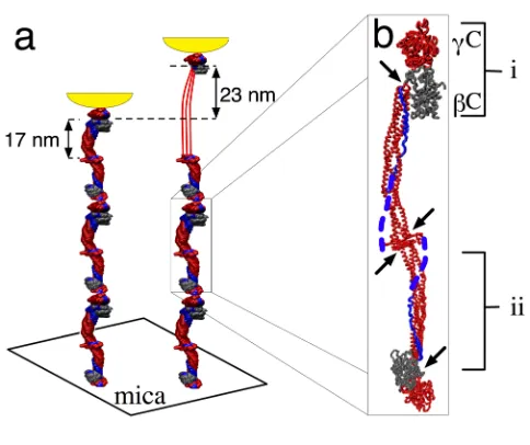
Figure 1: (a) Schematic of the pulling geometry. Fibrinogen oligomers adsorbed on mica were extended by the AFM tip shown in
yellow. If a coiled-coil is unfolded, the oligomer contour length increases by 23 nm. (b) Crystal structure of fibrinogen [6] with two
globular modules, "C and #C (i), at each end separated by triple-helical coiled-coils (ii) confined between clusters of disulphide bonds
(arrows). The force propagating coiled-coils, "C-modules, and the central region are shown in red. The C-terminal portions of the A!
31
mg/ml factor XIII solution (46 U/mg, glycerol/water, 0.5 mM EDTA and 2 mM CaCl2)
was treated with 2 U/ml human thrombin (American Diagnostica Inc., Greenwich, CT)
for 1 hour at room temperature, and the reaction was stopped by addition of hirudin (10
ATU/ml final concentration). Formation of single-stranded fibrinogen oligomers via
crosslinking between "Gln398 and "Lys406 of the "C-modules was corroborated by
transmission electron microscopy (TEM) (Fig. 2a-e) and the presence of the "-"-chain
band in SDS-PAGE of reduced samples of the fibrinogen preparation (Fig. 2f). To
separate non-ligated monomers from oligomers, 0.5 ml of the cross-linked fibrinogen
preparation was applied to a 1.5x15 cm Sepharose CL 6B column equilibrated with 20
mM HEPES buffer (pH 7.4) containing 100 mM NaCl and 3 mM CaCl2. As judged from
TEM, the fraction collected in the void volume contained only about 6% fibrinogen
monomers and 94% di-, tri-, tetra-, and pentamers (Fig. 2g).
For TEM, preparations of cross-linked fibrinogen were diluted with a volatile
buffer (50 mM ammonium formate, pH 7.4, 25% glycerol) to a concentration of 20-40
µg/ml, immediately sprayed onto freshly cleaved mica, and rotary shadowed with
tungsten in a vacuum evaporator as previously described [7]. Prepared specimens were
observed in a FEI 400 electron microscope at 80 kV and 60,000! magnification in many
different areas of the preparations to obtain a random sample.
For the AFM experiments, 50 µl of a 50 µg/ml solution of the oligomerized
fibrinogen were pipetted onto freshly cleaved mica and allowed to adsorb for ten minutes
before being rinsed gently with buffer. Force-extension curves were collected using a
32
Figure 2: Transmission electron micrographs of fibrinogen monomers (a), di- (b), tri- (c), tetra- (d), and pentamers (e). The scale bar
represents 50 nm. (f) The presence of the "-" band in SDS-PAGE confirms that the oligomers observed by TEM were covalently
crosslinked. (g) Histograms of degree of oligomerization are plotted as the logarithm of the frequency per 1000. The fractionated
sample contains only 6% monomer (black bars, n = 291) compared to the unfractionated sample that contains 85% monomer (gray
33
Results and Discussion
When unfolded under force, fibrinogen oligomers gave rise to a periodic sawtooth pattern
(Fig. 3a) with a length and regularity that was not observed in control experiments on
monomers. Since the unfolding geometry is specified by "-"-crosslinking, the observed
sawtooth patterns are most likely due to unfolding of either the coiled-coils or the
globular C-terminal portions of the "-chains. Each coiled-coil consists of 111 or 112
amino acid residues of the A!-, B"-, and "-chains, which, when fully unfolded, form a
thread 40 nm long (assuming a contour length per residue of 0.36 nm) (Fig. 1a)
corresponding to an expected peak-to-peak length of 23 nm (unfolded minus 17 nm
folded length), in good agreement with the experimental data (Fig. 3c). In contrast, the
C-terminal "-chains each consist of 215 amino acid residues (not including the disulfide
loops or the chain beyond the first crosslinking site) with an expected peak-to-peak
length of 77 nm, significantly bigger than the average unfolding length observed in our
experiments. The central region could also unfold but it is highly constrained by disulfide
bonds and does not seem to contribute.
Finally, we performed a Monte Carlo simulation that reproduces both the
observed force extension curves and the peak force histogram. Reasonable agreement
was obtained assuming negligible refolding and using a persistence length of 0.8 nm, an
unfolding rate at zero force of 0.03 s-1, and a transition state distance of 0.31 nm. These
parameters are in the same range as those observed previously for unfolding ubiquitin, a
globular protein [8]. It is interesting to note the difference between the unfolding of the
34
Figure 3: (a) Force-extension curves observed for fibrinogen oligomers (black) with simulated overlays (blue). Traces have been
offset for clarity. (b) Histogram of peak forces (bars) is well approximated by the simulated data (blue line). (c) Histogram of
35
For myosin II, a force plateau is observed at 20 pN as opposed to the sawtooth behavior
observed here for fibrinogen with an average peak force of 94 pN. The same two-state
unfolding model has been shown to account for both force plateaus and sawtooth patterns
in force extension curves by changing two parameters: the length of the unit that unfolds
in a two-state manner and that unit’s refolding rate [10]. There are several reasons to
expect that these parameters are different for myosin and fibrinogen. The coiled-coils in a
fibrinogen oligomer are divided into short segments 17 nm in length, whereas in myosin
the coiled-coil is unbroken for 150 nm. Furthermore, the coiled-coils of fibrinogen are (i)
interrupted by “stutters,” (ii) contain a kink in the middle, and (iii) are in fact partly
quadruple-helical [6]. This structure is significantly more complex than the coiled-coil of
myosin II and it is possible that when it partly unfolds, the remainder is sufficiently
destabilized to appear to unfold cooperatively on experimental timescales. Such an
observation is not unprecedented: a helical linker has already been observed to propagate
cooperative unfolding between adjacent globular domains in spectrin [11]. Given these
structural differences and its more complex topology, fibrinogen’s refolding rate indeed
seems likely to be considerably slower than that of myosin II.
The C-terminal part of fibrinogen’s A!-chain that forms the fourth strand of the
quadruple-helical portion of the coiled-coil (shown in blue, Fig. 1b) is known to interact
via the !C-domain with the central region [6]. This interaction could contribute to the
mechanical stability of fibrinogen and may also reduce the refolding rate. This possible
36
without these domains or by proteolytically cleaving them from oligomers prepared from
wild type fibrinogen.
Conclusions
This study identifies a new functional property of fibrinogen and suggests that the
coiled-coil is more than a passive structural element of this molecule. Coiled-coiled-coil unfolding
could account for up to a 2-fold strain in the recently observed large extensibility of fibrin
fibers [12] but its role in the macroscopic properties of fibrin gels [1] remains to be
determined. The constraints provided by our results will likely serve as a useful input for
multiscale modeling efforts that will ultimately be required to fully understand blood clot
37
Chapter 3
Summary
Multiscale Mechanics of Fibrin Polymer: Gel Stretching with Protein Unfolding and Loss of Water
Blood clots and thrombi consist primarily of a mesh of branched fibers made of the
protein fibrin. We propose a molecular basis for the remarkable extensibility and negative
compressibility of fibrin gels based on the structural and mechanical properties of clots at
the network, fiber, and molecular levels. The force required to stretch a clot initially rises
linearly and is accompanied by a dramatic decrease in the clot volume. These
macroscopic changes are accompanied by fiber alignment and bundling following forced
protein unfolding. Constitutive models are developed to integrate observations at spatial
scales that span six orders of magnitude and indicate that gel extensibility and expulsion
of water are both manifestations of protein unfolding, which is not apparent in other
matrix proteins such as collagen.
Introduction
Fibrin clots are proteinaceous gels that polymerize in the blood as a consequence of
biochemical cascades at sites of vascular injury. This meshwork, together with platelets,
stops bleeding and supports active contraction during wound healing[1, 2]. Fibrin also
provides a scaffold for thrombi, clots that block blood vessels and cause tissue damage,
38
To maintain hemostasis while minimizing the impact of thrombosis, fibrin must have
suitable stiffness and plasticity [4] but also sufficient permeability so that the network can
be effectively decomposed (lysed) by proteolytic enzymes [5, 6]. This is challenging
because open scaffolds would be expected to break at low strains—as is true for collagen
gels [7]. To address how fibrin clots are both permeable and highly extensible, we studied
fibrin structures across multiple spatial scales, from whole clots to single fibers and single
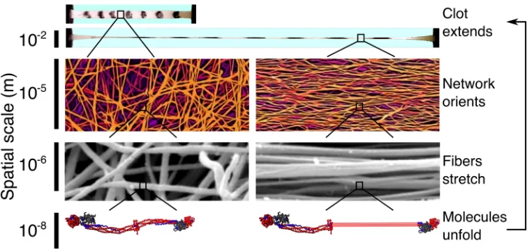
molecules (Fig.1).
Results and Discussion
Fibrin clots were made from purified human fibrinogen under conditions that resulted in
the formation of long straight fibers similar to those found in physiological clots. To
simplify the interpretation, the clots were covalently ligated using a transglutaminase
(blood clotting factor XIIIa) as naturally occurs in the blood, which prevents the
protofibrils from sliding past one another, thus eliminating persistent creep [8].
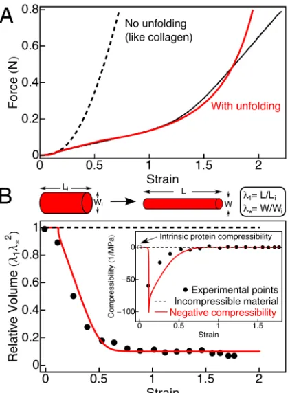
Measurements of the extensibility of 2 mm diameter fibrin clots (Fig. 2A) showed
that they could be stretched to over three times their relaxed length before breaking with
an average stretch of 2.7 ± 0.15 fold (n = 6). This is comparable to the single-fiber
extensibility observed when a fibrin fiber is laterally stretched with an atomic force
microscope [9]. Qualitatively, the resulting force-strain curve for fibrin is similar to those
observed for rubbers and other materials made from flexible chains [10]. However, for
fibrin clots, which are made of longer straighter fibers than the thermally fluctuating
39
Figure 1: Blood clots are highly extensible supramolecular protein polymers formed from well-separated, relatively straight and stiff fibers around 200 nm in diameter. When stretched, the fiber network aligns in the direction of the applied strain and the individual
fibers stretch, forcing the fibrin monomers that make up the fibers to extend. Ultimately, it is this molecular unfolding that allows clots
to stretch so far. Thus, understanding fibrin clot mechanics requires knowledge of the mechanical response and the corresponding
40
Figure 2: (A)Representative force-extension curve of a cylindrical fibrin clot reaching a 3-fold longitudinal stretch. The average stretch before breaking was 2.7 ± 0.15 fold (mean ± SEM, n = 6). As the strain (stretched length/initial length - 1) increases, the force
on the clot increases linearly until a strain of approximately 1.2, at which point the sample hardens and enters a new regime with a
steeper slope (black). The force-extension curve (black) is fit using a constitutive model that takes clot microstructure and protein
unfolding into account (red line). Without molecular unfolding (like collagen (7)), the model (dashed line) rapidly diverges from the
experimental data (black). (B)The relative clot volume decreases with strain (black points) in contrast to the behavior of an
incompressible material (dashed line). This decrease is predicted using the same model and parameters used to fit the force-extension
data (red line) demonstrating that the volume decrease occurs in parallel with molecular extension (see supplement, equation (29)). A
decreasing volume with stretch corresponds to a negative compressibility (inset), which indicates that there is a source of free energy
to drive contraction, possibly due to fiber bundling when hydrophobic side chains aggregate and bury after exposure during unfolding.
The negative compressibility is a property of the network. Proteins in solution have been observed to have intrinsic compressibilities
41
is wrong by seven orders of magnitude [4]. New models are therefore needed to
understand clot mechanics.
In addition to the large extensibility of fibrin clots, these gels also displayed a
dramatic decrease in volume upon stretching (supplementary video), unlike most rubbery
materials. This unusual effect is quantified in Fig. 2B where the lateral contraction of the
gel !* allows one to calculate the relative volume (!!!*2), which is plotted as function of
strain (black circles) and contrasted with a volume-conserving incompressible material
(dashed line). The shrinkage of the stretched clot was due to water expulsion, as
confirmed by an approximately ten-fold increase in the protein content in clots at a strain
of 2 (Fig. 5B). This protein concentrating effect, or syneresis, is mechanically induced
and corresponds to a negative compressibility for the gel (Fig. 2B, inset); the intrinsic
compressibility of proteins is usually positive and small, approximately 2 x 10⁻⁴ MPa-1
(open circle) [11]. This effect might be related to the phenomenon of negative normal
stress observed for networks of semiflexible polymers, because even though fibrin fibers
are relatively stiff, it is still possible that they buckle more easily than they stretch, thus
leading to an effective inward force [12]. However, our data below support an alternative
explanation in which the volume change is associated with protein unfolding in stretched
fibrin fibers.
To understand what makes fibrin so different from other highly extensible
polymers including rubbers and hydrogels, we quantified the structural changes that
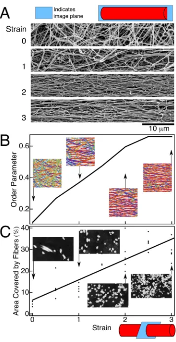
occur in stretched clots at the network and fiber levels. Unstrained clots imaged using
42
orientation (Fig. 3A, top image). When strain is applied (Fig. 3A, lower images), the
fibers begin to align and the network orientational order parameter increases gradually
from 0.1 to 0.7 at a strain of around 2.5 (Fig. 3B).
Transmission electron microscopy of transverse sections through fibrin clots at
increasing strain (Fig. 3C, inset images) provides a clear picture of how the fibers become
thinner, closer together, and bundle. The area occupied by fibers increases from about 5%
to 24% at a strain of 2 (Fig. 3C, graph). This increase is smaller than expected given the
macroscopic shrinkage of the entire clot (supplement, pp. 15-16) indicating that the
volume decrease is a combined result of fiber bundling and a decrease in fiber diameter
from 185 ± 36 nm to 74 ± 16 nm (mean ± SD, p << 0.0001, Student’s t-test). Similar
transmission electron micrographs (although without corresponding scanning electron
micrographs) of fiber ordering have been reported [13], but quantifying the strain
dependence of these observations proves important below in constraining possible
models. Furthermore, knowing the stress applied and the fractional cross-section
occupied by fibers allows an estimate of the force per protofibril of approximately 75 pN
at a strain of 2. Given the small loading rate used here, this force is large enough to result
in significant unfolding of fibrin [14]. Protein unfolding is generally associated with
increased exposure of hydrophobic groups that would tend to interact (e.g. bundle) and
expel water, as observed here.
The half-staggered packing of fibrin (Fig. 4A) leads to a ~22 nm repeat that can
be measured using small angle X-ray scattering and this can be used as a readout of the
43
Figure 3: Structural changes in stretched fibrin clots at the network and fiber levels. Scanning electron micrographs of stretched clots (A) show how the fibrin fibers align with strain. (B) These scanning electron micrographs are segmented using a Laplace of Gaussian
filter that determines which pixels are fibers and which are background and also calculates the orientation " at each fiber pixel. The
inset images show the results of the segmentation with the color at each pixel corresponding to that pixel’s orientation. These data are
summarized as an orientational order parameter <cos(2")> that can range between 0 for randomly oriented fibers and 1 for perfectly
aligned fibers. Data points are averages of five fields of view taken randomly over the surface of the clot at each strain. The order
parameter was fairly uniform across samples with an average standard error of 0.03. (C) Transmission electron micrographs of
transverse sections through unstretched and stretched clots show fiber bundling (inset images). The plot shows the total cross-sectional
area covered by fibers (black points on the plot, each representing a randomly chosen field of view), which is used to calculate the
force per fiber as a function of strain from the total force applied to the sample. Inset images are 4 µm across. At least 10 scanning and
44
Figure 4: Structural changes in stretched fibrin clots at the molecular level. (A) Schematic of a fibrin protofibril showing the half-staggered pattern that leads to a characteristic 22 nm repeat. A fibrin monomer within the protofibril is shown in red. (B) Small angle
X-ray scattering from fibrin clots leads to a clear first order peak (white arrow, upper inset) and to 3rd and 4th order peaks. The plots
show the peak shape as a function of the wave vector q = 2!/d, which increases radially from the center. The thick lines are fits to the
data using the sum of an exponential and a Gaussian with the exponential alone (thin lines) shown for comparison. The width of the
peak increases with stretch, which can be understood in terms of a two-state like extension of fibrin molecules that introduce defects
into the sample. (C) This effect is quantified as the Scherrer length L, which decreases with increasing strain (red). The decrease is
more rapid for samples that were pre-aligned in a magnetic field during polymerization (blue), which implies network alignment
accommodates some strain and delays unfolding. The transition is reversible when samples are allowed to relax, as indicated by the
blue arrow. When samples are not ligated using factor XIIIa, protofibril sliding becomes important and unfolding is decreased. In all
cases, the peak position remains relatively constant, implying that there is no gradual lengthening of the whole population of fibrin
monomers. Instead, there remains a population that is not unfolded and maintains a fairly constant spacing. Error bars are standard
deviations of the distribution of L determined from fits using the bootstrap method. This bootstrap error is similar to the average
standard error of 15 nm calculated by averaging over results from four samples. (D) This behavior is captured by the constitutive
45
not change significantly as the clot is stretched (Fig. 4B), ruling out a gradual extension
of molecules during stretch, which would otherwise increase this spacing, as suggested
by others [16]. The marked increase in peak width we observed indicates an increase in
disorder consistent with an increasing number of molecules unfolding in response to the
large strain. This increase in disorder is captured by the decrease in the Scherrer length L
(Fig. 4C): the length over which the 22 nm repeat is correlated [17]. A decrease in L
indicates that the average size of the regions containing a consistent 22 nm repeat
becomes smaller due to intervening regions of unfolded molecules. To control for the
effect on L of fiber alignment that accompanies strain, we made magnetically pre-aligned
samples in which the fibers were already oriented along the direction of applied strain. In
this case, the decrease in L occurs at lower strains since fiber alignment cannot take up as
much of the applied strain (Fig. 4C, blue points). This change is reversible when fibers
are allowed to relax (Fig. 4C, blue arrow). Similarly, to control for fiber sliding as an
alternative mechanism of strain accommodation, we stretched samples that were not
covalently ligated using factor XIIIa. Without ligation, protofibrils slide with respect to
each other instead of stretching, and unfolding is suppressed (Fig. 4C, black points). This
behavior is expected for two-state extension in which some molecules extend completely
while others remain folded, consistent with our earlier single-molecule observations of
the forced unfolding of coiled-coils in fibrinogen oligomers [14]. A more gradual
unfolding has been observed in molecular dynamics simulations [18], but the coiled-coil
spanning #C region is missing from published crystal structures of fibrinogen and is