Validating DNA Extraction Protocols for Bentonite Clay
Katja Engel,
aSara Coyotzi,
aMelody A. Vachon,
aJennifer R. McKelvie,
bJosh D. Neufeld
aaDepartment of Biology, University of Waterloo, Waterloo, Ontario, Canada bNuclear Waste Management Organization, Toronto, Ontario, Canada
ABSTRACT
Bentonite clay is an integral component of the engineered barrier
sys-tem of deep geological repositories (DGRs) that are planned for the long-term
stor-age of high-level radioactive waste. Although nucleic acid extraction and analysis
can provide powerful qualitative and quantitative data reflecting the presence,
abun-dance, and functional potential of microorganisms within DGR materials, extraction of
microbial DNA from bentonite clay is challenging due to the low biomass and
ad-sorption of nucleic acids to the charged clay matrix. In this study, we used
quantita-tive PCR, gel fingerprinting, and high-throughput sequencing of 16S rRNA gene
am-plicons to assess DNA extraction efficiency from natural MX-80 bentonite and the
same material “spiked” with
Escherichia coli
genomic DNA. Extraction protocols were
tested without additives and with casein and phosphate as blocking agents.
Al-though we demonstrate improved DNA recovery by blocking agents at relatively
high DNA spiking concentrations, at relatively low spiking concentrations, we
de-tected a high proportion of contaminant nucleic acids from blocking agents that
masked sample-specific microbial profile data. Because bacterial genomic DNA
asso-ciated with casein preparations was insufficiently removed by UV treatment, casein
is not recommended as an additive for DNA extractions from low-biomass samples.
Instead, we recommend a kit-based extraction protocol for bentonite clay without
additional blocking agents, as tested here and validated with multiple MX-80
ben-tonite samples, ensuring relatively high DNA recoveries with minimal contamination.
IMPORTANCE
Extraction of microbial DNA from MX-80 bentonite is challenging due
to low biomass and adsorption of nucleic acid molecules to the charged clay matrix.
Blocking agents improve DNA recovery, but their impact on microbial community
profiles from low-biomass samples has not been characterized well. In this study, we
evaluated the effect of casein and phosphate as blocking agents for quantitative
re-covery of nucleic acids from MX-80 bentonite. Our data justify a simplified
frame-work for analyzing microbial community DNA associated with swelling MX-80 bentonite
samples within the context of a deep geological repository for used nuclear fuel. This
study is among the first to demonstrate successful extraction of DNA from Wyoming
MX-80 bentonite.
KEYWORDS
Wyoming MX-80, bentonite, clay, bacteria, DNA extraction, casein,
phosphate
B
entonite clay from the Benton Shale near Rock River (Wyoming) is currently being
considered for use within the engineered barrier system of a deep geological
repository (DGR) for long-term management of high-level radioactive waste (used nuclear
fuel). In the Canadian design, the used nuclear fuel will be stored in steel containers for
mechanical stability. These containers will be coated with copper to resist corrosion
(1–4). Under anoxic DGR conditions, sulfide is considered the primary concern for
copper corrosion potential (5) by either diffusion from the surrounding environment or
production by sulfate-reducing bacteria (SRB) in the DGR itself. To prevent
microbio-CitationEngel K, Coyotzi S, Vachon MA,
McKelvie JR, Neufeld JD. 2019. Validating DNA extraction protocols for bentonite clay. mSphere 4:e00334-19.https://doi.org/10.1128/ mSphere.00334-19.
EditorHideyuki Tamaki, National Institute of
Advanced Industrial Science and Technology
Copyright© 2019 Engel et al. This is an
open-access article distributed under the terms of theCreative Commons Attribution 4.0 International license.
Address correspondence to Josh D. Neufeld, [email protected].
"Validating DNA extraction protocols for bentonite clay" from the lab of @joshdneufeld recommends a protocol that can be used to profile microbial DNA in MX-80 bentonite clay samples, with implications for the
microbiology of deep geological repositories.
Received8 May 2019
Accepted10 October 2019
Published
Applied and Environmental Science
30 October 2019
on September 8, 2020 by guest
http://msphere.asm.org/
logically influenced corrosion, the used nuclear fuel containers will be surrounded by
highly compacted bentonite (i.e., smectite/montmorillonite-rich swelling clay). The high
swelling pressure and low water activity of bentonite, when saturated, contributes to an
environment that greatly reduces the activity and survival of microorganisms, including
SRB (6–10). Furthermore, compacted bentonite can reduce the diffusion of sulfide to
the copper surface, limiting corrosion of the engineered container (9, 11).
Character-izing the microorganisms within bentonite using dependent and
culture-independent approaches is an important step for evaluating corrosion potential within DGR
design components.
Amplicon-based and “meta-omic” methods (e.g., metagenomics and
metatranscrip-tomics) offer enormous potential for assessing the presence and abundance of
micro-organisms and evaluating their potential activity within a barrier environment.
Al-though such methods are dependent on the recovery of nucleic acids from the charged
clay matrix, the extraction of microbial DNA from bentonite clay is challenging due to
its low biomass and ability to adsorb nucleic acids. Whereas culturing methods show
the presence of microorganisms in clay materials, DNA extractions have failed using
various methods (12–15). Indeed, no or very small amounts of DNA have been
recov-ered even from spiked clay samples (12, 16). Nonetheless, Chi Fru and Athar (17)
extracted DNA from MX-80 bentonite using an optimized phenol-chloroform method and
generated a clone library for analysis. Lopez-Fernandez and colleagues (18) extracted
DNA from Spanish bentonite deposits with up to 96% montmorillonite using a gentle
sodium dodecyl sulfate (SDS) lysis method with polyethylene glycol precipitation and
a final purification using a silica-based column. More recently, Liu and colleagues (19)
extracted DNA from Chinese bentonite deposit with 75% montmorillonite (20) using a
silica spin column-based kit, including a bead beating cell lysis step. Although
Lopez-Fernandez et al. (18) and Liu et al. (19) used high-throughput sequencing to measure
bacterial community composition and diversity, no such data are available for the
natural Wyoming MX-80 bentonite being considered for a deep geological repository
of used nuclear fuel, and no prior studies validated DNA extraction protocols for use
with bentonite clay.
Adsorption of nucleic acids to clay surfaces is primarily dependent on electrostatic
forces on the negatively charged surface of montmorillonite clay (21, 22). Cations
mediate the nucleic acid-clay complex, and divalent cations are more efficient than
monovalent cations in this process (23, 24). Adsorption of DNA to montmorillonite clay
surfaces therefore increases with decreasing pH (21, 25). At pH values below 5,
protonation of the amino groups associated with nucleic acid bases may increase the
attraction between positively charged DNA groups and negatively charged
montmo-rillonite clay surfaces (22). Binding is also affected by the physical properties of the DNA.
Linear DNA adsorbs more than supercoiled plasmids, likely due to molecule density and
the increased availability of free phosphate groups in linearized DNA (26, 27). Although
lower molecular weights reduce binding of DNA to clay, guanine-cytosine content has
no detectable influence on adsorption (28).
Numerous DNA extraction methods from clay-rich materials have been published
using commercial kits (12, 16, 29–33) and/or phenol-chloroform-based protocols (34–
37). Previous studies demonstrate that nucleic acid extraction yields can be increased
by using “blocking agents” that prevent DNA binding to clay or desorb it from clay by
changing the DNA structure or competing for binding sites. These blocking agents
include ATP, bovine serum albumin (BSA), casein, natural and synthetic DNA, RNA,
deoxynucleoside triphosphates (dNTPs), ethanol, Ficoll, lactose, NaCl, phosphates,
poly-vinylpyrrolidone, and skim milk (16, 31, 32, 37–41). Proteins, such as BSA, bind to
negatively charged clay surfaces via electrostatic and other interactions, preventing
DNA from binding (40). Although casein and skim milk have been used extensively,
these additives introduce various concentrations of contaminating DNA (31, 32).
Nat-ural and synthetic nucleic acids improve recovery and are potentially preferable to
biological materials for minimizing bacterial DNA contaminants (31, 41). Phosphate
groups bind competetively with DNA to clay and successfully decrease the amount of
on September 8, 2020 by guest
http://msphere.asm.org/
nucleic acid adsorbed on clay minerals (16, 28, 37, 38). However, added nucleic acids
may outcompete sample-specific signals in shotgun metagenomics and
metatranscrip-tomics, especially for low-biomass samples.
Successful desorption of nucleic acids from clay using blocking agents has been
demonstrated previously, but evaluations of the quantitative recovery of nucleic acids
at various starting concentrations, and the subsequent impact of those blocking agents
on microbial community profiles, have not been characterized well. In this study, we
used quantitative PCR, gel fingerprinting, and high-throughput 16S rRNA gene
se-quencing to assess DNA extraction efficiency from natural MX-80 bentonite, with and
without the addition of blocking agents and
Escherichia coli
genomic DNA. This study
is among the first to demonstrate successful extraction of DNA from natural Wyoming
MX-80 bentonite samples and is unique in providing an experimental validation for the
recommended protocol. Given that bentonite clay is considered a proposed
engi-neered barrier component of DGRs for many countries, the ability to monitor
micro-organisms within clay samples is critical for experiments that assess microbial growth
under DGR-like conditions.
RESULTS AND DISCUSSION
Initial characterization of Wyoming MX-80 bentonite.
Prior to assessments of
DNA recovery in the presence of blocking agents, we generated baseline data for a
representative bentonite sample with both dependent and
cultivation-independent approaches. The numbers of cultivated aerobic and anaerobic bacteria in
the Wyoming MX-80 bentonite samples were 2.7
⫻
10
2and 3.3
⫻
10
1CFU per g (dry
weight)
⫺1, respectively. Using a most probable number (MPN) method, 3.3
⫻
10
1MPN
g (dry weight)
⫺1SRB were detected. Previous studies reported cultivable aerobes
ranging from 10
2to 10
5CFU g (dry weight)
⫺1and anaerobes from 10
1to 10
4CFU g (dry
weight)
⫺1(6, 42, 43). Similar to our results, usually no or low numbers of culturable SRB
were detected (6, 42–45). Variations among reported estimates of cultured bacteria in
dry MX-80 bentonite are likely due to batch heterogeneity, coupled with production
and storage differences. A high number of viable but nonculturable (VBNC) bacteria
were shown previously in natural and compacted Wyoming MX-80 bentonite based on
phospholipid fatty acid (PLFA) analysis. Under increasing water activity conditions,
culturability increased by orders of magnitude (6). Others hypothesized that dry MX-80
bentonite powder extracts water from bacterial cells, leaving them in a desiccated state
(7). The revival of SRBs indigenous to MX-80 bentonite was dependent on incubation
temperatures above 40°C (45).
Genomic DNA extracted from 50 mg (dry weight) natural bentonite using the
PowerSoil DNA isolation kit was below the detection limit of the Qubit fluorometric
assay but nonetheless yielded 16S rRNA gene amplicons following PCR amplification.
Quantitative PCR detected 8.9
⫻
10
516S rRNA gene copies in 50 mg MX-80 bentonite
(dry weight) (Fig. 1). Due to the swelling of bentonite in the extraction buffer, 50 mg
was the maximum amount of sample material that could be used with the PowerSoil
DNA isolation kit. To increase DNA yield, we also extracted from 2 g bentonite using the
PowerMax DNA isolation kit. In doing so, we obtained 1.3
⫾
0.6 ng DNA per g natural
MX-80 bentonite, as quantified by the Qubit fluorometric assay, which corresponds to
8.7
⫻
10
416S rRNA gene copies per 50 mg (dry weight). The amount of DNA recovered
based on fluorometric assay or quantitative PCR and their cell abundance estimates
were higher by orders of magnitudes than those with the cultivation-based
ap-proaches. However, caution should accompany this comparison, because not all
mi-croorganisms may grow under the selected conditions and extracellular DNA could
result in an overestimated cell abundance through qPCR analysis.
DNA extraction yield of spiked bentonite samples.
Extraction of microbial DNA
from MX-80 bentonite is challenging, presumably due to adsorption of DNA to the
charged clay matrix. To examine the efficiency of DNA extraction with the PowerSoil
DNA isolation kit (Mo Bio), clay samples were spiked prior to extraction with high-purity
genomic DNA from
Escherichia coli
K-12 strain W3110 of up to 30-kb fragment size (see
on September 8, 2020 by guest
http://msphere.asm.org/
Table S1 and Fig. S2 in the supplemental material). Without the addition of bentonite
clay, the PowerSoil DNA isolation kit recovered 55% of the input DNA (Fig. 2). Recovery
was not improved by spiking the DNA after the beating step (Table S1), although the
extracted DNA was less sheared (Fig. S2). Kit-associated loss of DNA likely occurred
during binding to the silica membrane or subsequent elution, as reported elsewhere
(46). When
E. coli
DNA was incubated with 50 mg MX-80 bentonite (dry weight),
recovery decreased by 68% compared to that from the “DNA only” control (Fig. 2),
presumably due to adsorption of DNA to MX-80 bentonite. Consistent with this
observation, 60% to 80% of chromosomal
Bacillus subtilis
DNA can be adsorbed to
montmorillonite in water (22, 28).
Influence of blocking agents on DNA extraction yield.
To examine the effect of
casein and phosphate on DNA extraction yield, we first tested a relatively high spiking
concentration of 0.7 to 1
g genomic DNA per 50 mg MX-80 bentonite (dry weight).
The addition of 10, 50, or 100
mol phosphate per g MX-80 bentonite (dry weight) did
not increase the recovery of DNA significantly compared to that of the spiked MX-80
bentonite control (see Fig. S3A). Concentrations of
⬎
100
mol phosphate decreased
DNA recovery (Fig. S3), presumably due to excess phosphates competing for binding to
the silica-based membrane; phosphate-silanol interactions are important for
double-stranded DNA binding (47). Although previous studies showed that phosphates desorb
DNA from clay-rich materials (37, 38), our tested concentrations did not improve
recovery with the PowerSoil DNA isolation kit. When Direito et al. (16) substituted the
bead beating buffer of the PowerSoil DNA isolation kit with 1 M phosphate, they
0
102 104 106 108
Detected
16S rR
NA
gene copies
R² = 0.97 R² = 0.66 R² = 0.78 DNA only
MX-80 bentonite
DNA+MX-80
A
0
102 104 106 108
102 104 106 108
R² = 0.94 R² = 0.97 R² = 1.00
Detected
u
idA
gene
copies
Input gene copies
B
DNA only DNA+MX-80 DNA+MX-80+CP
DNA+MX-80+CP
FIG 1 Recovery of DNA from 50 mg (dry weight) MX-80 bentonite spiked with serially dilutedEscherichia
colistrain W3110 genomic DNA. Recovery was assessed using quantitative PCR targeting bacterial 16S rRNA genes (A) or the single-copyE. coli-specificuidAgene (B). Bacterial 16S rRNA genes recovered and quantified from native 50 mg natural MX-80 bentonite (dry weight) is indicated with an orange triangle in panel A. The theoretical optimal DNA recovery (100%) is indicated with dashed lines. Error bars represent standard deviations from two biological replicates.
on September 8, 2020 by guest
http://msphere.asm.org/
detected only a small increase in yield when extracting from a spiked montmorillonite
sample, likely because phosphates were already contained within the proprietary kit
solutions. Considering the competitive interactions between phosphate and DNA for
binding to the silica membrane, the ratio of phosphate to clay material is essential for
a successful extraction. Any excess phosphate not bound by clay might prevent DNA
from binding to the silica membrane and reduce recovery (Fig. S3B). Because the ideal
ratio of phosphate to clay will change based on clay type and concentration, phosphate
might not be a universal choice for silica membrane-based extraction protocols.
The addition of 400 mg casein per g MX-80 bentonite (dry weight) increased the
DNA yield to almost DNA-only control levels (Fig. 2). The application of casein at 40 mg
per g MX-80 bentonite (dry weight) did not show the same effect (Fig. S3C). The
addition of both 400 mg casein and 100
mol phosphate (CP) recovered DNA-only
control levels of DNA (Fig. 2). The proportional increased DNA yield in the presence of
casein was in agreement with previous publications (31, 39).
We also tested a phenol-chloroform-based extraction method modified from Lever
and colleagues (37) on spiked bentonite samples and found a higher DNA recovery
than with the commercial kit (see Fig. S4). The importance of pyrophosphate as a
blocking agent for clay-based extractions was apparent, because DNA recovered
without blocking agent addition was very low (Fig. S4). Previous results indicated that
DNA extraction efficiency from environmental samples was higher with
phenol-chloroform-based methods than with the commercial kit protocols (37, 48, 49). The
choices of DNA extraction method and 16S rRNA gene region influence microbial
diversity profiling (50–53), emphasizing the importance of selecting validated and
consistent protocols for research projects that require downstream data comparisons.
Although DNA recovery was higher with the phenol-chloroform-based extractions, we
used the PowerSoil isolation kit for all subsequent extractions to avoid use of
organic solvents and increase scalability for automation of extractions from many
samples (54, 55).
DNA extraction efficiency from MX-80 bentonite at various nucleic acid spiking
concentrations.
Given that nearly complete recovery of relatively high nucleic acid
concentrations from clay was possible using 400 mg casein and 100
mol phosphate
per g MX-80 bentonite (dry weight), we also assessed the efficiency of selected blocking
agents at various nucleic acid concentrations using the PowerSoil DNA isolation kit.
Genomic
E. coli
DNA was diluted serially from 10
9to 10
316S rRNA gene copies and then
a
b
c
b
a
0
10 20 30 40 50 60DN A only
MX80+D NA
MX80+D NA+C
MX80+D NA+P
MX80+D NA+CP
DNA
recov
ery
(%
)
FIG 2 Effect of casein and phosphate (CP) on recovery ofEscherichia colistrain W3110 genomic DNA
(700 ng) from 50 mg (dry weight) MX-80 bentonite (1:6 slurry) using the PowerSoil DNA isolation kit (Mo Bio). Casein and phosphate were supplemented at 400 mg and 100mol per g MX-80 bentonite (dry weight), respectively. DNA was quantified using the Qubit dsDNA HS assay kit (Invitrogen). Error bars represent the standard deviations from duplicates. Different lowercase letters above bars indicate significant differences (Tukey’s honestly significant difference [HSD],P⬍0.05).
on September 8, 2020 by guest
http://msphere.asm.org/
added to 50 mg MX-80 bentonite (dry weight). Total genomic DNA was estimated using
quantitative PCR (qPCR) targeting bacterial 16S rRNA genes or
E. coli
-specific
uidA
genes. Both qPCR analyses showed that additives increased genomic DNA extraction
recovery for the first dilution steps (Fig. 1). Although qPCR data targeting 16S rRNA
genes for the DNA-only extracts were consistent with the theoretical optimal DNA
recoveries, extracts with MX-80 bentonite and MX-80 plus CP no longer responded
linearly after the fourth dilution step (Fig. 1A). Because the 16S rRNA gene primers
quantify total bacterial DNA, any DNA extracted from additives, kit reagents, and/or
MX-80 bentonite will also be quantified. The proportion of DNA from those sources will
increase with lower
E. coli
genomic DNA concentrations and exceed concentrations of
the spiking DNA. Indeed, we detected 8.9
⫻
10
516S rRNA gene copies in 50 mg MX-80
bentonite (dry weight). Thus, we conclude that MX-80 bentonite-derived DNA
domi-nated the four highest genomic DNA spiking dilutions (i.e., the four lowest spiking DNA
concentrations) (Fig. 1A).
To circumvent clay-specific DNA quantification, we targeted the
uidA
gene specific
to
E. coli
templates. The qPCR data show that additives improved recovery even at the
lowest spiking concentrations (Fig. 1B). Although recovery for DNA-only controls and
DNA plus MX-80 plus CP was expected to be similar based on results from high spiking
concentrations (Fig. 2), extractions with additives had higher recovery for DNA-only
treatments at most spiking concentrations. Milk and casein can be contaminated with
up to 30%
E. coli
(56); however, no
uidA
genes were detected in casein and MX-80
bentonite extracts alone (data not shown). Others have reported that casein can
increase PCR efficiency (57) but this effect was not seen in our 16S rRNA gene qPCR
assay (Fig. 1A). Thus, it is unclear why the DNA-only amplification yields were lower
than for DNA plus MX-80 plus CP treatment samples.
Both denaturing gradient gel electrophoresis (DGGE) and high-throughput
sequenc-ing of 16S rRNA genes demonstrated that, as expected, a ssequenc-ingle DGGE band dominated
extracts from all MX-80 bentonite samples that were spiked with a serial dilution of
E.
coli
genomic DNA (Fig. 3). Consistent with qPCR data of MX-80 bentonite extracts
showing
⬃
10
5bacterial template copies per 50 mg (dry weight), “background” MX-80
bentonite DGGE patterns and 16S rRNA gene amplicon sequencing profiles were visible
FIG 3 Microbial community profiles of spiked and natural MX-80 bentonite samples. Denaturing gradient gel electrophoresis (DGGE) profiles of the V3 regions
of the bacterial 16S rRNA genes at various spiking concentrations (top) and taxonomic distributions in each sample based on high-throughput V3-V4 amplicon sequencing of 16S rRNA genes (bottom). Only operational taxonomic units (OTUs) at or above 5% relative abundance are shown. The OTUs with same classification were pooled and are indicated with asterisks. Casein and phosphate (CP) were added to spiked MX-80 bentonite samples (109to 10316S rRNA gene copies) for adsorption prevention. DNA was extracted from all samples using the PowerSoil DNA isolation kit, except one sample of natural MX-80 bentonite was also extracted with the PowerMax DNA isolation kit (indicated by #).
on September 8, 2020 by guest
http://msphere.asm.org/
when
⬃
10
5copies of
E. coli
genomic DNA, or less, were spiked prior to DNA extraction.
In addition to demonstrating quantitative and qualitative consistency when generating
sample-specific profiles, we here demonstrate reproducible MX-80 bentonite microbial
profiles. Although cultivation-based approaches are commonly used for characterizing
montmorillonite clay (12, 42, 45), here, we demonstrate that molecular approaches can
be effective with a high number of PCR cycles and careful assessment of reagent and
kit controls. In addition, our results provide a proof-of-principle methodological
ap-proach for assessing the amount of spiked control DNA that would be required to
generate quantitative data for 16S rRNA gene sequencing, as proposed recently for use
with soil microbial survey work (58).
Evaluation of reagent and laboratory contaminants.
A total of 4 DNA extraction
kit controls and 10 no-template controls were included in high-throughput amplicon
sequencing, even though no amplification was detected using gel electrophoresis.
Nested no-template controls (NTCs) that were prepared in single tubes (NTC1) had only
an average of 73 reads (see Table S2). The NTCs that were included within the 96-well
sample plate (NTC2) had higher average read counts (1,586 reads), presumably a result
of well-to-well contamination as demonstrated previously (59, 60). However, read
counts for NTCs were still relatively low. All DNA extraction kit controls were located
within the 96-well plate and had average read counts similar to those of NTC2 (1,585
reads). No common taxa were identified in all controls, except for an operational
taxonomic unit (OTU) affiliated with
Escherichia
in NTC2 and kit controls (see Fig. S5),
likely arising from well-to-well contamination. The presence of this OTU in NTC1 may be
caused by cross-contamination of indexed primers, mixed clusters on the flow cell, and
demultiplexing error; however, very low read counts for NTC1 (15 to 214 reads)
suggests that the error rate was negligible. At relatively low spiking concentrations (10
4and 10
316S rRNA gene copies) of the DNA-only control, only 29.2% of reads were
associated with the dominant
Escherichia
-associated OTU, on average, and most reads
were associated with common reagent and laboratory contaminants such as
Coryne-bacteriaceae
,
Staphylococcus
, and
Delftia
(Fig. 3). We demonstrated previously that low
template concentrations affect the 16S rRNA gene profile reproducibility (61). In
addition, when Lazarevic et al. (62) extracted DNA from 10
5and 10
4cells using a spin
column kit, only 27.5% to 37.5% of reads were related to the input strains. Salter et al.
(63) reported that 16S rRNA gene sequencing of extracts from 10
3cells were dominated
by contaminants and only 5% to 30% of reads were sample specific. As highlighted
previously (63, 64), quantifying sample microbial biomass can help gauge
contamina-tion risk. Because our qPCR data indicated that MX-80 bentonite extracts contained
⬃
10
5clay-specific 16S rRNA gene copies per 50 mg (dry weight) of sample, we
expected a low proportion of detectable reagent contamination relative to clay-specific
sequences.
Effect of DNA contamination from casein on low spiking concentration
sam-ples.
We tested the recovery of DNA from MX-80 bentonite that was spiked with
various nucleic acid concentrations. Here, we determined the impact of additives on
16S rRNA gene microbial community profiles using DGGE and high-throughput
se-quencing. Both DGGE and sequencing approaches detected a high proportion of
casein-related contaminant sequences at spiking concentrations of
⬍
10
516S rRNA
gene copies per extraction (Fig. 3). Two bands dominated the DGGE profiles of CP
extracts, and their presence increased in MX-80 bentonite samples as DNA spiking
concentration decreased (Fig. 3, top). Approximately 45% of OTUs in casein extract
sequence data were associated with
Streptococcus
(Fig. 3, bottom), and these lactic acid
bacteria are prevalent in milk samples (65). In MX-80 bentonite samples with low DNA
spiking concentrations (10
4and 10
316S rRNA gene copies), 23% to 30% of OTUs were
associated with
Streptococcus
. Other contaminants originating from casein were
asso-ciated with
Tumebacillus
,
Bacillus drentensis
,
Lactococcus
, and
Lactobacillus
, accounting
for 30% to 37% of reads at low spiking concentrations. On average, only 18% of reads
were associated with spiking DNA or the six most abundant OTUs identified in natural
on September 8, 2020 by guest
http://msphere.asm.org/
MX-80 bentonite. Without additives, 33% of reads were associated with those
sample-specific OTUs (supplemental OTU table [see “Data availability” paragraph below]). As
mentioned previously, at low spiking concentrations of the DNA-only control, only 29%
of reads were associated with the dominant
Escherichia
OTU, and the majority of reads
were associated with laboratory and reagent contamination. Despite large proportions
of reagent contaminants in samples spiked with 10
4and 10
316S rRNA gene copies,
MX-80 bentonite samples nonetheless grouped with extracts from natural MX-80
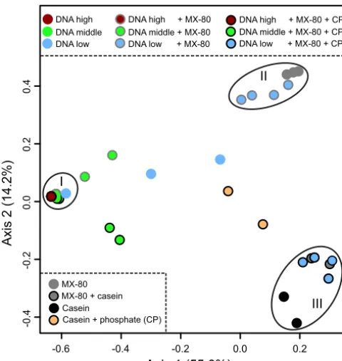
bentonite when additives were absent (Fig. 4). In the presence of CP, the same samples
grouped with casein extracts. Only samples with more than 10
616S rRNA gene copies
grouped distinctly from additive samples.
DNA extraction protocol verification.
We detected increased nucleic acid
recov-eries from MX-80 bentonite using casein and phosphate, but casein-associated
con-taminant DNA was detected at low spiking concentrations. Any additive increases the
risk of contamination, and we therefore recommend the PowerSoil and PowerMax DNA
isolation kits without additional blocking agents for future MX-80 bentonite DNA
extractions. To verify the suitability of the extraction protocol for MX-80 bentonite
samples, we extracted DNA from five additional Wyoming MX-80 bentonite samples
from different production dates (Table 1). The amount of DNA recovered from those
samples was consistent, with 1.8 to 2.5 ng per g (dry weight) bentonite (Table 1).
High-throughput 16S rRNA gene sequencing of DNA extracted from natural Wyoming
MX-80 bentonite samples showed that detected microbial communities differed based
on production date (Fig. 5A). The MX-80 bentonite samples produced in June 2015
were associated with 25.8% to 37.9% of the sequences associated with
Thiobacillus
sp.
(Fig. 5B), which are likely Gram-negative sulfur-oxidizing bacteria (66). Other batches
were characterized by OTUs affiliated with the
Holophagae
(phylum
Acidobacteria
) and
members of the
Gammaproteobacteria
.
Casein MX-80
DNA middle + MX-80 + CP DNA low + MX-80 + CP DNA high + MX-80 + CP
Casein + phosphate (CP) MX-80 + casein
DNA middle + MX-80 DNA low + MX-80 DNA high + MX-80
DNA low DNA high DNA middle
I
II
III
Axis 1 (55.0%)
-0.6 -0.4 -0.2 0.0 0.2
-0.4
-0.2
0
.0
0.2
0
.4
Axis 2 (14.2%)
FIG 4 A principal-coordinate analysis (PCoA) ordination plot based on Bray-Curtis distance metrics
showing grouping of samples (rarefied to 1,174 reads) based on high spiking concentration (group I), MX-80 bentonite (group II), or the presence of casein (group III). Samples were spiked with either 109to 107(DNA high), 106to 105(DNA middle), or 104to 103(DNA low) 16S rRNA gene copies. The presence of casein and phosphate (CP) in spiked extracts is indicated by black outlines around circles. Most samples with spiking concentrations of⬎105are contained within group I. The PCoA ordination plot was generated excluding one replicate of the 104DNA-only dilution series due to few reads for that sample.
on September 8, 2020 by guest
http://msphere.asm.org/
Few previous studies have demonstrated successful recovery of DNA from natural
bentonite clay. Although Chi Fru and Athar (17) extracted DNA from Wyoming MX-80
bentonite, they identified primarily
Bacillus
spp. in the associated clone library. Only 3%
of reads in our high-throughput sequencing results from all six Wyoming-MX-80 samples
were associated with the
Bacillus
genus. Both Lopez-Fernandez et al. (18) and Liu et al.
(19) used high-throughput sequencing to assess bacterial diversity in DNA extracts
from natural bentonite deposits in Spain and China. Common bacteria, such as those
affiliated with the
Acidobacteria
and
Paracoccus
, were identified (18, 67); however, the
dominant microorganisms detected were distinct for each deposit.
Conclusions.
We evaluated the effect of casein and phosphate as blocking agents
for quantitative recovery of nucleic acids from MX-80 bentonite at various starting
concentrations and detected increased recovery of nucleic acids from bentonite using
casein and phosphate (Fig. 1 and 3). However, predominantly casein-associated
con-taminant DNA was detected at spiking concentrations of
⬍
10
516S rRNA gene copies
(Fig. 2). Although we have previously used 30 min of UV light to decontaminate
additives successfully (68), this treatment was insufficient to remove background DNA
contamination from casein; therefore, casein is not recommended as a blocking agent
for DNA extraction from low-biomass clay samples. Synthetic nucleotides as blocking
agents can be designed to minimize interference with gene-specific studies, but they
might affect downstream analyses such as shotgun metagenomics. Nucleic acid
con-tamination in any blocking agent will reduce sample-specific signal in low-biomass
samples, and decontamination might not remove all contaminants or might negatively
TABLE 1Summary of analyzed Wyoming MX-80 samplesIDa Lot no.
Production
date (mo-yr) DNA (ng/g)
B01 065275768 06-2015 1.3
B02 065275772 06-2015 2.2
B03 116315319 11-2016 2.5
B04 037324182 03-2017 2.2
B05 037324184 03-2017 2.2
B06 037324190 03-2017 1.8
aID, identifier.
Axis 1 (34.7%)
Axis 2 (10
.1%)
−0.2 0.0 0.2 0.4
−
0
.3
−
0
.2
−
0
.1
0.0
0
.1
0.2
B02 (06-2015)
B04 (03-2017) B05 (03-2017) B06 (03-2017) B01 (06-2015)
B03 (11-2016) Bentonite (production date)
A
B
OTU taxonomi
c
af
filiation
FIG 5 Grouping of natural Wyoming MX-80 bentonite samples based on production dates in a principal-coordinate analysis (PCoA; Bray-Curtis dissimilarity
metric) ordination plot (A) and microbial community profiles generated using high-throughput V4-V5 16S rRNA gene amplicon sequencing (B). Only operational taxonomic units (OTUs) at or above 3% relative abundance are shown, and numbers in the bubbles represent the relative abundance (%) of each OTU in the corresponding library. Lowercase a or b after each sample name distinguishes duplicate extractions from the same sample batch. For an additional description of each sample, see Table 1.
on September 8, 2020 by guest
http://msphere.asm.org/
affect PCR efficiency (69, 70). Reagent and laboratory contaminations are inevitable and
require extensive postrun analysis, as demonstrated here and elsewhere (59).
Because any additive increases the risk of further contamination, we recommend the
PowerSoil and PowerMax DNA isolation kits without additional blocking agents for
future MX-80 bentonite DNA extractions. Indeed, the high sensitivity of PCR with the
equivalent of 50 cycles and subsequent fingerprinting or high-throughput sequencing
analyses suggest that some additional loss of DNA is a reasonable trade-off in order to
minimize methodological customization and the potential for introducing additional
sources of contamination. We successfully extracted DNA from six Wyoming MX-80
bentonite samples and generated high-throughput 16S rRNA gene profiles. Together,
our results provide a simplified framework for analyzing microbial community DNA
associated with swelling MX-80 bentonite samples within the context of a DGR for used
nuclear fuel. Although transcriptomics would focus on active members of the
commu-nity in bentonite clays, it will be very challenging to extract RNA successfully from
Wyoming MX-80 bentonite.
MATERIALS AND METHODS
Extraction of genomicE. coliDNA and copy number calculation.Escherichia coliK-12 strain
W3110 was grown in 200 ml LB medium overnight at 37°C and 180 rpm. Cells were harvested by centrifugation at 7,000⫻gfor 10 min. A 2-g cell pellet was used for nucleic acid extraction with the PowerMax DNA isolation kit (Mo Bio Laboratories, Carlsbad, CA, USA) according to the manufacturer’s instructions. Genomic DNA concentration was determined using the Qubit dsDNA High Sensitivity (HS) assay kit (Invitrogen, Carlsbad, CA, USA) with fluorescence measured on a FilterMax F5 MultiMode plate reader (Molecular Devices, San Jose, CA, USA) at excitation and emission wavelengths of 485 and 525 nm, respectively. The quality of extractedE. coliDNA was assessed using a NanoDrop 2000 spectrophotom-eter (Thermo Scientific, Waltham, MA, USA) and pulsed-field gel electrophoresis (PFGE) using a CHEF Mapper XA apparatus (Bio-Rad, Hercules, CA, USA). The PFGE gel was run at 14°C for 16 h at 5.5 V/cm with a 1- to 6-s linear pulse in a 1% agarose gel. The gels were stained using GelRed (Biotium, Fremont, CA, USA) and visualized with an AlphaImager HP System (ProteinSimple, San Jose, CA, USA).
The spike samples of genomicE. coliDNA were prepared as 10-fold serial dilutions ranging from 9.8⫻108to 9.8⫻10216S rRNA gene copies per extraction usingE. coliK-12 strain W3110 genome size (71) and the following equation (72): 16S rRNA gene copies⫽ 7 operons ⫻(DNA amount [g] ⫻ 6.02⫻1023[copies/mol])/(4.65⫻106bp⫻650 g/mol/bp). For simplicity, 9.8⫻108to 9.8⫻10216S rRNA gene copies are reported as 109to 103throughout, respectively.
Wyoming MX-80 bentonite.Six Wyoming MX-80 bentonite samples from different production
dates were analyzed in this research (Table 1). The MX-80 bentonite sample B01 was used for the spiking experiments. This sample was processed on 3 June 2015 (lot 065275768) by Caldic Canada (Mississauga, ON, Canada). Moisture content and pH were determined by Caldic Canada as 18.5% and 9.96, respec-tively. The MX-80 bentonite consists of approximately 74% to 90% montmorillonite (73, 74), causing swelling on water uptake. The swelling index was determined with 28 ml/2 g (ASTM D5890-06) for oven-dried (24 h at 105°C) bentonite.
Enumeration of naturally occurring bacteria in Wyoming MX-80 bentonite.Enumeration of
cultivable bacteria in bentonite was conducted as previously described (6), with several modifications. Bentonite suspensions were prepared by slowly adding 2 g bentonite powder (18.5% moisture content) to 18 ml liquid medium while it was continuously agitated using a vortex to avoid clumping. The bentonite suspension was further mixed in a rotating incubator at 15 rpm for 30 min at room temper-ature. For the enumeration of cultivable aerobic and anaerobic bacteria, the bentonite suspension and 10-fold serial dilutions were prepared in R2A medium (M1687; HiMedia Laboratories, West Chester, PA, USA). Serial dilutions were plated on R2A agar plates, in triplicates, and incubated under oxic or anoxic conditions at 30°C for 7 or 28 days, respectively. A 5-tube most probable number (MPN) method was used for the enumeration of cultivable sulfate-reducing bacteria incubated under anoxic conditions at 30°C for 4 weeks. The bentonite suspension and 10-fold serial dilutions were prepared in media. Sulfate-reducing bacteria were quantified in Triple Pack medium (M803; HiMedia Laboratories) and assessed for black ferrous sulfide (FeS) precipitation, signifying hydrogen sulfide production and, consequently, sulfate reduction.
Large-scale DNA extraction from natural Wyoming MX-80 bentonite.Total genomic DNA from
natural bentonite powder was extracted using the PowerMax DNA isolation kit (Mo Bio Laboratories). A total of 2 g of bentonite powder was slowly added to 15 ml PowerBead solution while being agitated. After addition of lysis solution, the tube was incubated at 65°C for 30 min before bead beading for 10 min at 30 Hz (Mixer Mill MM 400; Retsch, Germany). The remainder of the extraction was carried out according to the manufacturer’s instructions. Purified DNA was eluted in 2 ml of 10 mM Tris. Nucleic acids were precipitated using 4l/ml Co-Precipitant linear polyacrylamide (Bioline, Germany), 0.1 volumes of 5 M NaCl (prepared in 0.2-m filter-sterilized PCR water [HyClone HyPure Water]; GE Healthcare Life Sciences, Logan, UT, USA), 1 volume of isopropanol (high-pressure liquid chromatography [HPLC] grade), and stored at⫺20°C overnight. Precipitated DNA was pelleted by centrifugation at 13,000⫻gfor 30 min and then washed with 80% ethanol (HPLC grade), air dried, and eluted in 150l of 10 mM Tris. Aliquots were
on September 8, 2020 by guest
http://msphere.asm.org/
frozen at⫺20°C until PCR analysis. A control extraction without any sample was carried out in parallel to assess potential contamination from kit reagents.
DNA extraction from natural and spiked Wyoming MX-80 bentonite.Genomic DNA was
ex-tracted from natural and spiked MX-80 bentonite samples using the PowerSoil DNA isolation kit (Mo Bio Laboratories). A control extraction without any sample was carried out in parallel to assess reagent contamination. Powdered MX-80 bentonite was used to prepare a 1:6 slurry (solid/liquid ratio) in sterile nuclease and nucleic acid-free PCR water (GE Healthcare Life Sciences). As described above, bentonite suspensions were prepared by slowly adding bentonite powder to liquid with vortex agitation to avoid clumping. The MX-80 slurry was spiked withE. coliDNA at various concentrations, mixed by inversion, and incubated in a rotating incubator at 15 rpm for 30 min at room temperature to enable DNA binding to the clay matrix; note that we did not assess the specific proportion of DNA adsorbed to MX-80 bentonite. Genomic DNA extractions from 0.3-ml slurries (50 mg MX-80 bentonite [dry weight]) were performed in duplicates according to the manufacturer’s instructions with several modifications. Spe-cifically, after the addition of lysis buffer, samples were incubated at 65°C for 30 min to enhance lysis and desorption of DNA from clay matrix. Bead beating was conducted in the FastPrep-24 instrument (MP Biomedicals, Solon, OH, USA) at 5.5 m/s for 45 s. All supernatant was transferred at every step for maximum nucleic acid recovery. Total DNA was quantified using a Qubit dsDNA High Sensitivity assay kit (Invitrogen).
Blocking agents.To assess a representative subset of many possible blocking agents commonly
used in DNA extractions, we tested casein (catalog number C7078; 400 mg per g MX-80 bentonite [dry weight]; Sigma) and sodium pyrophosphate (catalog number S6422; 100mol PO43⫺per g MX-80 bentonite [dry weight], 0.4 M; Sigma) for our bentonite clay nucleic acid extractions. Because Ikeda and colleagues (31) tested both casein and BSA on DNA extractions of Andisol soil samples and found that casein was successful in extracting DNA from more Andisol soil samples than BSA, we selected casein as a representative protein-based blocking agent for this study. To remove potential nucleic acid contam-ination from casein, as reported previously (31), we added casein (20 mg) to the Mo Bio bead beating solution and UV treated the tube for 30 min on a 302-nm UV transilluminator. Phosphate was prepared in PCR water and UV treated in the same way. We chose UV illumination as the method for decontam-ination, because we have used it successfully to remove contaminants from additives in DNA extractions (68). We tested casein and phosphates on spiked and natural MX-80 bentonite samples with the PowerSoil DNA isolation kit using the protocol and modifications described above. We did not test the use of nucleic acids as blocking agents, because they would coextract with low-biomass target DNA and influence downstream applications, such as metagenomics. It was shown by Jacobsen and colleagues (41) that a commercially available blocking reagent increased the yield from low-biomass clay subsoil without introducing contaminating DNA. However, we have not tested this blocking agent in this research.
Denaturing gradient gel electrophoresis analysis.The V3 regions of bacterial 16S rRNA genes
were amplified using primer pair 341F-GC and 518R (75). The 50-l PCR mix contained 1⫻ThermoPol buffer, 0.2M each primer, 200M dNTPs, 30g BSA, 1.25 UTaqDNA polymerase (New England Biolabs, Ipswich, MA, USA), and 2l of template. The PCR was performed in two rounds by adding 1l of amplification product from the first PCR (PCR1) into the second PCR (PCR2). The PCR amplifications were performed as follows: 95°C for 3 min, 35 (PCR1) or 15 (PCR2) cycles of 95°C for 15 s, 55°C for 30 s, and 68°C for 30 s, and a final extension of 68°C for 7 min. Equal amounts of PCR amplicons were separated on a 10% (wt/vol) polyacrylamide gel with a denaturant gradient ranging from 30% to 70% to a maximum of 20l for samples with low PCR yield. The gels were run as previously described (76) for 15 h at 85 V in a DGGEK-2401 denaturing gradient gel electrophoresis (DGGE) system (C.B.S. Scientific Company, San Diego, CA, USA), stained with SYBR green I DNA stain (Invitrogen) for 1 h, and scanned using the Molecular Imager Pharos FX Plus (Bio-Rad).
Quantitative PCR.Genomic DNA in spiked and natural MX-80 bentonite extracts was quantified by
targeting multicopy bacterial 16S rRNA genes using primers 341F/518R and the single-copyE. coli -specific uidA gene (coding for -D-glucuronidase) using primers uidA405F/uidA405R (77). All PCR amplifications were performed in duplicates. For theuidAgene PCR, the 15-l reaction volume contained 1⫻SsoAdvanced Universal SYBR green Supermix (Bio-Rad), 0.3M each primer, 7.5g BSA, and 2l of template. The 16S rRNA gene PCR was performed in 10-l reaction volumes with PCR components in concentrations as listed above, without the addition of BSA. The PCRs were performed on a CFX96 Real-Time PCR detection system (Bio-Rad). ForuidAgene amplification, PCR conditions were 98°C for 3 min followed by 40 cycles of 98°C for 15 s and 60°C for 60 s. For 16S rRNA genes, the PCR conditions were 98°C for 3 min followed by 40 cycles of 98°C for 15 s and 55°C for 30 s. PurifiedE. coligenomic DNA was used as a standard template for both PCR protocols. Amplification efficiencies ranged from 90.3% to 99.6%, and all coefficients of determination (R2) exceeded 0.995. Starting DNA copy numbers were calculated from the linear regression equation of each standard curve.
Amplification of 16S rRNA genes and Illumina sequencing.The V3-V4 regions of the 16S rRNA
genes were amplified using universal prokaryotic dual-indexed primers Pro341F and Pro805R (78). In addition to the unique 6-bp index sequence for sample multiplexing, each primer contained Illumina flow cell binding and sequencing sites (79). The PCR was set up in a PCR workstation using ISO 5 HEPA-filtered air (AirClean Systems, Creedmoor, NC, USA). The surface was cleaned with UltraClean Lab Cleaner (Mo Bio) and treated with UV light irradiation for 15 min. In addition, tubes, 96-well plates, PCR water, and BSA were UV treated on a 302 nm transilluminator (ProteinSimple, San Jose, CA, USA) for 15 min. The 25-l PCR mixture contained 1⫻ThermoPol buffer, 0.2M forward primer, 0.2M reverse primer, 200M dNTPs, 15g BSA, 0.625 UTaqDNA polymerase (New England Biolabs), and 2l of
on September 8, 2020 by guest
http://msphere.asm.org/
template (up to 10 ng). Each PCR was prepared in triplicates, with two rounds by adding 1l product from PCR1 into PCR2. The PCRs were performed as follows: 95°C for 3 min, 35 (PCR1) or 15 (PCR2) cycles of 95°C for 30 s, 55°C for 30 s, and 68°C for 1 min, and a final extension of 68°C for 7 min. Equal quantities of PCR2 amplicons were pooled (average of 10l per sample). Twenty microliters of DNA extraction blanks and no-template controls (NTCs) was included, even though no amplicon was visible in a stained agarose gel. The pooled 16S rRNA gene amplicons were excised from an agarose gel and purified using a Wizard SV Gel and PCR Clean-Up system (Promega, Madison, WI, USA). A 5-pM library containing 5% PhiX control library (Illumina, San Diego, CA, USA) was sequenced on a MiSeq instrument (Illumina) using a 2⫻250-cycle MiSeq reagent kit v2 (Illumina Canada, Vancouver, BC, Canada). For comparison of six natural Wyoming MX-80 bentonite samples, the V4-V5 region of 16S rRNA genes was amplified using universal prokaryotic primers 515F-Y (80) and 926R (81) according to the method described above but at an annealing temperature of 50°C.
Illumina sequence analysis.All MiSeq reads were demultiplexed using MiSeq Reporter software
(version 2.5.0.5; Illumina). Reads were assembled using the paired-end assembler for Illumina sequences (PANDAseq, version 2.8) (82) with a quality threshold of 0.9, 8-nucleotide minimum overlap, and 300-nucleotide minimum assembled read length. Assembled reads were analyzed using Quantitative Insights Into Microbial Ecology (QIIME version 1.9.0) (83), managed by automated exploration of microbial diversity (AXIOME version 1.5) (84). Sequences were clustered using UPARSE algorithm USE-ARCH version 7.0.1090 (85) at 97% identity and aligned with the Python Nearest Alignment Space Termination tool (PyNAST version 1.2.2) (86). All representative sequences were classified using the Ribosomal Database Project (RDP version 2.2) (87) with a stringent confidence threshold (0.8). Taxonomy was assigned using the SILVA database release 128 (88). Chimeric sequences were filtered with UCHIME (89). Bubble plots showing taxonomy profiles were created using the ggplot2 package (90) in R v.3.4.4 using operational taxonomic unit (OTU) tables generated by AXIOME.
Data availability.All sequences were deposited into European Nucleotide Archive (https://www.ebi
.ac.uk/ena) with study accession number PRJEB29317. Supplemental OTU tables were deposited at
10.5281/zenodo.3459859and10.5281/zenodo.3459870.
SUPPLEMENTAL MATERIAL
Supplemental material for this article may be found at
https://doi.org/10.1128/
mSphere.00334-19
.
TEXT S1
, PDF file, 0.1 MB.
FIG S1
, PDF file, 0.3 MB.
FIG S2
, PDF file, 2.3 MB.
FIG S3
, PDF file, 0.1 MB.
FIG S4
, PDF file, 0.1 MB.
FIG S5
, PDF file, 0.2 MB.
TABLE S1
, PDF file, 0.1 MB.
TABLE S2
, PDF file, 0.1 MB.
REFERENCES
1. Keech PG, Vo P, Ramamurthy S, Chen J, Jacklin R, Shoesmith DW. 2014. Design and development of copper coatings for long term storage of used nuclear fuel. Corros Eng Sci Technol 49:425– 430.https://doi.org/ 10.1179/1743278214Y.0000000206.
2. Ottosson M, Boman M, Berastegui P, Andersson Y, Hahlin M, Korvela M, Berger R. 2017. Copper in ultrapure water, a scientific issue under debate. Corros Sci 122:53– 60.https://doi.org/10.1016/j.corsci.2017.03.014. 3. Hedin A, Johansson AJ, Lilja C, Boman M, Berastegui P, Berger R,
Ot-tosson M. 2018. Corrosion of copper in pure O2-free water? Corros Sci 137:1–12.https://doi.org/10.1016/j.corsci.2018.02.008.
4. King F, Lilja C. 2011. Scientific basis for corrosion of copper in water and implications for canister lifetimes. Corros Eng Sci Technol 46:153–158.
https://doi.org/10.1179/1743278210Y.0000000002.
5. King F, Lilja C, Vähänen M. 2013. Progress in the understanding of the long-term corrosion behaviour of copper canisters. J Nucl Mater 438: 228 –237.https://doi.org/10.1016/j.jnucmat.2013.02.080.
6. Stroes-Gascoyne S, Hamon CJ, Maak P, Russell S. 2010. The effects of the physical properties of highly compacted smectitic clay (bentonite) on the culturability of indigenous microorganisms. Appl Clay Sci 47: 155–162.https://doi.org/10.1016/j.clay.2008.06.010.
7. Masurat P, Eriksson S, Pedersen K. 2010. Microbial sulphide production in compacted Wyoming bentonite MX-80 underin situconditions relevant to a repository for high-level radioactive waste. Appl Clay Sci 47:58 – 64.
https://doi.org/10.1016/j.clay.2009.01.004.
8. Motamedi M, Karland O, Pedersen K. 1996. Survival of sulfate reducing
bacteria at different water activities in compacted bentonite. FEMS Micro-biol Lett 141:83– 87.https://doi.org/10.1111/j.1574-6968.1996.tb08367.x. 9. Bengtsson A, Pedersen K. 2017. Microbial sulphide-producing activity in
water saturated Wyoming MX-80, Asha and Calcigel bentonites at wet densities from 1500 to 2000 kg m⫺3. Appl Clay Sci 137:203–212.https://
doi.org/10.1016/j.clay.2016.12.024.
10. Pedersen K. 2010. Analysis of copper corrosion in compacted bentonite clay as a function of clay density and growth conditions for sulfate-reducing bacteria. J Appl Microbiol 108:1094 –1104.https://doi.org/10 .1111/j.1365-2672.2009.04629.x.
11. Pedersen K, Bengtsson A, Blom A, Johansson L, Taborowski T. 2017. Mobility and reactivity of sulphide in bentonite clays – implications for engineered bentonite barriers in geological repositories for radioactive wastes. Appl Clay Sci 146:495–502.https://doi.org/10.1016/j.clay.2017.07 .003.
12. Stone W, Kroukamp O, Moes A, McKelvie J, Korber DR, Wolfaardt GM. 2016. Measuring microbial metabolism in atypical environments: ben-tonite in used nuclear fuel storage. J Microbiol Methods 120:79 –90.
https://doi.org/10.1016/j.mimet.2015.11.006.
13. Poulain S, Sergeant C, Simonoff M, Le Marrec C, Altmann S. 2008. Microbial investigations in Opalinus clay, an argillaceous formation under evaluation as a potential host rock for a radioactive waste repository. Geomicrobiol J 25:240 –249.https://doi.org/10.1080/01490450802153314.
14. Stroes-Gascoyne S, Schippers A, Schwyn B, Poulain S, Sergeant C, Simo-noff M, Le Marrec C, Altmann S, Nagaoka T, Mauclaire L, McKenzie J, Daumas S, Vinsot A, Beaucaire C, Matray JM. 2007. Microbial community
on September 8, 2020 by guest
http://msphere.asm.org/
analysis of Opalinus clay drill core samples from the Mont Terri Under-ground Research Laboratory, Switzerland. Geomicrobiol J 24:1–17.
https://doi.org/10.1080/01490450601134275.
15. Urios L, Marsal F, Pellegrini D, Magot M. 2012. Microbial diversity of the 180 million-year-old Toarcian argillite from Tournemire, France. Appl Geochemistry 27:1442–1450.https://doi.org/10.1016/j.apgeochem.2011 .09.022.
16. Direito SOL, Marees A, Röling W. 2012. Sensitive life detection strategies for low-biomass environments: optimizing extraction of nucleic acids adsorbing to terrestrial and Mars analogue minerals. FEMS Microbiol Ecol 81:111–123.https://doi.org/10.1111/j.1574-6941.2012.01325.x. 17. Chi Fru E, Athar R. 2008. In situbacterial colonization of compacted
bentonite under deep geological high-level radioactive waste repository conditions. Appl Microbiol Biotechnol 79:499 –510. https://doi.org/10 .1007/s00253-008-1436-z.
18. Lopez-Fernandez M, Cherkouk A, Vilchez-Vargas R, Jauregui R, Pieper D, Boon N, Sanchez-Castro I, Merroun ML. 2015. Bacterial diversity in bentonites, engineered barrier for deep geological disposal of radioac-tive wastes. Microb Ecol 70:922–935.https://doi.org/10.1007/s00248-015 -0630-7.
19. Liu H, Dang X, Zhang H, Dong J, Zhang Z, Wang C, Zhang R, Yuan Y, Ren Y, Liu W, Zhuang D, Yang Z, Duan Z, Li Y, Zuo Y, Chai D. 2019. Microbial diversity in bentonite, a potential buffer material for deep geological disposal of radioactive waste. IOP Conf Ser Earth Environ Sci 227:22010.
https://doi.org/10.1088/1755-1315/227/2/022010.
20. Sun D, Sun W, Fang L. 2014. Swelling characteristics of Gaomiaozi bentonite and its prediction. J Rock Mech Geotech Eng 6:113–118.
https://doi.org/10.1016/j.jrmge.2014.01.001.
21. Cai P, Huang Q, Zhang X, Chen H. 2006. Adsorption of DNA on clay minerals and various colloidal particles from an Alfisol. Soil Biol Biochem 38:471– 476.https://doi.org/10.1016/j.soilbio.2005.05.019.
22. Khanna M, Stotzky G. 1992. Transformation ofBacillus subtilisby DNA bound on montmorillonite and effect of DNase on the transforming ability of bound DNA. Appl Environ Microbiol 58:1930 –1939. 23. Franchi M, Ferris JP, Gallori E. 2003. Cations as mediators of the
adsorp-tion of nucleic acids on clay surfaces in prebiotic environments. Orig Life Evol Biosph 33:1–16.https://doi.org/10.1023/A:1023982008714. 24. Beall GW, Sowersby DS, Roberts RD, Robson MH, Lewis LK. 2009. Analysis
of oligonucleotide DNA binding and sedimentation properties of mont-morillonite clay using ultraviolet light spectroscopy. Biomacromolecules 10:105–112.https://doi.org/10.1021/bm800970v.
25. Greaves MP, Wilson MJ. 1969. The adsorption of nucleic acids by mont-morillonite. Soil Biol Biochem 1:317–323.https://doi.org/10.1016/0038 -0717(69)90014-5.
26. Poly F, Chenu C, Simonet P, Rouiller J, Monrozier LJ. 2000. Differences between linear chromosomal and supercoiled plasmid DNA in their mechanisms and extent of adsorption on clay minerals. Langmuir 16: 1233–1238.https://doi.org/10.1021/la990506z.
27. Gallori E, Bazzicalupo M, Dal Canto L, Fani R, Nannipieri P, Vettori C, Stotzky G. 1994. Transformation ofBacillus subtilisby DNA bound on clay in non-sterile soil. FEMS Microbiol Ecol 15:119 –126.https://doi.org/10 .1111/j.1574-6941.1994.tb00236.x.
28. Pietramellara G, Franchi M, Gallori E, Nannipieri P. 2001. Effect of mo-lecular characteristics of DNA on its adsorption and binding on homo-ionic montmorillonite and kaolinite. Biol Fertil Soils 33:402– 409. 29. Arlinger J, Bengtsson A, Edlund J, Eriksson L, Johansson J, Lydmark S,
Rabe L, Pedersen K. 2013. Prototype repository – Microbes in the re-trieved outer section. SKB P-13–16. International Atomic Energy Agency, Vienna, Austria.
30. Dineen SM, Aranda R, Anders DL, Robertson JM. 2010. An evaluation of commercial DNA extraction kits for the isolation of bacterial spore DNA from soil. J Appl Microbiol 109:1886 –1896. https://doi.org/10.1111/j .1365-2672.2010.04816.x.
31. Ikeda S, Tsurumaru H, Wakai S, Noritake C, Fujishiro K, Akasaka M, Ando K. 2008. Evaluation of the effects of different additives in improving the DNA extraction yield and quality from Andosol. Microbes Environ 23: 159 –166.https://doi.org/10.1264/jsme2.23.159.
32. Takada-Hoshino Y, Matsumoto N. 2004. An improved DNA extraction method using skim milk from soils that strongly adsorb DNA. Microb Environ 19:13–19.https://doi.org/10.1264/jsme2.19.13.
33. Tournier E, Amenc L, Pablo AL, Legname E, Blanchart E, Plassard C, Robin A, Bernard L. 2015. Modification of a commercial DNA extraction kit for safe and rapid recovery of DNA and RNA simultaneously from soil,
without the use of harmful solvents. MethodsX 2:182–191.https://doi .org/10.1016/j.mex.2015.03.007.
34. Aerts S, Jacops E, Dewel A. 2009 Microbial activity around the connect-ing gallery. SCK-CEN-ER-61. SCK-CEN, Mol, Belgium.
35. Hurt RA, Robeson MS, Shakya M, Moberly JG, Vishnivetskaya TA, Gu B, Elias DA. 2014. Improved yield of high molecular weight DNA coincides with increased microbial diversity access from iron oxide cemented sub-surface clay environments. PLoS One 9:e102826.https://doi.org/10 .1371/journal.pone.0102826.
36. Boivin-Jahns V, Ruimy R, Bianchi A, Daumas S, Christen R. 1996. Bacterial diversity in a deep-subsurface clay environment. Appl Environ Microbiol 62:3405–3412.
37. Lever MA, Torti A, Eickenbusch P, Michaud AB, Šantl-Temkiv T, Jørgensen BB. 2015. A modular method for the extraction of DNA and RNA, and the separation of DNA pools from diverse environmental sample types. Front Microbiol 6:476.https://doi.org/10.3389/fmicb.2015.00476. 38. Yankson KK, Steck TR. 2009. Strategy for extracting DNA from clay soil
and detecting a specific target sequence via selective enrichment and real-time (quantitative) PCR amplification. Appl Environ Microbiol 75: 6017– 6021.https://doi.org/10.1128/AEM.00211-09.
39. Saeki K, Sakai M, Kunito T. 2012. Effect of␣-casein on DNA adsorption by Andosols and by soil components. Biol Fertil Soils 48:469 – 474.https:// doi.org/10.1007/s00374-011-0640-7.
40. Yu WH, Li N, Tong DS, Zhou CH, Lin CX, Xu CY. 2013. Adsorption of proteins and nucleic acids on clay minerals and their interactions: a review. Appl Clay Sci 80 – 81:443– 452.https://doi.org/10.1016/j.clay.2013.06.003.
41. Jacobsen CS, Nielsen TK, Vester JK, Stougaard P, Nielsen JL, Voriskova J, Winding A, Baldrian P, Liu B, Frostegård Å, Pedersen D, Tveit AT, Sven-ning MM, Tebbe CC, Øvreås L, Jakobsen PB, Blazewicz SJ, Hubablek V, Bertilsson S, Hansen LH, Cary SC, Holben WE, Ekelund F, Bælum J. 2018. Inter-laboratory testing of the effect of DNA blocking reagent G2 on DNA extraction from low-biomass clay samples. Sci Rep 8:5711.https:// doi.org/10.1038/s41598-018-24082-y.
42. Jalique DR, Stroes-Gascoyne S, Hamon CJ, Priyanto DG, Kohle C, Evenden WG, Wolfaardt GM, Grigoryan AA, McKelvie J, Korber DR. 2016. Cultur-ability and diversity of microorganisms recovered from an eight-year old highly-compacted, saturated MX-80 Wyoming bentonite plug. Appl Clay Sci 126:245–250.https://doi.org/10.1016/j.clay.2016.03.022.
43. Grigoryan AA, Jalique DR, Medihala P, Stroes-Gascoyne S, Wolfaardt GM, McKelvie J, Korber DR. 2018. Bacterial diversity and production of sulfide in microcosms containing uncompacted bentonites. Heliyon 4:e00722.
https://doi.org/10.1016/j.heliyon.2018.e00722.
44. Pedersen K, Motamedi M, Karnland O, Sandén T. 2000. Mixing and sulphate-reducing activity of bacteria in swelling, compacted bentonite clay under high-level radioactive waste repository conditions. J Appl Microbiol 89: 1038 –1047.https://doi.org/10.1046/j.1365-2672.2000.01212.x.
45. Masurat P, Eriksson S, Pedersen K. 2010. Evidence of indigenous sulphate-reducing bacteria in commercial Wyoming bentonite MX-80. Appl Clay Sci 47:51–57.https://doi.org/10.1016/j.clay.2008.07.002. 46. Katevatis C, Fan A, Klapperich CM. 2017. Low concentration DNA
extrac-tion and recovery using a silica solid phase. PLoS One 12:e0176848.
https://doi.org/10.1371/journal.pone.0176848.
47. Shi B, Shin YK, Hassanali AA, Singer SJ. 2015. DNA binding to the silica surface. J Phys Chem B 119:11030 –11040.https://doi.org/10.1021/acs .jpcb.5b01983.
48. Luna GM, Dell’Anno A, Danovaro R. 2006. DNA extraction procedure: a critical issue for bacterial diversity assessment in marine sediments. Environ Microbiol 8:308 –320.https://doi.org/10.1111/j.1462-2920.2005 .00896.x.
49. Hwang C, Liu W-T, Andersen GL, Ling F, LeChevallier MW. 2012. Evalu-ation of methods for the extraction of DNA from drinking water distri-bution system biofilms. Microbes Environ 27:9 –18. https://doi.org/10 .1264/jsme2.me11132.
50. Teng F, Darveekaran Nair SS, Zhu P, Li S, Huang S, Li X, Xu J, Yang F. 2018. Impact of DNA extraction method and targeted 16S-rRNA hyper-variable region on oral microbiota profiling. Sci Rep 8:16321.https://doi .org/10.1038/s41598-018-34294-x.
51. Abusleme L, Hong BY, Dupuy AK, Strausbaugh LD, Diaz PI. 2014. Influ-ence of DNA extraction on oral microbial profiles obtained via 16S rRNA gene sequencing. J Oral Microbiol 6:23990.https://doi.org/10.3402/jom .v6.23990.
52. Cruaud P, Vigneron A, Lucchetti-Miganeh C, Ciron PE, Godfroy A, Cambon-Bonavita M-A. 2014. Influence of DNA extraction method, 16S rRNA targeted hypervariable regions, and sample origin on microbial