1535-9778/02/$04.00⫹0 DOI: 10.1128/EC.1.3.469–480.2002
Copyright © 2002, American Society for Microbiology. All Rights Reserved.
GTPase-Activating Proteins for Cdc42
Gregory R. Smith,† Scott A. Givan,‡ Paul Cullen, and George F. Sprague Jr.*
Institute of Molecular Biology, University of Oregon, Eugene, Oregon 97403-1229Received 10 December 2001/Accepted 20 March 2002
The Rho-type GTPase, Cdc42, has been implicated in a variety of functions in the yeast life cycle, including septin organization for cytokinesis, pheromone response, and haploid invasive growth. A group of proteins called GTPase-activating proteins (GAPs) catalyze the hydrolysis of GTP to GDP, thereby inactivating Cdc42. At the time this study began, there was one known GAP, Bem3, and one putative GAP, Rga1, for Cdc42. We identified another putative GAP for Cdc42 and named it Rga2 (Rho GTPase-activating protein 2). We confirmed by genetic and biochemical criteria that Rga1, Rga2, and Bem3 act as GAPs for Cdc42. A detailed characterization of Rga1, Rga2, and Bem3 suggested that they regulate different subsets of Cdc42 function. In particular, deletion of the individual GAPs conferred different phenotypes. For example, deletion ofRGA1, but notRGA2orBEM3, caused hyperinvasive growth. Furthermore, overproduction or loss of Rga1 and Rga2, but not Bem3, affected the two-hybrid interaction of Cdc42 with Ste20, a p21-activated kinase (PAK) kinase required for haploid invasive growth. These results suggest Rga1, and possibly Rga2, facilitate the interaction of Cdc42 with Ste20 to mediate signaling in the haploid invasive growth pathway. Deletion ofBEM3resulted in cells with severe morphological defects not observed inrga1⌬orrga2⌬strains. These data suggest that Bem3 and, to a lesser extent, Rga1 and Rga2 facilitate the role of Cdc42 in septin organization. Thus, it appears that the GAPs play a role in modulating specific aspects of Cdc42 function. Alternatively, the different phenotypes could reflect quantitative rather than qualitative differences in GAP activity in the mutant strains.
In the yeastSaccharomyces cerevisiae, the Cdc42 protein (33)
is a member of the Rho subfamily of the Ras superfamily of GTPases, which act as molecular switches and regulate many cellular processes (1, 34). Like all GTPases, Cdc42 can exist in two states, a GTP-bound, active state and a GDP-bound, in-active state. The cycling of Cdc42 between these states is con-trolled by two sets of proteins: guanine nucleotide exchange factors (GEFs) and GTPase-activating proteins (GAPs). There is one GEF for Cdc42 in yeast, called Cdc24 (62, 67, 69). At the time this study began, there was one protein with demon-strated biochemical GAP activity for Cdc42, Bem3 (68), and one protein with potential GAP activity based on the presence of a 170-amino-acid GAP domain, Rga1 (14, 63). Although Bem3 and Rga1 are fairly similar in their GAP domains, they are divergent over the remainder of their protein sequences.
Rga1 contains two tandemLin-11,Isl-1,Mec-3(LIM) domains
(23, 35, 63, 66). LIM domains bind zinc ions and are thought to mediate protein-protein interactions (2, 18, 45). In contrast, Bem3 contains a Pleckstrin homology (PH) domain. PH do-mains are thought to play roles in membrane localization and protein-protein interactions (40).
Cdc42 is required for polarization of the actin cytoskeleton, for cytokinesis, and for morphological changes that accompany activation of some signal transduction pathways, specifically the pheromone response pathway and the filamentous growth pathway (for review, see reference 32). Several proteins, aside
from Cdc24, Rga1, and Bem3, that bind to Cdc42 have been identified and are presumed to be effectors of various Cdc42 functions. These proteins include Ste20 and Cla4, two protein kinases (17, 61), and Gic1 and Gic2, two proteins of similar sequence but as-yet-undefined biochemical activity (7, 13). Ste20 is required for operation of the pheromone and filamen-tation pathways (38, 53, 56), Cla4 plays ill-defined roles in septin organization and cytokinesis (4, 9, 17, 41), and together Gic1 and Gic2 are required to polarize the actin cytoskeleton (5).
The finding that there is more than one potential GAP for Cdc42 raises the possibility that each GAP may regulate Cdc42 activity with respect to only a subset of its biological roles. To explore this possibility, we sought to identify the full comple-ment of GAPs for Cdc42 and examine the phenotypic conse-quences of the absence of one or more of these GAPs. Here we report the identification of a third potential GAP, Rga2, which has strong sequence similarity to Rga1. We demonstrate that Rga1 and Rga2 have biochemical GAP activity for Cdc42. Finally, we provide evidence that strains lacking Rga1, Rga2, and Bem3 display distinct phenotypes, supporting the idea that they influence Cdc42 activity in different biological settings.
MATERIALS AND METHODS
Strains, plasmids, and microbiological techniques.The yeast strains used are listed in Table 1. Gene deletions were constructed by PCR and confirmed by PCR and phenotyping when applicable. In all cases, the entire coding region was replaced with the indicated marker. Yeast and bacterial strains were propagated using standard methods (8, 57, 59). Yeast extract-peptone-dextrose and synthetic dextrose media were prepared as previously described (58). Yeast transforma-tions were performed using modificatransforma-tions of the LiOAc method (12, 24). Bac-terial transformations, DNA preparations, and plasmid constructions were per-formed by standard methods (59). Growth capabilities at various temperatures were measured by spotting equal volumes of serial dilutions of mid-log phase cultures to agar plates and incubating at the indicated temperatures.
* Corresponding author. Mailing address: Institute of Molecular Biology, University of Oregon, Eugene, OR 97403-1229. Phone: (541) 346-5883. Fax: (541) 346-4854. E-mail: [email protected] .edu.
† Present address: Department of Biomolecular Chemistry, Univer-sity of Wisconsin, Madison, WI 53706.
‡ Present address: Center for Gene Research and Biotechnology, Oregon State University, Corvallis, OR 97331.
469
on September 8, 2020 by guest
http://ec.asm.org/
Two-hybrid analysis.Two-hybrid analysis (22) of interactions between the panel of GTPases and Rga1, Rga2, and Bem3 was performed with strain EGY40
(26). To assess-galactosidase activity, strain EGY40 containing theLexAop
-lacZreporter plasmid pSH18-34 (26) was transformed with a pEG202-derived
LexA DNA-binding domain (DBD) plasmid and a pJG4-5-derived Gal4 activa-tion domain (AD) plasmid. At least three separate isolates were grown to mid-log phase in rich medium containing 2% galactose and quantitated as pre-viously described (63). Most of the LexA DBD fusions in pEG202 and Gal4 AD
fusions in pJG4-5 were previously described (63). pJG4-5-RGA2was constructed
using gap repair (42) of the entireRGA2coding region andEcoRI-cut pJG4-5
(26). pEG202-RGA2was constructed by ligating anXhoI-EcoRI fragment of
pJG4-5-RGA2intoXhoI-EcoRI-cut pEG202 (26). The expression of two-hybrid
constructs was confirmed by Western blotting techniques (59) using␣-LexA
DBD and␣-Gal4 AD antibodies, respectively (52).-Galactosidase assays were
used to determine the relative strength of the interactions and were performed essentially as described previously (29).
Additional two-hybrid analysis was performed with the PJ69-4A strain (30).
pGS38 and pGS39 were constructed by ligating theEcoRI-XhoI fragments from
pEG202-CDC42G12V,C188Sand pEG202-CDC42Q61L,C188S(63), respectively, into
the Gal4 activation domain fusion vector pGAD-C(2) (30), which was cut with
EcoRI andSalI. pSL2682 contains a truncation ofSTE20(encoding amino acids
1 to 565) cloned into pOBD.CYH2(64) was a kind gift from Megan Keniry. The
expression of these fusion proteins was confirmed by Western blot analysis using
a␣-Gal4 DBD monoclonal antibody (52). pGS40 was constructed by ligating a
KpnI-HindIII fragment from YEp13-RGA1containing the coding region and
1,500 nucleotides upstream ofRGA1toKpnI-HindIII-cut YEp352 (28). pGS41
was constructed by ligating aSacI-XhoI fragment from YEp13-RGA2containing
the coding region and 800 nucleotides upstream ofRGA2 toSacI-XhoI-cut
YEp352. pGS42 was constructed by ligating aSacI-PstI fragment from
YEp13-BEM3containing the coding region and 800 nucleotides upstream ofBEM3to
SacI-PstI cut YEp352.-Galactosidase assays were used to determine the
rela-tive strength of the interactions and were performed essentially as described previously (29).
Protein purification.The GTPases Cdc42 and Rsr1 (Bud1) were purified as
glutathioneS-transferase (GST) fusion proteins using hybrid genes present in
the plasmid pGEX-5X-1 (47). pGEX-5X-1-Cdc42C188S and
pGEX-5X-1-Cdc42Q61L,C188Swere gifts from David Mitchell, and pGEX-Rsr1 was kindly
provided by Hay-Oak Park (49).Escherichia colicells (DH5␣) were grown to
mid-log phase, protein expression was induced with 1 mM IPTG (Sigma) for 2 h, and cells were lysed by two passages through a French press. The lysate was centrifuged at 5,000 rpm in an SS-34 Sorvall rotor for 10 min to pellet unbroken cells and cellular debris. Swollen glutathione agarose beads were added to the
supernatant, incubated at 4°C for 2 h, washed, and frozen at⫺80°C in GTPase
storage buffer (20 mM Tris-HCl [pH 7.5], 1 mM dithiothreitol [DTT], 100M
GTP, 1⫻protease inhibitors cocktail [Roche], and 15% glycerol).
The GAP domains of Rga1, Rga2, and Bem3 were purified as maltose binding protein (MBP) fusions. These fusions were expressed from plasmids based on
pMAL-c2 (25). pGS53 was constructed by addition ofSalI andHindIII sites to
the ends of anRGA1PCR product using a plasmid containing theRGA1coding
region as a template. This PCR product was digested and ligated toSalI-Hin
dIII-cut pMAL-c2. pGS54 was constructed by addition ofSalI andPstI sites to the
ends of anRGA2PCR product by using a plasmid containing theRGA2coding
region as a template. This PCR product was digested and ligated toSalI-PstI-cut
pMAL-c2. pGS55 was constructed by addition ofEcoRI andSalI sites to the ends
of aBEM3PCR product by using a plasmid containing theBEM3coding region
as a template. This PCR product was digested and ligated toEcoRI-SalI-cut
pMAL-c2. Bacterial protein expression and cell lysis were performed essentially as described above for the GST fusions, except proteins were purified using an amylose column (44) and eluted with Column Buffer II (20 mM Tris-HCl [pH 7.4], 1 mM EDTA, 200 mM NaCl, 10 mM maltose). In all cases, standard sodium dodecyl sulfate-polyacrylamide gel electrophoresis separation and Coomasie staining confirmed protein expression and purification (59).
Biochemistry.Purified GST, GST-Cdc42C188S, GST-Cdc42Q61L,C188S, or
GST-Rsr1 was incubated with⬃2Ci of [␥-32P]GTP or [␣-32P]GTP (New England
Nuclear Life Sciences) at room temperature for 10 min in reaction buffer (20
mM Tris-HCl [pH 7.5], 1 mM DTT, 100M GTP). Then, 2 nmol of GTP was
added to each reaction mixture. Each reaction mixture contained one of the following: 25.6 pmol of GST-Cdc42 (C188S), 21.3 pmol of GST-Cdc42 (Q61L, C188S), or 71.6 pmol of GST-Rsr1. As a result, 5.7% of the GST-Cdc42 (C188S)
bound [␣-32P]GTP, 5.9% of the GST-Cdc42 (C188S) bound [␥-32P]GTP, and
7.6% of the GST-Rsr1 bound [␥-32P]GTP. Glutathione agarose beads bound
with a [␥-32P]GTP- or [␣-32P]GTP-loaded GTPase were added to micro-spin
columns (Bio-Rad) and washed six times with 250l of wash buffer (20 mM
Tris-HCl [pH 7.5], 1 mM DTT, 5 mM MgCl2). The beads were resuspended,
removed from the column, and added to⬃2g (28.6 pmol) of MBP, MBP-Rga1,
MBP-Rga2, or MBP-Bem3. The mixtures were incubated at room temperature for the indicated time, added to a micro-spin column, spun, and washed. Then,
100l of elution buffer (1% sodium dodecyl sulfate–20 mM EDTA) was added
to the column and incubated at 65°C for 15 min to denature the protein. The samples were centrifuged to collect the released nucleotides. The reaction buffer, wash buffer, and eluate were counted in a scintillation counter. The ratio of bound to released counts was used to determine GAP activity.
Microscopy.Standard microscopic techniques were used and cells were
exam-ined using a 100⫻objective. Methods for staining with rhodamine phalloidin to
visualize F-actin, Calcofluor to visualize chitin deposits in bud scars, and DAPI
(4⬘,6⬘-diamidino-2-phenylindole) for visualization of DNA were performed
es-sentially as previously described (51). Septins were visualized by
immunofluo-rescence using a␣-Cdc3 antibody (36; purified by April Goehring) followed by
fluorescence microscopy.
FUS1-lacZassays.Cells containing aFUS1-lacZreporter construct integrated
atmfa2⌬(6) were grown to mid-log phase at 30°C in yeast
extract-peptone-dextrose medium.-Galactosidase assays were performed as described
previ-ously (31).
Invasive growth assays.The plate-washing assay was performed as previously described (56). The single cell assay was also performed essentially as previously described (16). For some experiments, cells were scraped from plates using 4 ml
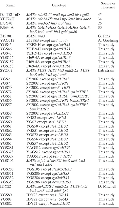
TABLE 1. Yeast strains
Strain Genotype Source orreference
DJTD2-16D MAT␣cdc42-1tsura3 trpl leu2 his4 gal2 45a YEF24H MAT␣cdc24-Htsura3 trpl leu2 his4 ade2 34
EGY40 MAT␣ura3-52 his3 trpl leu2 26
PJ69-4A MATaGALI-HIS3 GAL2-ADE8
GAL7-lacZ leu2 ura3 his3 gal4 gal80 30
⌺1278B MAT␣ura3 G. Fink
YAG512 ⌺1278B excepthis3::ura3- A. Goehring
YGS45 YEF24H exceptrga1::HIS3 This study
YGS46 YEF24H exceptrga2::HIS3 This study
YGS47 YEF24H exceptbem3::HIS3 This study
YGS156 PJ69-4A exceptrga1::URA3 This study
YGS157 PJ69-4A exceptrga2::URA3 This study
YGS158 PJ69-4A exceptbem3::URA3 This study
SY2002 MATaFUS1::HIS3 his3 mfa2-⌬1::
FUS1-lacZ adel leu2 trpl ura3 Lab strain
YGS2 SY2002 exceptrga1::URA3 This study
YGS7 SY2002 exceptrga2::TRP1 This study
YGS50 SY2002 exceptbem3::TRP1 This study
YGS72 SY2002 exceptrga1::URA3 rga2::TRP1 This study YGS51 SY2002 exceptrga1::URA3 bem3::TRP1 This study YGS56 SY2002 exceptrga2::TRP1 bem3::TRP1 This study YGS57 SY2002 exceptrga1::URA3 rga2::TRP1
bem3::TRP1 This study
YGS58 SY2002 exceptste4::LEU2 This study
YGS59 YGS2 exceptste4::LEU2 This study
YGS60 YGS7 exceptste4::LEU2 This study
YGS61 YGS50 exceptste4::LEU2 This study
YGS62 YGS51 exceptste4::LEU2 This study
YGS63 YGS72 exceptste4::LEU2 This study
YGS64 YGS56 exceptste4::LEU2 This study
YGS65 YGS57 exceptste4::LEU2 This study
YGS281 YAG512 exceptrga1::HIS3 This study
YGS328 YAG512 exceptrga2::HIS3 This study
YGS329 YAG512 exceptbem3::HIS3 This study
YGS105 MATamfa2-⌬1::FUS1-lacZ his3 leu2
trp1 ura3 ade1 This study
YGS286 YGS105 exceptste20::URA3 This study
YGS351 YGS286 exceptrga1::HIS3 This study
YGS352 YGS286 exceptrga2::HIS3 This study
YGS353 YGS286 exceptbem3::HIS3 This study
IDY22 MAT␣cla4::TRP1 mfa2-⌬1::FUS1-lacZ
leu2 ura3 ade2 ade3 lys2 D. Mitchell
YGS80 IDY22 exceptrga1::URA3 This study
YGS81 IDY22 exceptrga2::URA3 This study
YGS82 IDY22 exceptbem3::LEU2 This study
on September 8, 2020 by guest
http://ec.asm.org/
of distilled water, concentrated by centrifugation, resuspended in 20l of water, and visualized by microscopy. For other experiments, a coverslip was placed
directly on the agar medium and cells were visualized by microscopy at 100⫻.
RESULTS
Rga2 is putative Rho-GTPase-activating protein for Cdc42.
In an effort to find putative GAPs for Cdc42, we performed a homology search of the Saccharomyces Genome Database (http://genome-www.stanford.edu/Saccharomyces/) by using the BLAST algorithm. Searching for proteins similar to Rga1 identified seven proteins that contained a potential Rho-GAP motif: Ydr379w, Bem3, Lrg1, Rgd1, Bem2, Sac7, and Bag7 (Fig. 1A). Many of these proteins have been previously de-scribed as GAPs for a variety of Rho-type GTPases. Bem3 was shown to be a GAP for Cdc42 (68). Rgd1 was demonstrated to have GAP activity for Rho3 and Rho4, but not for Cdc42 (3, 20). The putative Rho-GAP, Lrg1, acts as a GAP for Rho1
during 1,3--glucan synthesis (65). Bem2 was shown to be a
GAP for Rho1, but not for Cdc42 (67, 68). Sac7 is thought to act as a GAP for Rho1 (60). Since Rgd1, Lrg1, Bem2, Sac7, and Bag7 have been at least partially characterized as being involved in other processes, they were not pursued as putative GAPs for Cdc42.
One of the open reading frames identified, YDR379W,
showed a high degree of similarity toRGA1across the entire
protein sequence and was renamed RGA2for
Rho-GTPase-activating protein 2.RGA2encodes a 1,009-amino-acid protein
that contains two tandem LIM domains and a Rho-GAP do-main (Fig. 1B). Rga1 and Rga2 share particularly high levels of identity in these regions. Rga2 also has sequence similarity to Bem3 over the GAP domain, but Bem3 does not contain LIM domains (Fig. 1B). Due to its high sequence similarity to Rga1 and, to a lesser extent, to Bem3, Rga2 was designated a puta-tive GAP for Cdc42p.
Genetic support for Rga2 as a GAP for Cdc42.To investi-gate the possibility that Rga2 is indeed a GAP for Cdc42, we
first asked whether RGA2showed the same genetic
interac-tions withCDC42andCDC24as doRGA1andBEM3(63). We
reasoned that an increase in GAP activity for Cdc42 should reduce the amount of active Cdc42 in the cell, thereby lowering
the restrictive temperature of a temperature-sensitiveCDC42
mutant. Consistent with this reasoning, overexpression of
RGA2lowered the restrictive temperature of acdc42-1strain
(data not shown), as was observed forRGA1andBEM3(63).
Conversely, we expected that a reduction in GAP activity would reduce the requirement for Cdc24 (GEF) activity. In
FIG. 1. Protein sequence alignments of Rho-GAPs in yeast. (A) Protein alignment of putative GAP motifs for Rho-type GTPases in the yeast S.cerevisiae. Consensus residues are shaded for residues shared by seven or eight of the proteins and unshaded for residues shared by four to six of the proteins.ⴱⴱ, 61 omitted residues for Sac7. (B) A graphical representation of the known and putative GAPs for Cdc42. The numbers correspond to amino acid positions in the protein sequence. I, identity; S, similarity; LIM, tandem LIM domains; PH, a pleckstrin homology domain; Rho-GAP, consensus GAP motif as shown in panel A.
on September 8, 2020 by guest
http://ec.asm.org/
accord with this expectation, deletion ofRGA2orBEM3
in-creased the restrictive temperature of acdc24-H strain (data
not shown), just as previously shown for RGA1 (63). Thus,
genetic evidence supports the hypothesis that Rga2, as well as Rga1 and Bem3, are GAPs for Cdc42.
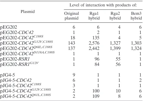
As a second test of the possibility that Rga2 is a GAP for Cdc42, we investigated the two-hybrid interactions between Rga2 and known GTPases of the Rho subfamily. Rga2 inter-acted with activated forms (G12V and Q61L) but not an inac-tive form (D118A) of Cdc42, as had previously been seen for Rga1 and Bem3 (63; Table 2). Rga2 failed to interact with Rho1, Rho2, Rho3, or Rho4, including activated versions of these proteins that could not be prenylated (data not shown). In addition, Rga1 and Rga2, but not Bem3, interacted with the GTPase, Rsr1, in both its wild-type and activated (G12V) forms (Table 2). This interaction pattern is distinct from the pattern seen for GTPases and their GAPs: GAPs usually in-teract preferentially with the active form of the GTPase. Rsr1 is a Ras-type GTPase involved in bud site selection and has been shown to interact with Cdc24 (49). Together, these ex-periments support the hypothesis that Rga1, Rga2, and Bem3 are all GAPs for Cdc42 and also suggest that Rga1 and Rga2 may have a connection with Rsr1.
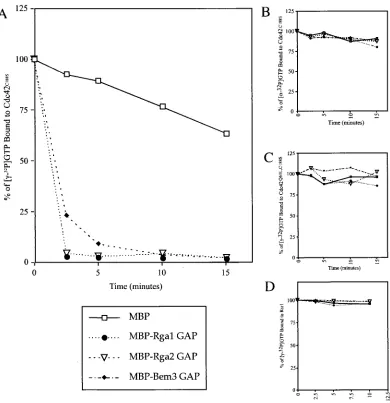
Rga1 and Rga2 have in vitro biochemical GAP activity.To expand on these genetic studies, we sought to determine whether Rga1 and Rga2 have biochemical GAP activity toward Cdc42; that is, we asked whether they accelerate GTP hydro-lysis by Cdc42. A fusion protein containing the Bem3 GAP domain joined to GST was previously shown to catalyze the hydrolysis of GTP to GDP when the nucleotide was bound to
Cdc42 (68). GST-Cdc42C188S was purified using glutathione
agarose beads and bound to [␥-32P]GTP (see Materials and
Methods), and the GAP domains of Rga1, Rga2, and Bem3 were purified as MBP fusions. When mixed with the
[␥-32P]GTP-loaded GST-Cdc42C188S, all three GAP domains
catalyzed the hydrolysis of GTP to GDP (Fig. 2A). After only 2.5 min, the GAP domains of Rga1 and Rga2 had catalyzed the
hydrolysis of over 94% of the GTP on Cdc42 (summary of multiple data sets). In contrast, incubation with MBP alone resulted in hydrolysis of less than 20% of the GTP after 2.5 min. Incubation with the Bem3 GAP domain resulted in
hy-drolysis of⬃75% of the GTP bound to Cdc42 after 2.5 min.
Thus, it appears that Rga1, Rga2, and Bem3 all catalyze GTP hydrolysis on Cdc42. However, Rga1 and Rga2 may do so more efficiently than Bem3.
To ensure that the loss of counts from the beads was due to GTP hydrolysis and not simply dissociation of the bound
nu-cleoside triphosphate, purified Cdc42C188S was bound to
[␣-32P]GTP instead of [␥-32P]GTP and then incubated with the
GAPs. If Rga1, Rga2, and Bem3 were to cause dissociation of
GTP rather than hydrolysis to GDP, [␣-32P]GTP would be
released in this experiment, resulting in a loss of bound radio-activity. In fact, very little radioactivity was released, indicating
that [␣-32P]GTP did not dissociate from Cdc42C188Swhen
in-cubated with the GAP domains of Rga1, Rga2, or Bem3 (Fig. 2B). The GAP assay was also performed with
GST-Cdc42Q61L,C188S. Since the Q61L substitution locks Cdc42 in
its GTP-bound, active state (70), this mutant should not be capable of GTP hydrolysis. Indeed, incubation with Rga1,
Rga2, or Bem3 did not cause hydrolysis of [␥-32P]GTP when
bound to Cdc42Q61L, C188S(Fig. 2C). These results show that
the GAP domains of Rga1, Rga2, and Bem3 catalyze the hydrolysis of GTP by Cdc42.
Motivated by the two-hybrid results discussed above (Table 2), biochemical GAP assays were performed to ask whether the GAP domain of Rga1, Rga2, and Bem3 could accelerate GTP hydrolysis by the bud-site selection GTPase, Rsr1. The presence of the Rga1, Rga2, or Bem3 GAP domains did not catalyze the hydrolysis of GTP bound to GST-Rsr1 (Fig. 2D). Thus, Rga1, Rga2, and Bem3 are GAPs for Cdc42 and, based on two-hybrid and biochemical evidence, it seems unlikely that they act as GAPs for Rsr1, Rho1, Rho2, Rho3, or Rho4.
Deletion of GAPs for Cdc42 alters cell morphology.Why are there three GAPs for Cdc42? One possibility is that Rga1, Rga2, and Bem3 are responsible for regulating distinct subsets of Cdc42 function. To explore this possibility, we examined the
phenotypes conferred by deletion ofRGA1,RGA2, andBEM3,
singly and in combination. We first observed cellular morphol-ogy, which, for ease of quantification, was divided into five classes: normal, elongated, peanut, fingered, and misshapen. Elongated cells were defined as cells whose length is greater than 1.5 times but less than three times, their width. Peanut cells appeared as two round or elongated cells with a smooth connection between them, reminiscent of two newly fused mat-ing cells. Cells of the fmat-ingered class had multiple growth pro-jections or had a length greater than three times their width. Misshapen cells were defined as cells of aberrant morphology that did not fit in any of the other classes. As previously
re-ported (63), somerga1⌬cells displayed an elongated cell
mor-phology (40% versus 9% for wild type; Table 3). No
morpho-logical aberrations were observed forrga2⌬cells. Interestingly,
a small percentage of thebem3⌬cells (8% versus⬍3% for the
wild type; Table 3) displayed severe morphological defects (peanut, fingered, and misshapen cells). The combination of
rga1⌬ and bem3⌬ deletions resulted in a high percentage
(60%) of cells with severe morphological defects. Deletion of
TABLE 2. Two-hybrid interactions of Rga1, Rga2, and Bem3, with Cdc42 and Rsr1a
Plasmid
Level of interaction with products of: Original
plasmid hybridRga1 hybridRga2 hybridBem3
pEG202 6 6 4 6
pEG202-CDC42 1 2 1 1
pEG202-CDC42C188S 18 135 4 5
pEG202-CDC42G12V,C188S 143 2,576 1,327 1,303 pEG202-CDC42Q6IL,C188S 137 2,442 1,399 1,324
pEG202-CDC42D118A,C188S 1 1 1 1
pEG202-RSR1 1 96 55 1
pEG202-RSR1G12V 1 84 56 1
pJG4-5 9 1 1 1
pJG4-5-CDC42 6 1 1 2
pJG4-5-CDC42C188S 3 1 1 1
pJG4-5-CDC42G12V,C188S 2 100 10 6
pJG4-5-CDC42Q61L,C188S 2 109 8 6
aProducts of plasmid pEG202 and its constructs interacted with products of
pJG4-5 and its constructs and vice versa.-Galactosidase activity was
deter-mined as described in Materials and Methods. The reported values are given in Miller units and are an average of three independent determinations.
on September 8, 2020 by guest
http://ec.asm.org/
rga2⌬from the single and double mutants increased slightly the severity of cell morphology defects in all cases.
Despite the high percentage of severe morphologies
ob-served inrga1⌬rga2⌬bem3⌬mutants, these cells were viable.
However, therga1⌬bem3⌬andrga1⌬rga2⌬bem3⌬deletion
strains both showed a growth defect at 30°C when compared to wild-type and single mutant strains (data not shown). The viability of the triple mutant may indicate that there are
addi-FIG. 2. GAP domains of Rga1, Rga2, and Bem3 display biochemical GAP activity for Cdc42, but not for Rsr1. The relative percentage of [␥-32P]GTP bound to GST-Cdc42C188S(A), [␣-32P]GTP bound to GST-Cdc42C188S(B), [␥-32P]GTP bound to GST-Cdc42Q61L, C188S(C), and [␥-32P]GTP bound to GST-Rsr1 when incubated with⬃2g of purified MBP, MBP-Rga1 GAP, MBP-Rga2 GAP, or MBP-Bem3 GAP (D). The ratio of released counts (32P
i) to counts still bound to protein is plotted as a function of time. (A) Shown is a representative graph from multiple data sets.
TABLE 3. Quantification of cell morphologies for deletions of the GAPs for Cdc42a
Strain Genotype % Normal % Elongated % Fingered % Peanut % Misshapen
SY2002 Wild type 90 9 ⬍1 ⬍1 ⬍1
YGS2 rga1⌬ 59 40 ⬍1 ⬍1 ⬍1
YGS7 rga2⌬ 90 9 ⬍1 ⬍1 ⬍1
YGS50 bem3⌬ 73 18 3 4 1
YGS51 rga1⌬rga2⌬ 49 49 ⬍1 ⬍1 ⬍1
YGS72 rga1⌬bem3⌬ 14 26 21 34 5
YGS56 rga2⌬bem3⌬ 90 21 4 10 3
YGS57 rga1⌬rga2⌬bem3⌬ 13 14 16 51 4
aAll strains are in the SY2002 background. More than 400 cells were counted for each strain.
on September 8, 2020 by guest
http://ec.asm.org/
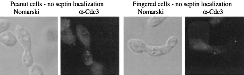
FIG. 3. Septin localization in normal, peanut, and fingered cells. Septins were visualized in the YGS57 (rga1⌬rga2⌬bem3⌬) strain. Nomarski optics was used to examine cell morphology and an␣-Cdc3 antibody was used to reveal septins. Localization patterns are shown for round, peanut, and fingered cells. See Table 4 for quantitation.
on September 8, 2020 by guest
http://ec.asm.org/
tional proteins with GAP activity for Cdc42 or that the inher-ent GTPase activity of Cdc42 is sufficiinher-ent for viability (27, 67). The localization of actin, chitin, and septin rings was exam-ined in peanut and fingered cells. Such cells had defects in chitin localization as revealed by Calcofluor staining. In par-ticular, these cells showed diffuse staining around the periph-ery in contrast to the rings of staining seen at previous bud sites in normal cells (data not shown). Immunofluorescence
micros-copy using the␣-Cdc3 antibody to highlight septins revealed
defects in the peanut and fingered cells (Fig. 3). Septins nor-mally localize as a double ring at the bud neck (19; for review, see reference 21; Fig. 3). However, the septins rings were improperly localized in some peanut and fingered cells (Fig. 3). For cells with large buds, 43% of peanut cells and 68% of fingered cells failed to properly localize their septins rings, compared to only 4% of round cells in the same strain (Table 4). In most of the cells displaying mislocalized septins, a single septin ring had formed on one side of the bud neck. Because many of these cells had not yet undergone nuclear division, the mother cell could be identified by the presence of the only nucleus. In all cases, this single septin ring was located on the daughter side of the bud neck (data not shown). Finally, no defects in actin localization were detected in the GAP mutants (data not shown). Thus, peanut and fingered cells were found to have defects in the organization of septins (Fig. 3; Table 4) and chitin (data not shown), but not actin (data not shown).
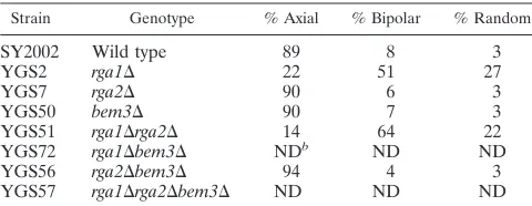
Bud site selection defects in GAP deletion strains.Wild-type haploid cells are round and bud in an axial pattern in which each new bud is adjacent to the site of the previous bud (10, 11). Using Calcofluor to visualize bud scars, we observed
non-axial budding patterns forrga1⌬cells. In toto, 78% of cells in
arga1⌬deletion strain displayed a nonaxial budding pattern
(Table 5; see also reference 63): 51% of therga1⌬cells had a
bipolar pattern in which buds were seen at both poles of the cell and 27% of cells showed a random budding pattern with bud scars at either or both poles and in the middle of the cell
(Table 5). In contrast,rga2⌬, bem3⌬, andrga2⌬bem3⌬cells
exhibited an axial pattern with all of the bud scars at one pole
(Table 5). Therga1⌬rga2⌬double mutant cells had a budding
pattern similar to that ofrga1⌬cells (Table 5). Budding
pat-terns forrga1⌬bem3⌬andrga1⌬rga2⌬bem3⌬cells could not
be accurately determined due to the aberrant cell morpholo-gies and diffuse chitin staining (see above). Thus, Rga1 has a distinct function in bud site selection that is not shared by Rga2 or Bem3.
Activation of FUS1-lacZreporter in GAP deletion strains.
RGA1was first identified in a screen for negative regulators of
the pheromone response pathway (63). It was shown thatste4⌬
rga1⌬cells activated aFUS1-lacZreporter construct (63). We
determined that deletion of RGA2 or BEM3 did not cause
activation of the FUS1-lacZ reporter (Table 6). Thus, the
rga1⌬ deletion appears to be unique in its ability to affect
FUS1-lacZ expression. Deletion of RGA2 in a rga1⌬ strain
caused a slight increase in FUS1-lacZ expression (38.9 for
ste4⌬rga1⌬versus 45.5 forste4⌬rga1⌬rga2⌬). However,
de-letion of all three GAP genes caused an eightfold increase in expression (Table 6). Thus, although Rga1 has a primary role
in controlling the activation ofFUS1-lacZ, Rga2, and Bem3
must also have a negative regulatory role, albeit a modest one.
Roles of Rga1, Rga2, and Bem3 in haploid invasive growth.
We also examined the roles of Rga1, Rga2, and Bem3 in haploid invasive growth, a process involving Cdc42 (46). These
experiments were done with the⌺1278b strain (provided by G.
Fink), a strain that exhibits more vigorous agar invasion than other lab strains. Two techniques were used to assess filamen-tous growth: the plate-washing assay (56), which assesses agar invasion, and the single-cell assay (16), which provides detailed information on cell morphology and budding patterns. By
the plate-washing assay, rga1⌬ cells displayed hyperinvasive
growth, whereasrga2⌬andbem3⌬cells invaded the agar at a
level similar to wild-type cells (Fig. 4). On glucose-rich
me-dium, rga1 mutant cells showed a propensity to bud at the
distal pole (17%), whereasbem3 (2% distal),rga2(1%), and
wild-type (1%) cells did not (Fig. 4b). Therga1mutant also had
an elongated cell morphology in glucose-rich medium; 45% of the cells were elongated compared to less than 1% for
wild-type and therga2mutant. Thebem3mutant also had elongated
cells (18%) on this medium, but the cells were not as elongated
TABLE 4. Quantification of septin localizationa
Strain Cell type % with normallocalization % with mislo-calization localization% with no
SY2002 Round 99 ⬍1 ⬍1
YGS57 Round 96 2 2
YGS57 Peanut 57 34 9
YGS57 Fingered 33 36 32
aSeptin staining was performed as described in the Materials and Methods.
Only cells with large buds were counted. More than 200 cells were counted for each class.
TABLE 5. Budding patterns of GAP deletion strainsa
Strain Genotype % Axial % Bipolar % Random
SY2002 Wild type 89 8 3
YGS2 rga1⌬ 22 51 27
YGS7 rga2⌬ 90 6 3
YGS50 bem3⌬ 90 7 3
YGS51 rga1⌬rga2⌬ 14 64 22
YGS72 rga1⌬bem3⌬ NDb ND ND
YGS56 rga2⌬bem3⌬ 94 4 3
YGS57 rga1⌬rga2⌬bem3⌬ ND ND ND
aBud scars were visualized using Calcofluor as described in Materials and
Methods. Only cells with three of more scars were counted. More than 200 cells were counted for each strain.
bND, not determined. Accurate counts could not be determined due to diffuse
chitin staining and abnormal cell morphologies
TABLE 6. Activation ofFUS1-lacZby deletion of GAPs for Cdc42
Strain Genotypea FUS1-lacZexpressionb
YGS58 ste4⌬ 1.2⫾0.1
YGS59 ste4⌬rga1⌬ 38.9⫾2.5
YGS60 ste4⌬rga2⌬ 1.8⫾0.5
YGS61 ste4⌬bem3⌬ 1.3⫾0.1
YGS62 ste4⌬rga1⌬rga2⌬ 45.5⫾2.8
YGS63 ste4⌬rga1⌬bem3⌬ 30.3⫾3.7
YGS64 ste4⌬rga2⌬bem3⌬ 3.9⫾1.2
YGS65 ste4⌬rga1⌬rga2⌬bem3⌬ 396.1⫾20.5
aAll strains are in the SY2002 background.
b-Galactosidase activity was determined as described in Materials and
Meth-ods. The reported values are given in 100⫻Miller units and are averages
(⫾standard deviation) of three determinations.
on September 8, 2020 by guest
http://ec.asm.org/
asrga1cells. On glucose-limited medium,rga1cells were
hy-perelongated compared to wild-type cells or rga2 and bem3
mutants, and the rga1 cells displayed a more exaggerated
unipolar budding pattern. Thus, among the GAPs, Rga1 ap-pears to play a unique role in regulating haploid invasive growth.
Genetic interactions between GAP proteins and Cdc42 ef-fectors.The data presented above suggest that the GAPs for Cdc42 play unique roles in septin organization (Bem3) and haploid invasive growth (Rga1). The p21-activated kinase (PAK) kinases Ste20 and Cla4 both interact with Cdc42 and are known to be important for these processes (17, 61). Ste20 is required for haploid invasive growth (56) whereas Cla4
in-fluences septin organization (4, 17). Furthermore,ste20⌬and
cla4⌬ deletions are synthetically lethal, implying that these
kinases share an essential activity (4, 17). It seemed plausible that these PAK kinases could link the GAPs for Cdc42 to the processes they mediate.
If the GAPs for Cdc42 facilitate the interaction between active Cdc42 and Ste20 or Cla4, the GAPs might exhibit ge-netic interactions with the protein kinases. We examined the
effect ofrga1⌬,rga2⌬, andbem3⌬deletions onste20⌬orcla4⌬
strains. The rga1⌬ cla4⌬ and rga2⌬ cla4⌬ strains displayed
synthetic temperature sensitivity at 37°C (Fig. 5A). The cells arrested with fattened bud necks, and some cells had multiple projections emanating from the cell body (data not shown). In
contrast, cells of thecla4⌬strain, thebem3⌬cla4⌬strain, and
all GAP single deletions grew well at this temperature (Fig. 5A; data not shown). Since Rga1, Rga2, and Ste20 each
dis-played synthetic interactions withcla4⌬deletions, we
hypoth-esize that they may play roles in the same cellular function.
Moreover, because BEM3did not show genetic interactions
withcla4⌬, we infer that the GAPs can have specific functions
with respect to effectors for Cdc42. However, we cannot rule out the possibility that these phenotypic differences reflect quantitative rather than qualitative differences in GAP activity.
RGA1, RGA2, and BEM3 also showed genetic interactions
withste20⌬. In this case, loss of any individual GAP for Cdc42 resulted in synthetic temperature sensitivity, though the effect
may be more pronounced forbem3⌬(Fig. 5B). These findings
suggest that the GAPs may coordinate Cla4 activity, perhaps via Cdc42.
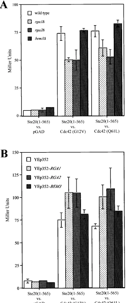
Deletion or overexpression ofRGA1,RGA2, andBEM3 af-fects interaction of Cdc42 and Ste20.To explore further the hypothesis that Rga1, Rga2, and Bem3 play differential roles in regulating Cdc42 function, we investigated the effects of their deletion or overexpression on the two-hybrid interaction be-tween Cdc42 and Ste20. Specifically, the interaction bebe-tween two activated forms of Cdc42 (G12V, C188S and Q61L, C188S) and a truncated form of Ste20 (provided by M. Keniry) was compared under a variety of conditions. The interaction was measured for four different strains: PJ69-4A, YGS156
(PJ69-4A rga1⌬), YGS157 (PJ69-4A rga2⌬), and YGS158
(PJ69-4Abem3⌬) (Fig. 6A). Deletion ofRGA1orRGA2
de-creased expression of thelacZreporter, suggesting a decrease
in the interaction between Ste20 and Cdc42. Loss ofBEM3had
no significant effect. This experiment suggests that Rga1 and Rga2 may facilitate the interaction between Ste20 and active Cdc42. In a complementary experiment, the interaction be-tween Ste20 and active Cdc42 was compared in strains over-expressing the GAPs for Cdc42. The PJ69-4A strain was
trans-formed with YEp352, pGS40 (YEp352-RGA1), pGS41
(YEp352-RGA2), or pGS42 (YEp352-BEM3). Overexpression
ofRGA1 orRGA2 increased the expression of the lacZ
re-porter, suggesting an increased interaction between Ste20 and the activated forms of Cdc42 (Fig. 6B). Together, these data suggest that Rga1 and Rga2 facilitate the two-hybrid interac-tion between Ste20 and Cdc42.
DISCUSSION
We have presented evidence that there are three distinct GAPs for Cdc42: Rga1, Rga2, and Bem3. Each of these pro-teins exhibited two-hybrid interactions with Cdc42, and genetic
FIG. 4. Haploid invasive growth phenotypes of strains lacking the Cdc42 GAPs. (A) Cells of the⌺1278b background were grown for 4 days at 30°C on synthetic medium. Plates were washed with water and rubbed with a wet finger. (B) Single-cell invasive growth assay. Equal concentrations of wild-type (wt),rga1,rga2, andbem3cells were spread onto SCD (⫹Glu) or SC (⫺Glu) medium, incubated for 16 h at 25°C, and photographed at 100⫻by placing a coverslip directly on the agar medium. All images were taken at the same scale. Bar, 30m.
on September 8, 2020 by guest
http://ec.asm.org/
experiments argued that their activity influences the amount of active Cdc42 in the cell. Moreover, each protein was shown to have GAP activity; that is, the ability to accelerate the hydro-lysis of GTP bound to Cdc42. The loss of individual GAPs conferred distinct phenotypes, suggesting that each GAP reg-ulates a subset of Cdc42 functions. Hence, these studies com-plement and extend previous studies that demonstrated that Bem3 has biochemical GAP activity against Cdc42 (68) and that suggested that Rga1 was a Cdc42 GAP (63).
Bem3 and, to a lesser extent, Rga1 and Rga2, play roles in septin organization. Phenotypic analysis suggests that Bem3 may moderate the function of Cdc42 in morphogenesis. This
conclusion is supported by the observation thatbem3⌬strains
produce peanut and fingered cells whereasrga1⌬ and rga2⌬
strains do not (Table 3). However, the double and triple
dele-tions, especially the rga1⌬ bem3⌬ and rga1⌬ rga2⌬ bem3⌬
strains, produce many more peanut and fingered cells than the single deletions, suggesting that Rga1 and Rga2 play a lesser role in the formation of these morphologies. Cells exhibiting the peanut and fingered morphologies were shown to have mislocalized septin rings and occasionally display no septin
localization, thus linking loss ofBEM3and, to a lesser extent,
RGA1andRGA2, to septin ring organization.
The finding that ste20⌬ rga1⌬, ste20⌬ rga2⌬, and ste20⌬
bem3⌬cells display synthetic temperature sensitivity (Fig. 5B)
also supports the idea that Bem3, Rga1, and Rga2 play roles in septin organization and/or bud neck morphogenesis. This sug-gestion is based on two pieces of information: Cla4 is involved
in the process of bud neck morphogenesis (4, 9, 41) andcla4⌬
and ste20⌬ are synthetically lethal (17). Thus, proteins that participate in the same process as Cla4 might also display
synthetic interactions with ste20⌬, as seen for deletions of
RGA1,RGA2, andBEM3. Thebem3⌬ste20⌬strain may have
a slightly greater defect than therga1⌬ste20⌬orrga2⌬ste20⌬
strains (see Fig. 5B), suggesting that Bem3 may play a more central role in mediating the function of Cdc42 and Cla4 in cytokinesis.
Unpublished results from E. Bi and J. Pringle also support the hypothesis that Rga1, Rga2, and Bem3 play roles in septin organization. They showed the level of expression of Cdc42
GAPs affects the temperature sensitivity of a cdc12-6strain.
Cdc12 is a component of the septin ring (41). Overexpression
ofCLA4,RGA1,RGA2, orBEM3, but notSTE20, raises the
restrictive temperature of acdc12-6strain. In addition,
dele-tion of any of the GAPs for Cdc42, but particularlybem3⌬,
lowers the restrictive temperature of acdc12-6strain (E. Bi
and J. R. Pringle, unpublished data). Thus, GAP function for Cdc42 appears to play a positive role in septin organization. Furthermore, immunolocalization showed that Rga1, Rga2, and Bem3 localize to the bud neck during late anaphase and early telophase (E. Bi and J. R. Pringle, unpublished data), consistent with a function in septin ring organization.
Finally, mutational analysis of Cdc42 also supports the hy-pothesis that Bem3 mediates the interaction of Cdc42 with Cla4. Point mutations in the effector domain of Cdc42 lead to a variety of phenotypes, often including abnormal cell mor-phologies and mislocalization of actin, chitin, and/or septins (37, 54, 55). Furthermore, some of these Cdc42 alleles display altered interactions with Cla4 and Bem3, but interactions with Rga1, Rga2, and Ste20 are not affected (54; T. J. Richman and
D. I. Johnson, unpublished data). Thus, mutational analyses provide additional support to link Bem3 and Cla4 in septin ring organization.
Role of Rga1 and Rga2 in haploid invasive growth and in regulating interaction between Cdc42 and Ste20.The
pheno-types of the rga1⌬ deletion strain imply a role for Rga1 in
haploid invasive growth. In particular, loss of Rga1 causes cells to display hyperinvasion, to elongate, and to exhibit a nonaxial budding pattern, each a characteristic of invasive growth (43).
In addition, activation of theFUS1-lacZreporter, previously
thought to be specific for pheromone response activation (63), may actually reflect activation of the Ste12/Tec1 transcription factor complex, another characteristic of haploid invasive growth (15, 48). This explanation is supported by the observa-tion that deleobserva-tion of the pheromone response pathway scaffold, Ste5, which has no role in invasive growth but is important for
pheromone response, does not affect theFUS1-lacZactivation
in anrga1⌬strain (63). Moreover, diminished interaction
be-FIG. 5. Temperature-dependant growth of strains lacking a PAK kinase and one of the Cdc42 GAPs. (A) Aliquots of 10l of a 1:100 dilution of IDY22 (cla4⌬), YGS80 (rga1⌬ cla4⌬), YGS81 (rga2⌬ cla4⌬), or YGS82 (bem3⌬cla4⌬) mid-log phase liquid cultures were spotted onto rich plates and incubated at the indicated temperatures. (B) Aliquots of 10l of a 1:1,000 and a 1:10,000 dilution of YGS 286 (ste20⌬), YGS 351 (rga1⌬ste20⌬), YGS 352 (rga2⌬ste20⌬), and YGS 353 (bem3⌬ste20⌬) mid-log phase liquid cultures were spotted onto rich plates and incubated at the indicated temperatures.
on September 8, 2020 by guest
http://ec.asm.org/
tween Ste20 and active Cdc42 had little effect on pheromone response pathway signaling and mating but did result in de-creased invasive growth (39, 50). Together, these findings sup-port the idea that Rga1 affects haploid invasive growth, possi-bly by mediating the interaction of active Cdc42 with Ste20.
A role for Rga1 in mediating the interaction between Cdc42 and Ste20 is also supported by the synthetic temperature
sen-sitivity ofrga1⌬andcla4⌬mutations (Fig. 5). Sincecla4⌬and
ste20⌬are synthetically lethal (17), it would follow that a pro-tein involved in the same process as Ste20 might also display
synthetic interactions with cla4⌬. Interestingly, rga2⌬ cla4⌬
cells, but notbem3⌬cla4⌬cells, also display synthetic
temper-ature sensitivity, suggesting that Rga2 might also play a role in mediating the interaction between Cdc42 and Ste20. These phenotypic differences may reflect different quantitative con-tributions to overall GAP activity by Rga1, Rga2, and Bem3. However, the phenotypic differences could also indicate that the GAPs have qualitatively different roles in orchestrating Cdc42 activity. Consistent with the proposed role of Rga1 and Rga2 as facilitators of the interaction between Cdc42 and
Ste20, deletion ofRGA1 orRGA2 decreased the two-hybrid
interaction between activated forms of Cdc42 with Ste20,
whereasRGA1orRGA2overexpression increased this
inter-action. These experiments suggest that Rga1 and Rga2 facili-tate the interaction between Cdc42 and Ste20. This function would appear to be distinct from the GAP activity of Rga1 and Rga2 since these two-hybrid experiments were performed with versions of Cdc42 locked in the GTP-bound, active form (70). How do Rga1 and Rga2 facilitate this interaction? Given that Rga1 and Rga2 have not been observed to interact with Ste20 by two-hybrid analysis (63; data not shown), perhaps these two GAPs do not directly promote an interaction between Ste20 and Cdc42. Rather, perhaps Rga1 and Rga2 prevent Cdc42 from interacting with other effectors or perhaps they recruit some unidentified protein that aids in this process.
ACKNOWLEDGMENTS
We thank April Goehring, Megan Keniry, Hay-Oak Park, Phil James, Brian Stevenson, Betsy Ferguson, Dave Mitchell, Erica Gole-mis, Gerald Fink, and Doug Johnson for providing advice, strains, and/or plasmids. Thanks to Erfei Bi, John Pringle, Tammy Richman, and Doug Johnson for sharing results before publication. We also thank David Rivers, Hilary Kemp, Jesse Dillon, and Phil Kinsey for helpful comments and suggestions.
This work was supported by research (GM-30027 to G.F.S) and training (GM-07413 to G.R.S., HD-07348 to S.A.G., and GM-19888 to P.C.) grants from the U.S. Public Health Service and by a fellowship from the American Heart Association (AHA1206352) to P.C.
REFERENCES
1.Adams, A. E., D. I. Johnson, R. M. Longnecker, B. F. Sloat, and J. R. Pringle.
1990.CDC42andCDC43, two additional genes involved in budding and the
establishment of cell polarity in the yeastSaccharomyces cerevisiae. J. Cell
Biol.111:131–142.
2.Archer, V. E., J. Breton, I. Sanchez-Garcia, H. Osada, A. Forster, A. J. Thomson, and T. H. Rabbitts.1994. Cysteine-rich LIM domains of LIM-homeodomain and LIM-only proteins contain zinc but not iron. Proc. Natl.
Acad. Sci. USA91:316–320.
3.Barthe, C., G. de Bettignies, O. Louvet, M. F. Peypouquet, C. Morel, F. Doignon, and M. Crouzet.1998. First characterization of the geneRGD1in
the yeastSaccharomyces cerevisiae. C. R. Acad. Sci. Ser. III321:453–462.
4.Benton, B. K., A. Tinkelenberg, I. Gonzalez, and F. R. Cross.1997. Cla4p, a Saccharomyces cerevisiaeCdc42p-activated kinase involved in cytokinesis, is
activated at mitosis. Mol. Cell. Biol.17:5067–5076.
5.Bi, E., J. B. Chiavetta, H. Chen, G. C. Chen, C. S. Chan, and J. R. Pringle.
2000. Identification of novel, evolutionarily conserved Cdc42p-interacting
FIG. 6. Effects of deletion or overexpression of Cdc42 GAPs on two-hybrid interaction between Ste20 and activated versions of Cdc42. (A) The interaction of Ste20 residues 1 to 565 fused to the Gal4 DBD (pSL2682) with three versions of the Gal4 transcription activation domain—GAD itself and GAD fused either to Cdc42G12V,C188S (pGS38) or to Cdc42Q61L,C188S(pGS39)—in four separate strains were compared: PJ69-4A, YGS156 (rga1⌬), YGS157 (rga2⌬), and YGS158 (bem3⌬). Expression of thelacZreporter in three separate isolates was measured. (B) The same two-hybrid interactions investigated above were compared for strain PJ69-4A transformed with four different plasmids: YEp352, pGS40 (YEp352-RGA1), pGS41 (YEp352-RGA2), and pGS42 (YEp352-BEM3). The expression of thelacZreporter was measured for three separate isolates in four independent trials. The combined normalized data were graphed.
on September 8, 2020 by guest
http://ec.asm.org/
proteins and of redundant pathways linking Cdc24p and Cdc42p to actin
polarization in yeast. Mol. Biol. Cell.11:773–793.
6.Boone, C., N. G. Davis, and G. F. Sprague, Jr.1993. Mutations that alter the third cytoplasmic loop of the a-factor receptor lead to a constitutive and
hypersensitive phenotype. Proc. Natl. Acad. Sci. USA90:9921–9925.
7.Brown, J. L., M. Jaquenoud, M. P. Gulli, J. Chant, and M. Peter.1997. Novel Cdc42-binding proteins Gic1 and Gic2 control cell polarity in yeast. Genes
Dev.11:2972–2982.
8.Burke, D., D. Dawson, and T. Stearns.2000. Methods in yeast genetics. Cold Spring Harbor Laboratory Press, Plainview, N.Y.
9.Carroll, C. W., R. Altman, D. Schieltz, J. R. Yates, and D. Kellogg.1998. The septins are required for the mitosis-specific activation of the Gin4 kinase.
J. Cell Biol.143:709–717.
10.Chant, J., and I. Herskowitz.1991. Genetic control of bud site selection in yeast by a set of gene products that constitute a morphogenetic pathway. Cell
65:1203–1212.
11.Chant, J., M. Mischke, E. Mitchell, I. Herskowitz, and J. R. Pringle.1995. Role of Bud3p in producing the axial budding pattern of yeast. J. Cell Biol.
129:767–778.
12.Chen, D. C., B. C. Yang, and T. T. Kuo.1992. One-step transformation of
yeast in stationary phase. Curr. Genet.21:83–84.
13.Chen, G. C., Y. J. Kim, and C. S. Chan.1997. The Cdc42 GTPase-associated
proteins Gic1 and Gic2 are required for polarized cell growth in
Saccharo-myces cerevisiae. Genes Dev.11:2958–2971.
14.Chen, G. C., L. Zheng, and C. S. Chan.1996. The LIM domain-containing Dbm1 GTPase-activating protein is required for normal cellular
morpho-genesis inSaccharomyces cerevisiae. Mol. Cell. Biol.16:1376–1390.
15.Cullen, P. J., J. Schultz, J. Horecka, B. J. Stevenson, Y. Jigami, and G. F. Sprague, Jr.2000. Defects in protein glycosylation causeSHO1-dependent
activation of aSTE12signaling pathway in yeast. Genetics155:1005–1018.
16.Cullen, P. J., and G. F. Sprague, Jr.2000. Glucose depletion causes haploid
invasive growth in yeast. Proc. Natl. Acad. Sci. USA97:13619–13624.
17.Cvrckova, F., C. De Virgilio, E. Manser, J. R. Pringle, and K. Nasmyth.1995. Ste20-like protein kinases are required for normal localization of cell growth
and for cytokinesis in budding yeast. Genes Dev.9:1817–1830.
18.Dawid, I. B., J. J. Breen, and R. Toyama.1998. LIM domains: multiple roles as adapters and functional modifiers in protein interactions. Trends Genet.
14:156–162.
19.DeMarini, D. J., A. E. Adams, H. Fares, C. De Virgilio, G. Valle, J. S. Chuang, and J. R. Pringle.1997. A septin-based hierarchy of proteins
re-quired for localized deposition of chitin in theSaccharomyces cerevisiaecell
wall. J. Cell Biol.139:75–93.
20.Doignon, F., C. Weinachter, O. Roumanie, and M. Crouzet.1999. The yeast Rgd1p is a GTPase activating protein of the Rho3 and Rho4 proteins. FEBS
Lett.459:458–462.
21.Field, C. M., and D. Kellogg.1999. Septins: cytoskeletal polymers or
signal-ling GTPases? Trends Cell Biol.9:387–394.
22.Fields, S., and O. Song.1989. A novel genetic system to detect
protein-protein interactions. Nature340:245–246.
23.Freyd, G., S. K. Kim, and H. R. Horvitz.1990. Novel cysteine-rich motif and
homeodomain in the product of theCaenorhabditis eleganscell lineage gene
lin-11. Nature344:876–879.
24.Gietz, R. D., R. H. Schiestl, A. R. Willems, and R. A. Woods.1995. Studies on the transformation of intact yeast cells by the LiAc/SS-DNA/PEG
pro-cedure. Yeast11:355–360.
25.Guan, C., P. Li, P. D. Riggs, and H. Inouye.1987. Vectors that facilitate the
expression and purification of foreign peptides inEscherichia coliby fusion
to maltose-binding protein. Gene67:21–30.
26.Gyuris, J., E. Golemis, H. Chertkov, and R. Brent.1993. Cdi1, a human G1
and S phase protein phosphatase that associates with Cdk2. Cell75:791–803.
27.Hart, M. J., K. Shinjo, A. Hall, T. Evans, and R. A. Cerione.1991. Identi-fication of the human platelet GTPase activating protein for the CDC42Hs
protein. J. Biol. Chem.266:20840–20848.
28.Hill, J. E., A. M. Myers, T. J. Koerner, and A. Tzagoloff.1986. Yeast/E. coli
shuttle vectors with multiple unique restriction sites. Yeast2:163–167.
29.Ijzerman, M. M., J. O. Falkinham III, and C. Hagedorn.1993. A liquid, colorimetric presence-absence coliphage detection method. J. Virol.
Meth-ods45:229–233.
30.James, P., J. Halladay, and E. A. Craig.1996. Genomic libraries and a host strain designed for highly efficient two-hybrid selection in yeast. Genetics
144:1425–1436.
31.Jarvis, E. E., D. C. Hagen, and G. F. Sprague, Jr.1988. Identification of a
DNA segment that is necessary and sufficient for␣-specific gene control in
Saccharomyces cerevisiae: implications for regulation of␣-specific anda
-spe-cific genes. Mol. Cell. Biol.8:309–320.
32.Johnson, D. I.1999. Cdc42: an essential Rho-type GTPase controlling
eu-karyotic cell polarity. Microbiol. Mol. Biol. Rev.63:54–105.
33.Johnson, D. I., C. W. Jacobs, J. R. Pringle, L. C. Robinson, G. F. Carle, and M. V. Olson.1987. Mapping of theSaccharomyces cerevisiae CDC3,CDC25, andCDC42genes to chromosome XII by chromosome blotting and tetrad
analysis. Yeast3:243–253.
34.Johnson, D. I., and J. R. Pringle. 1990. Molecular characterization of
CDC42, aSaccharomyces cerevisiaegene involved in the development of cell
polarity. J. Cell Biol.111:143–152.
35.Karlsson, O., S. Thor, T. Norberg, H. Ohlsson, and T. Edlund.1990. Insulin gene enhancer binding protein Isl-1 is a member of a novel class of proteins
containing both a homeo- and a Cys-His domain. Nature344:879–882.
36.Kim, H. B., B. K. Haarer, and J. R. Pringle.1991. Cellular morphogenesis in theSaccharomyces cerevisiaecell cycle: localization of theCDC3gene
prod-uct and the timing of events at the budding site. J. Cell Biol.112:535–544.
37.Kozminski, K. G., A. J. Chen, A. A. Rodal, and D. G. Drubin.2000. Functions
and functional domains of the GTPase Cdc42p. Mol. Biol. Cell11:339–354.
38.Leberer, E., D. Dignard, D. Harcus, D. Y. Thomas, and M. Whiteway.1992. The protein kinase homologue Ste20p is required to link the yeast phero-mone response G-protein beta gamma subunits to downstream signalling
components. EMBO J.11:4815–4824.
39.Leberer, E., C. Wu, T. Leeuw, A. Fourest-Lieuvin, J. E. Segall, and D. Y. Thomas.1997. Functional characterization of the Cdc42p binding domain of
yeast Ste20p protein kinase. EMBO J.16:83–97.
40.Liu, X., H. Wang, M. Eberstadt, A. Schnuchel, E. T. Olejniczak, R. P. Meadows, J. M. Schkeryantz, D. A. Janowick, J. E. Harlan, E. A. Harris, D. E. Staunton, and S. W. Fesik.1998. NMR structure and mutagenesis of the N-terminal Dbl homology domain of the nucleotide exchange factor
Trio. Cell95:269–277.
41.Longtine, M. S., H. Fares, and J. R. Pringle.1998. Role of the yeast Gin4p protein kinase in septin assembly and the relationship between septin
as-sembly and septin function. J. Cell Biol.143:719–736.
42.Ma, H., S. Kunes, P. J. Schatz, and D. Botstein.1987. Plasmid construction
by homologous recombination in yeast. Gene58:201–216.
43.Madhani, H. D., and G. R. Fink.1997. Combinatorial control required for
the specificity of yeast MAPK signaling. Science275:1314–1317.
44.Maina, C. V., P. D. Riggs, A. G. I. Grandea, B. E. Slatko, L. S. Moran, J. A. Tagliamonte, L. A. McReynolds, and C. Guan.1988. A vector to express and
purify foreign proteins inEscherichia coliby fusion to, and separation form,
maltose-binding protein. Gene74:365–373.
45.Michelsen, J. W., K. L. Schmeichel, M. C. Beckerle, and D. R. Winge.1993. The LIM motif defines a specific zinc-binding protein domain. Proc. Natl.
Acad. Sci. USA90:4404–4408.
45a.Miller, P. J., and D. I. Johnson.1997. Characterization of theSaccharomyces cerevvisiae cdc42-1 allele and new temperature-conditional-lethalcdc42
al-leles. Yeast13:561–572.
46.Mosch, H. U., T. Kohler, and G. H. Braus.2001. Different domains of the
essential GTPase Cdc42p required for growth and development of
Saccha-romyces cerevisiae. Mol. Cell. Biol.21:235–248.
47.Nilsson, B., L. Abrahmsen, and M. Uhlen.1985. Immobilization and purifi-cation of enzymes with staphylococcal protein A gene fusion vectors. EMBO
J.4:1075–1080.
48.O’Rourke, S. M., and I. Herskowitz.1998. The Hog1 MAPK prevents cross
talk between the HOG and pheromone response MAPK pathways in
Sac-charomyces cerevisiae. Genes Dev.12:2874–2886.
49.Park, H. O., E. Bi, J. R. Pringle, and I. Herskowitz.1997. Two active states of the Ras-related Bud1/Rsr1 protein bind to different effectors to determine
yeast cell polarity. Proc. Natl. Acad. Sci. USA94:4463–4468.
50.Peter, M., A. M. Neiman, H. O. Park, M. van Lohuizen, and I. Herskowitz.
1996. Functional analysis of the interaction between the small GTP binding
protein Cdc42 and the Ste20 protein kinase in yeast. EMBO J.15:7046–7059.
51.Pringle, J. R., R. A. Preston, A. E. Adams, T. Stearns, D. G. Drubin, B. K. Haarer, and E. W. Jones.1989. Fluorescence microscopy methods for yeast.
Methods Cell Biol.31:357–435.
52.Printen, J. A., and G. F. Sprague, Jr.1994. Protein-protein interactions in the yeast pheromone response pathway: Ste5p interacts with all members of
the MAP kinase cascade. Genetics138:609–619.
53.Ramer, S. W., and R. W. Davis.1993. A dominant truncation allele identifies
a gene,STE20, that encodes a putative protein kinase necessary for mating
inSaccharomyces cerevisiae. Proc. Natl. Acad. Sci. USA90:452–456. 54.Richman, T. J., and D. I. Johnson.2000.Saccharomyces cerevisiaeCdc42p
GTPase is involved in preventing the recurrence of bud emergence during
the cell cycle. Mol. Cell. Biol.20:8548–8559.
55.Richman, T. J., M. M. Sawyer, and D. I. Johnson.1999. The Cdc42p GTPase is involved in a G2/M morphogenetic checkpoint regulating the
apical-iso-tropic switch and nuclear division in yeast. J. Biol. Chem.274:16861–16870.
56.Roberts, R. L., and G. R. Fink.1994. Elements of a single MAP kinase
cascade inSaccharomyces cerevisiaemediate two developmental programs in
the same cell type: mating and invasive growth. Genes Dev.8:2974–2985.
57.Rose, A. B., and J. R. Broach.1990. Propagation and expression of cloned
genes in yeast: 2-microns circle-based vectors. Methods Enzymol.185:234–
279.
58.Rose, M. D., F. Winston, and P. Hieter.1990. Methods in yeast genetics. Cold Spring Harbor Laboratory Press, Plainview, N.Y.
59.Sambrook, J., E. F. Fritsch, and T. Maniatis.1989. Molecular cloning: a laboratory manual, 2nd ed. Cold Spring Harbor Laboratory Press, Cold Spring Harbor, N.Y.
60.Schmidt, A., M. Bickle, T. Beck, and M. N. Hall.1997. The yeast
on September 8, 2020 by guest
http://ec.asm.org/
tidylinositol kinase homologTOR2activatesRHO1andRHO2via the
ex-change factorROM2. Cell88:531–542.
61.Simon, M. N., C. De Virgilio, B. Souza, J. R. Pringle, A. Abo, and S. I. Reed.
1995. Role for the Rho-family GTPase Cdc42 in yeast mating-pheromone
signal pathway. Nature376:702–705.
62.Sloat, B. F., A. Adams, and J. R. Pringle.1981. Roles of theCDC24gene
product in cellular morphogenesis during theSaccharomyces cerevisiaecell
cycle. J. Cell Biol.89:395–405.
63.Stevenson, B. J., B. Ferguson, C. De Virgilio, E. Bi, J. R. Pringle, G. Am-merer, and G. F. Sprague, Jr.1995. Mutation ofRGA1, which encodes a putative GTPase-activating protein for the polarity-establishment protein
Cdc42p, activates the pheromone-response pathway in the yeast
Saccharo-myces cerevisiae. Genes Dev.9:2949–2963.
64.Uetz, P., L. Giot, G. Cagney, T. A. Mansfield, R. S. Judson, J. R. Knight, D. Lockshon, V. Narayan, M. Srinivasan, P. Pochart, A. Qureshi-Emili, Y. Li, B. Godwin, D. Conover, T. Kalbfleisch, G. Vijayadamodar, M. Yang, M. Johnston, S. Fields, and J. M. Rothberg.2000. A comprehensive analysis of
protein-protein interactions in Saccharomyces cerevisiae. Nature403:623–
627.
65.Watanabe, D., M. Abe, and Y. Ohya.2001. Yeast Lrg1p acts as a specialized
RhoGAP regulating 1,3-beta-glucan synthesis. Yeast18:943–951.
66.Way, J. C., and M. Chalfie.1988. mec-3, a homeobox-containing gene that
specifies differentiation of the touch receptor neurons inC.elegans. Cell
54:5–16.
67.Zheng, Y., R. Cerione, and A. Bender.1994. Control of the yeast bud-site
assembly GTPase Cdc42. J. Biol. Chem.269:2369–2372.
68.Zheng, Y., M. J. Hart, K. Shinjo, T. Evans, A. Bender, and R. A. Cerione.
1993. Biochemical comparisons of theSaccharomyces cerevisiaeBem2 and
Bem3 proteins. Delineation of a limit Cdc42 GTPase-activating protein
domain. J. Biol. Chem.268:24629–24634.
69.Ziman, M., and D. I. Johnson.1994. Genetic evidence for a functional
interaction between Saccharomyces cerevisiae CDC24andCDC42. Yeast
10:463–474.
70.Ziman, M., J. M. O’Brien, L. A. Ouellette, W. R. Church, and D. I. Johnson.
1991. Mutational analysis of CDC42Sc, aSaccharomyces cerevisiaegene that
encodes a putative GTP-binding protein involved in the control of cell
polarity. Mol. Cell. Biol.11:3537–3544.