C A S E R E P O R T
Open Access
Paediatric T-cell lymphoma of the appendix: a
case report
Yoshifumi Matsushita
1and Morishige Takeshita
2*Abstract
A 7-year-old boy with no history of malnutrition or diarrhoea complained of acute abdominal pain, was diagnosed with acute appendicitis, and underwent appendectomy. Histologically, a diffuse infiltrate of large atypical lymphoid cells was found in the entire appendiceal wall. Immunohistochemical examination revealed that the tumour cells expressed T-cell receptor (TCR)-βF1, CD3, CD4, CD25, cytotoxic-related protein TIA1 and granzyme-B, but were negative for CD8, Foxp3, CD20, CD30 and CD56. Polymerase chain reaction (PCR) revealed clonal bands of TCR-γ gene products in the tumour tissue. No anti-cytomegalovirus antibody-positive cells were detected. In situ hybridization revealed no nuclear signals of Epstein-Barr virus (EBV)-encoded RNA.Helicobacter pyloriinfection was detected in tumour tissue by anti-East Asian cytotoxin-associated gene (Cag) A antibody and PCR using its specific primers. The patient received chemotherapy and has remained in remission for 2 years. To the best of our
knowledge, only two cases of appendiceal T-cell non-Hodgkin lymphoma (NHL) have been reported, both in elderly patients. We believe that this is the first reported case of childhood CD4- and TIA1-positive cytotoxic T (Th1)-cell NHL in the appendix or gastrointestinal tract.Helicobacter pyloriinfection might be an initiator of atypical cytotoxic T-cell proliferation.
Virtual Slides:The virtual slide(s) for this article can be found here: http://www.diagnosticpathology.diagnomx.eu/ vs/1302380563830412.
Keywords:T-cell lymphoma, Appendix, Child
Background
The specification of non-Hodgkin lymphoma (NHL) in the “WHO Classification 2008” is based on recent pro-gress in immunohistochemical and genetic analysis and clinical findings, including prognostic data [1]. Among extranodal NHLs, the alimentary tract is the most fre-quently affected site [1,2]. In the small intestine, diffuse large B-cell NHL (DLBCL) is the most common subtype of lymphoma, followed by mucosa-associated lymphoid tissue (MALT) lymphoma in the elderly, while Burkitt’s lymphoma is the predominant childhood NHL. Appen-diceal involvement is extremely rare, constituting less than 1% of all small- and large-intestinal NHLs [2,3]. About 50 cases of appendiceal NHL have been previ-ously reported, but immunohistochemical and genetic examinations have been performed in only a limited
number of recent cases, including two T/natural killer (NK)-cell NHL cases [2-5]. Among intestinal T/NK-cell NHLs, enteropathy-associated T-cell lymphoma (EATL) has been frequently reported in the elderly [1,6-8]. Type I EATL is a CD4- and CD8-negative and CD30-positive large-cell lymphoma associated with coeliac disease, and is seen in northern Europe and the United States. Type II EATL is a CD56-positive and CD8-positive or -nega-tive medium-sized lymphoma that is less strongly asso-ciated with coeliac disease. There are several reports of nasal-type NK-cell lymphomas with Epstein-Barr virus (EBV) infection in intestine and colon, which are usually encountered in the elderly and rarely in children [7,9]. The present case is a CD3-, CD4- and TIA1-positive and CD30-negative (Th1) large-cell lymphoma without EBV infection. CD4-positive T/NK-cell lymphoma has occa-sionally been reported in the stomach, but is rarely seen in the intestine [10,11]. The intestinal bacterial florae,
Helicobacter pylori and Campylobacter jejuni, may be initiators of abnormal lymphocytic proliferation in the * Correspondence:[email protected]
2
Department of Pathology, Faculty of Medicine, Fukuoka University, Nanakuma 7-45-1, Jonan-ku, Fukuoka 814-0180, Japan
Full list of author information is available at the end of the article
stomach and intestine [12,13]. Here, we present what is, to the best of our knowledge, the first report of a child-hood case of appendiceal CD4-positive T-cell NHL and discuss the influence ofH. pyloriinfection.
Case presentation Clinical history
A 7-year-old boy was referred to our hospital with com-plaints of abdominal discomfort and high fever. Four days before admission, the patient complained of uneasiness and sneezing and was afebrile. On the following day, the patient complained of increasing abdominal pain and ap-petite loss. One day before admission, the symptoms wor-sened and the patient’s temperature rose to 39.1°C. There was no history of recurrent diarrhoea, malnutrition or fail-ure to thrive. On admission, the white blood cell count remained within normal limits, but C-reactive protein was elevated to a concentration of 3.6 mg/dL. An abdominal ultrasound revealed a mildly swollen appendix. In addition, a few mildly swollen lymph nodes, up to 10 mm in diameter, were seen in the mesentery. A diagnosis of acute appendicitis was made and appendectomy was per-formed on the second day of hospitalisation.
Material and methods Immunohistochemistry
The antibodies used in this study were as follows:
TCR-βF1 (Endogen, Rockford, IL, USA); CD3, CD5, CD7, CD8, CD25, CD56, CD57, and terminal deoxynucleotidyl trans-ferase (TdT) (Novocastra, Newcastle, UK); CD4 (MBL, Nagoya, Japan); Foxp3 (e-Bioscience, San Diego, CA, USA); TIA-1 (Immunotech, Marseille, France); Granzyme B (Chemicon, Temecula, CA, USA); CD20 (Nichirei, Tokyo, Japan); and CD79a, CD30, CD15, anaplastic lymph-oma kinase (ALK), myeloperoxidase, epithelial membrane
antigen (EMA), AE1/AE3 and anti-cytomegalovirus
(CMV) antibody (Dako Cytomation, Glostrup, Denmark). Anti-East AsianH. pylori CagA antibody was kindly pro-vided by Dr. T. Uchida, Department of Molecular Medicine, Oita University, Japan [14].
In situ hybridisation for detection of Epstein-Barr virus (EBV)-encoded RNAs
Tissue sections were digested with proteinase K and incubated in a solution of 50% formamide containing digoxigenin/biotin-labelled EBV-encoded RNA (EBER) oligonucleotide probes (Dako Cytomation). A peroxidase-conjugated anti-FITC antibody was applied to the sections to detect the hybridized probes.
Polymerase chain reaction (PCR) for T-cell receptor
(TCR)-γandH. pylori-associated genes
For evaluations of genes associated with TCR-γ and H.
pylori, DNA was extracted from paraffin-embedded
tumour sections. TCR-γ gene analysis was performed according to the BIOMED II PCR method [15].
The East Asian-type CagA genes were detected using pri-mer sets CAGJF/CAGTR and CAGTF/CAGJR, which yielded 222- and 293- to 299-bp products, respectively [16].
Results Pathology
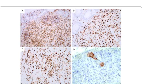
Macroscopically, the excised appendix was 4.0 × 0.8 cm in size, with an eroded mucosal layer and a haemorrhage on the serosal surface. However, there was no evidence of tumour formation or perforation. Microscopically, the mucosal layer appeared severely eroded with remnants of mucosal tissue. Intraepithelial lymphocytes (IELs) were not increased in number and lymphoid follicles with germinal centres were preserved (Figure 1A). Atyp-ical large lymphoid cells with round nuclei were found mainly in the submucosal and muscle layers (Figure 1B). Extensive and diffuse invasion by large atypical lympho-cytes can be seen in the eroded lesion. A severe histiocy-tic reaction involving many phagocyhistiocy-tic macrophages was noted in the area of tumour cell invasion (Figure 1C, 1D). No definite granuloma formation was detected in the sections examined. Based on these findings, malig-nant lymphoma, rather than epithelial tumour, was highly suspected. The ileal tissue excised during append-ectomy revealed oedematous mucosa and no infiltrating atypical cells.
Immunohistochemistry and genetic analysis
Immunohistochemical analysis revealed that the atypical tumour cells were positive for CD3 (Figure 2A),
TCR-βF1, CD4 (Figure 2B), CD5, CD7, CD25, cytotoxicity-related protein TIA-1 (Figure 2C) and granzyme B, but
were negative for TCR-CγM1, CD8, Foxp3, CD15,
Figure 1Histological features of the appendix. (A)Atypical lymphoid cells and preserved lymph follicle are in submucosal layer (H&E stain, ×100). (B)Intraepithelial lymphocytes are not increased, and abnormal tumour cell invasion is not prominent in the mucosal layer (H&E stain, ×200). Large atypical lymphocytes with small distinct nuclei diffusely infiltrate the (C) submucosal layer and (D) eroded lesion. Many reactive histiocytes are seen, mainly in (C) (H&E stain, ×400).
and 293 to 299 bp) of the East Asian CagA genes were detected by two probes (Figure 4).
Follow-up clinical data and history
Serum interleukin-2 receptor (sIL-2R) was 2,451 U/mL in a pre-surgical blood sample, which decreased to 1,089 U/mL 1 week after appendectomy. Anti-EBV antibodies had not increased, and anti-human T-lymphotropic virus-1 (HTLV-1) antibody was negative. No evaluation of serum anti-H. pylori antibodies was performed. The patient was transferred to another hospital with a paediatric
haematology facility for intensive chemotherapy, and has been in remission for 2 years.
Discussion
A study by Gustafsson et al. of 2,757 appendiceal tumours included 47 NHLs; immunological studies were performed in 11 cases and all were DLBCL. Several cases of appendiceal MALT-type lymphoma, mantle cell lymphoma and Burkitt’s lymphoma have been reported [17-19]. The present case was diagnosed as CD3-, CD4-, CD5-, CD7-, CD25- and TIA1-positive cytotoxic T-cell NHL. In childhood, ALK-positive anaplastic large cell lymphoma is a major type of CD4- and TIA1-positive cytotoxic T/NK-cell lymphoma. In the present case, this diagnosis was unlikely, because there was no expression of CD30, ALK and EMA [1]. Most intestinal T/NK-cell NHL are EATL, especially in the jejunum. Patients with EATL usually complain of diarrhoea, malnutrition and abdominal pain [1,6]. Type I EATL is a CD4- and CD8-negative and CD30-positive large-cell NHL. Type II EATL is a CD4-negative, CD8-positive or -negative and CD56-positive medium-sized NHL [1,8,11]. In addition, EATL expresses CD7 and TIA1, and is negative for CD4, CD5 and CD25. The current patient had no history of recurrent diarrhoea and malnutrition, which are both frequently found in cases of coeliac and Crohn’s diseases. Intraepithelial lymphocytes (IELs), which are typical for EATLs, were not found in the present case. Weiss et al. [20] reported on a 6-year-old patient with NK cell-like T-cell lymphoma restricted to the jejunum; the tumour cells were positive for CD3 and CD56 and negative for CD4, CD8 and CD30, and there was no EBV infection, similar to type II EATL. Considered together, these find-ings suggest that our patient’s lesion had clinicopatholo-gical and phenotypic characteristics different from those of EATL.
Primary T-cell NHL involving the appendix has previ-ously been reported in two elderly patients. Kitamura et al. [4] reported on a case of T/NK-cell NHL in an 84-year-old male. In their study, tumour cells expressed CD3, CD8 and granzyme-B, but were negative for EBV infection. Another case was a 45-year-old male who had received a renal transplant 17 years earlier and had sub-sequently developed CD56-positive nasal-type EBV-positive large T/NK-cell lymphoma [5]. The two previ-ously reported cases of appendiceal T-cell NHL occurred in adults. Therefore, the authors believe that this is the first reported case of childhood CD4- and TIA-1-positive cytotoxic T-cell lymphoma in the appendix, or, indeed, in the entire gastrointestinal tract.
This patient received cytotoxic treatment and has been in remission for 2 years. Chuang et al. [7] evaluated 24 cases of primary T-cell NHL and 6 cases of NK-cell NHL in the gastrointestinal tract [1]. According to their
Figure 3Detection of TCR-Vγ1f to -Jγ1.1/2.1 gene products. C1: nodal adult T-cell leukaemia/lymphoma (positive control); C2: non-neoplastic intestinal specimen (negative control); Pat.: patient sample. The 230 bp clonal band (TCR-VγtoJγ) is identified in lane C1 and in the patient sample.
report, using univariate and multivariate COX propor-tional hazard regression analysis, NK-cell lineage was associated with poor prognosis. EBV infection plays an important role in the progression of various NHLs [21]. We speculate that the early clinical stage and EBV-free status of the current patient predicted better prognosis. However, this is a single case and the follow-up period was limited. Identification of additional cases of intes-tinal T/NK-cell NHL and long-term follow-up is neces-sary in order to fully understand the clinical features of appendiceal T/NK-cell NHL.
In Japan, gastric carcinoma and MALT-type lymphoma have higher incidences compared with those occurring in other regions of the world [22]. It was strongly sug-gested that the East Asian CagA gene and protein have a great influence on the tumourigenesis of these two disorders [14,15]. Kiriya et al. [23] demonstrated that the T-cell reaction against the captured, round-shaped H.
pylori seen in dendritic cells of Peyer’s patches in the small intestine plays a critical role in H. pylori gastritis. CD4-positive T cells, including Th1 and regulatory T cells, are distributed in the gastric mucosa in H. pylori infection [24], and cases of primary CD4-, CD5-, CD25-and TIA1-positive cytotoxic T-cell lymphoma have been reported in the stomach [10]. Among CD4-positive T cells, neoplastic cells of the present case had phenotypic findings regarding TIA1 expression similar to those of Th1 effector cells [25]. Helicobacter pylori infection might play a role in abnormal proliferation of CD4-positive cytotoxic T (Th1) cells. However, although Küpeli et al. [26] in Turkey reported that 3 of 15 cases (20%) of childhood systemic NHL had serologicalH.
pyl-ori infection and that 2 cases were T-cell type ALCL, they suggested thatH. pylori infection was not an agent responsible for lymphomagenesis.
Conclusion
We present a rare paediatric case of appendiceal CD3-, CD4- and TIA1-positive cytotoxic T (Th1)-cell lymph-oma. Further studies are necessary to examine the rela-tionships between H. pylori infection, including the Asian variety, and NHL.
Competing interests
The authors declare that they have no conflicts of interest.
Authors’contributions
YM carried out initial pathological diagnosis of this case. YM and MT participated in the sequence alignment and drafted the manuscript. Both authors read and approved the final manuscript.
Acknowledgements
The authors thank Dr. Hiroaki Kumazawa, Department of Surgery, and Dr. Hiroshi Kobayashi, Department of Paediatrics, Chidoribashi Hospital, for patient care. Written informed consent was obtained from his family for publication and any accompanying images.
Author details 1
Laboratory of Pathology, Chidoribashi Hospital, 5-18-1 Chiyo, Hakata-ku, Fukuoka 812-8633, Japan.2Department of Pathology, Faculty of Medicine,
Fukuoka University, Nanakuma 7-45-1, Jonan-ku, Fukuoka 814-0180, Japan.
Received: 20 October 2012 Accepted: 13 December 2012 Published: 9 January 2013
References
1. Swerdlow SH, Campo E, Harris NL, Jaffe ES, Pileri SA, Stein H, Thiele J, Vardiman JW:WHO classification of tumours of haematopoietic and lymphoid tissues. Lyon: International Agency for Research on Cancer; 2008. 2. Gustafsson BI, Siddique L, Chan A, Dong M, Dorozdov I, Kidd M, Modlin IM:
Uncommon cancers of the small intestine, appendix and colon: An analysis of SEER 1973–2004, and current diagnosis and therapy.Int J Oncol2008,33:1121–1131.
3. O’Donnell ME, Badger SA, Beattie GC, Carson J, Garstin WIH:Malignant neoplasms of the appendix.Int J Colorectal Dis2007,22:1239–1248. 4. Kitamura Y, Ohta T, Terada T:Primary T-cell non-Hodgkin's malignant
lymphoma of the appendix.Pathol Int2000,50:313–317.
5. Ratuapli SK, Murarka S, Miller KA, Ferraro JC, Zafar H:Epstein-Barr virus-positive large T-cell lymphoma presenting an acute appendicitis 17 years after cadaveric renal transplant: a case report.J Med Case Reports2011,5:5.
6. Dalabie J, Holte H, Vose JM, Ullrich F, Jaffe ES, Savage KJ, Connors JM, Rimsza L, Harris NL, Müller-Hermelink K,et al:Enteropathy-associated T-cell lymphoma: clinical and histological findings from the international peripheral T-cell lymphoma project.Blood2011,118:148–155.
7. Chuang S-S, Chang S-T, Chuang W-Y, Huang W-T, Hsieh P-P, Tsou M-H, Liao Y-L, Lin S-H, Hsieh Y-H, Lu C-L,et al:NK-cell lineage predicts poor survival in primary intestinal NK-cell and T-cell lymphoma.Am J Surg Pathol2009,
33:1230–1240.
8. Takeshita M, Nakamura S, Kikuma K, Nakayama Y, Nimura S, Yao T, Urabe S, Ogawara S, Yonemasu H, Matsushita Y,et al:Pathological and
immunohistological findings and genetic aberrations of intestinal enteropathy-associated T-cell lymphoma in Japan.Histopathol2011,
58:395–407.
9. Sun J, Lu Z, Yang D, Chen J:Primary intestinal T-cell and NK-cell lymphomas: a clinicopathological and molecular study from China focused on type II enteropathy-associated T-cell lymphoma and primary NK-cell lymphoma.Mod Pathol2011,24:983–992.
10. Kawamoto K, Nakamura S, Iwashita A, Watanabe J, Oshiro Y, Nakayama Y, Nimura S, Kimura N, Aoyagi K, Yao T,et al:Clinicopathological characteristics of primary gastric T-cell lymphoma.Histopathol2009,
55:641–651.
11. Tse E, Gill H, Loong F, Kim SJ, Ng S-B, Tang T, Ko Y-H, Chng W-J, Lim S-T, Kim WS, Kwong Y-L:Type II enteropathy-associated T-cell lymphoma: A multicenter analysis from the Asia lymphoma study group.Am J Hematol
2012,87:663–668.
12. Memeo L, Jhang J, Hisshoosh H, Green PH, Rotterdam H, Bhagat G:
Duodenal intraepithelial lymphocytosis with normal villous architecture: common occurrence inH. pylorigastritis.Mod Pathol2005,18:1134–1144. 13. Lecuit M, Abachin E, Martin A, Poyart C, Pochart P, Suarez F, Bengoufa D,
Feuillard J, Lavergne A, Gordon JI,et al:Immunoproliferative small intestinal disease associated withCampylobacter jejuni.New Eng J Med
2004,350:239–248.
14. Uchida T, Kanada R, Tsukamoto Y, Hijiya N, Matsuura K, Yano S, Yokoyama S, Kishida T, Kodama M, Murakami K:Immunohistochemical diagnosis of the CagA-gene genotype ofHelicobacter pyloriwith anti-East Asian CagA-specific antibody.Cancer Sci2007,98:521–528.
15. van Dongen JJ, Langerak AW, Brüggemann M, Evans PA, Hummel M, Lavender FL, Delabesse E, Davi F, Schuuring E, García-Sanz R,et al:Design and standardization of PCR primers and protocols for detection of clonal immunoglobulin and T-cell receptor gene recombinations in suspect lymphoproliferations: report of the BIOMED-2 concerted action. BMH4-CT98-3936.Leukemia2003,17:2257–2317.
16. Yamaoka Y, Osato M, Sepulveda AR, Gutierrez O, Figura N, Kim JG, Kodama T, Kashima K, Graham DY:Molecular epidemiology ofHelicobacter pylori: separation ofH. pylorifrom East Asian and non-Asian countries.
17. Marte A, Sabatino MD, Cautiero P, Accardo M, Romano M, Parmeggiani P:
Unexpected finding of laparoscopic appendectomy: appendix MALT lymphoma in children.Pediatr Surg Int2008,24:471–473.
18. Rahimi K, Gologan A, Haliotis T, Lamoureux E, Chetty R:Gastrointestinal stromal tumor with autonomic nerve differentiation and coexistent mantle cell lymphoma involving the appendix.Int J Clin Exp Pathol2009,
2:608–613.
19. Khanna M, Buddhavarapu SR:Primary Burkitt’s lymphoma of the appendix presenting as acute appendicitis: A case report.Gastrointestinal Radiol
2008,2:9–14.
20. Weiss RI, Lazarus KH, Macon WR, Gulley ML, Kjeldsberg CR:Natural killer-like T-cell lymphoma in the small intestine of a child without evidence of enteropathy.Am J Surg Pathol1997,21:964–969.
21. Langer R, Geissinger E, Ruediger T, von Schilling C, Ott G, Mandl-Weber S, Quintanilla-Martinez L, Fend F:Peripheral T-cell lymphoma with progression to a clonally related, Epstein Barr virus+, cytotoxic, aggressive T-cell lymphoma.Am J Surg Pathol2010,34:1382–1387. 22. Hatakeyama M:Helicobacter pyloriand gastric carcinogenesis.
J Gastroenterol2009,44:239–248.
23. Kiriya K, Watanabe N, Nishio A, Okazaki K, Kido M, Saga K, Tanaka J, Akamatsu T, Ohashi S, Asada M,et al:Essential role of Peyer’s patches in the development ofHelicobacter-induced gastritis.Int Immunol2007,
19:435–446.
24. Riedel S, Kraft M, Kucharzik T, Pauels HG, Tiemann M, Steinbüchel A, Domschke W, Lügering N:CD4+ Th1-cells predominate in low-grade B-cell lymphoma of gastric mucosa-associated lymphoid tissue (MALT type).Scand J Gastroenterol2001,11:1198–1203.
25. Zaunders JJ, Dyer WB, Munier ML, Ip S, Liu J, Amyes E, Rawlinson W, De Rose R, Kent SJ, Sullivan JS,et al:CD127+, CCR5+, CD38+++ CD4+ Th1 effector cells are an early component of the primary immune response to vaccinia virus and precede development of inerleukin2+ memory CD4+ T cells.J Virol2006,80:1051–1061.
26. Küpeli S, Varan A, Demir H, Aydin B, Yüce A, Büyükpamukçu M:Association ofHelicobacter pyloriand childhood lymphoma.Pediatr Hematol Oncol
2007,29:301–304.
doi:10.1186/1746-1596-8-2
Cite this article as:Matsushita and Takeshita:Paediatric T-cell lymphoma of the appendix: a case report.Diagnostic Pathology20138:2.
Submit your next manuscript to BioMed Central and take full advantage of:
• Convenient online submission
• Thorough peer review
• No space constraints or color figure charges
• Immediate publication on acceptance
• Inclusion in PubMed, CAS, Scopus and Google Scholar
• Research which is freely available for redistribution