_____________________________________________________________________________________________________ *Corresponding author: E-mail: [email protected];
Caralluma dalzielii
Ethanolic Extract Prevents
High-fat-diet-induced Obesity in Mice
Dramane Pare
1*, Adama Hilou
1, Jotham Yhi-pênê N’DO
1,
Nogma Ernest Sombie
1, Samson Guenne
1and Noufou Ouedraogo
21Laboratory of Biochemistry and Applied Chemistry (LABIOCA), UFR/SVT, 09 BP 848,
Ouagadougou 09, University of Ouagadougou, Ouagadougou, Burkina Faso.
2
Research Institute for Health Sciences (IRSS), 03 BP 7192 Ouagadougou 03, Ouagadougou, Burkina Faso.
Authors’ contributions
This work was carried out in collaboration between all authors. Authors DP and AH designed the study, performed the statistical analysis, wrote the protocol and wrote the first draft of the manuscript. Authors JYPN, NES and SG managed the analyses of the study. Author NO managed the literature searches. All authors read and approved the final manuscript.
Article Information
DOI: 10.9734/IJBCRR/2018/44490 Editor(s): (1)Dr. Muhammad Farhan Jahangir Chughtai, Assistant Professor, Khwaja Fareed University of Engineering & Information Technology, Rahim Yar Khan, Pakistan.
Reviewers: (1)Nnadiukwu, Anthony Tochukwu, University of Port Harcourt, Nigeria. (2)Vetriselvan Subramaniyan, MAHSA University, Malaysia. (3)Ugwu Melvin Nnaemeka, Cross River University of Technology, Okuku Campus, Nigeria. Complete Peer review History:http://www.sciencedomain.org/review-history/28050
Received 05 August 2018 Accepted 23 October 2018 Published 01 January 2019
ABSTRACT
Aim: The purpose of this study is to determine the effect of C. dalzielii extract on weight, biochemical parameter and antioxidant enzymes of obese Mice induced.
Place and Duration of Study: Laboratory of Biochemistry and Applied Chemistry (LABIOCA), Research Institute for Health Sciences (IRSS).
Background: Obesity is a pathology that occurs as a result of energy imbalance and this metabolic pathology is dramatically increasing in developing countries and it is the cause of many morbidities.
Caralluma dalzielii is a medicinal plant traditionally used in northern Burkina Faso for weight management. The objective of this study was to determine the anti-obesity potential of the ethanolic extract of C. dalzielii.
Methods: The ethanol extract obtained by maceration was administered by gavage to NMRI mice for the determination of toxicity, the effect of the extract on weight gain, food intake and a
biochemical parameter of serum. The antioxidant and inhibitory activity of digestive enzymes (pancreatic lipase and trypsin), inflammatory enzymes (lipoxygenase and xanthine oxidase) were also determined.
Results: Caralluma dalzielii ethanolic extract has exhibited no toxicity (with an LD50 greater than 3000 mg / kg body weight). It caused a reduction of 7.1% on body weight of the animals treated at 400 mg/kg against an increase of 38.16% in the positive control. Animals in the control group showed a higher concentration of triglyceride and LDL-Cholesterol in serum than those that received the extract. Caralluma dalzielii extract has inhibited lipoxygenase at 65.75 ± 0.05% (at 1 mg/ml) greater than that of gallic acid used as a reference (54.87 ± 0.04%). It has also shown good inhibition potential on pancreatic lipase, trypsin and DPPH radical.
Conclusion: These results suggest that Carallumadalzielii extract may be a good candidate for the establishment of a phytomedicine in the management of obesity and its complications.
Keywords: Caralluma dalzielii; obesity; fat; food intake; lipase; inflammatory enzymes; antioxidant.
1. INTRODUCTION
Obesity is a chronic condition characterised by excess body fat that leads to increased body weight [1]. This pathology has become the fifth leading risk factor for global mortality, nearly three million people die each year. Long regarded as a problem of "developed" countries, it increases dramatically in countries with low or
average income for several years [2]. In these developing countries, urbanisation is
accompanied by a change in eating habits (more meat products, fats, salt and sweets) and a decline in physical activity (Individual or collective transport more accessible), resulting in an increase in the rate of obesity in these countries. In South Africa, 56% of women and 17% of adolescents are overweight or obese. Fifty-two percent (52%) of Cameroon's population is considered obese. Similar rates are found in Nigeria and Gambia, especially among women [3-4]. This disease is a metabolic disorder caused by environmental factors, genetic predisposition, changing dietary habits [5]. Obesity is considered as a chronic inflammatory state because adipocytes and preadipocytes are sources of proinflammatory cytokine production, including Tumor Necrosis Factor α (TNF-a), Interleukin-1 (IL-1) and Interleukin-6 (IL-6). TNF-α inhibits the activity of the chain polymerisation reaction, thereby increasing the interaction of electrons with oxygen to generate superoxide anion [6]. The increase of these molecules in the obese individuals would be responsible for the increase of the observed oxidative stress [7], obesity is most often associated with cardiovascular diseases (such as hypertension, cardiac insufficiency, stroke), type II diabetes, insulin resistance, dyslipidemia, and many cancers [8].
To treat diseases associated with obesity several strategies are used, such as surgical treatments, pharmaceutical drugs, phytotherapy, etc. Pharmaceutical drugs, most of which are from synthetic origin often have many side effects. That is why in recent years, some pharmaceuticals (Sibutral, Rimonabant, Isomeride, Ponderal and Xenical) have been withdrawn from the market [9]. Given the low accessibility and non-evident long-term safety of synthetic drugs to treat obesity, the search for new natural molecules is becoming a necessity. In herbal medicine, several species of plants are used against this pathology; namely, plants whose extracts possess activities that inhibit the activity of lipases, adipocyte differentiation, or those which increase thermogenesis and anorexia. In Burkina Faso, many medicinal plants are traditionally used by the population for the treatment of obesity and obesity-linked metabolic diseases [10].
The Caralluma genus is characterised by the presence of many pregnane glycosides [11]., these molecules isolated in many plants such as
Caralluma fimbriata, Hoodia gordonii are known for their anorectic and antiobesity activities [12-13].
Ethnobotanical studies have shown that
Caralluma dalzielii (present in northern Burkina) is used against otitis, asthma, malaria, hernia and urinary retention [14]. This plant is used in the Sahelian regions of West Africa (Nigeria, Senegal) as an antispasmodic and analgesic remedy [15]. In Mali its latex is used to treat wounds, the stems are ground and used as a toning remedy, against heart problems [16]. Studies have shown that ethanol extract of
exhibited good anti-hyperglycaemic activities in diabetic rats [17]. The purpose of this study is to determine the effect of C. dalzielii on high-fat-diet-induced obesity in Mice.
2. MATERIALS AND METHODS
2.1 Plant Material
The whole plant of Caralluma dalzielii was harvested in Gorom-gorom (Burkina Faso) locality located at 360Km on the Ouagadougou-kaya road (12°29'42.7 N, 1°24'1.2 W) in March 2018. The specie was authenticated by Professor MILLOGO R. Jeanne, a botanist at the UFR / SVT of the University I Professor Joseph Ki Zerbo - Burkina Faso. A herbarium was deposited in the UFR / SVT under identification codes ID-17052. Samples were dried under laboratory conditions away from the sun and then pulverised and stored in freezer bags for different extractions.
2.2 Preparation of Extract
The powder (50 g) of plant material Caralluma dalzielii was placed in bottles containing 500 ml of absolute ethanol. The bottles were subjected to mechanical stirring for 24 h at room temperature. The macerated were filtered and then concentrated in an evaporator equipped with a vacuum pump and then evaporated to dryness. These extracts preserved and used for different tests.
2.3 Experimental Animals
MNRI mice aged 5 to 6 weeks were obtained from the Pet Shop of Department of Animal Physiology at University Ouaga I Professor Joseph Ki Zerbo -Burkina Faso. Before the experiment, the animals were acclimatised to laboratory conditions for one week at the Animal Transit Room of Research Institute for Health Sciences (IRSS) Burkina Faso. The animals are placed 12 hours in the light; 12 hours in the dark and they had free access to food and water. All experimental animal protocols had complied with the instructions of the Institutional Animal Ethics Committee (directive 2010/63/EU on the protection of animals used for scientific purposes). Ethical approval code: 2010/63/EU, Date of approval: 20 October 2010. The institutional animal ethical guidelines were strictly observed. All authors hereby declare that "Principles of laboratory animal care were followed, as well as specific national laws where applicable.
2.4 Oral Acute Toxicity Study of the Plant Extract
The toxicity was determined according to the method described by OCDE [18].The mice were randomised into batches of 6 mice (females). The animals were pre-fasted for 12 hours, then the weight of each mouse was taken, and they received a batch dose of extract. The route of administration of the extracts was oral, Signs of toxicity (writhing, panic, moribund state, death) were noted by batch after 2 h, 24 h, 48 h, 72 h and the animals were kept under observation for two weeks.
2.5 Animals Treatment
The animals were divided into five (5) groups of six (6) mice each. A series of three doses (200, 400, 600 mg/kg body weight) of extract preparation was administered to the different groups of animals, with a negative control group that received only the vehicle (water) plus the standard food and a positive control that received the vehicle and hyperlipidic food. The extracts were administered for a volume not exceeding 200 µl. The animals were placed 12 hours in the light; 12 hours in the dark and they had free access to food and water. They were treated for 30 days [19].
Group 1: Standard Food Group 2: Hyperlipidic Food
Group 3: Hyperlipidic Food + 200 mg / kg body weight of the extract
Group 4: Hyperlipidic Food + 400 mg / kg body weight of the extract
Group 5: Hyperlipidic Food + 600 mg / kg body weight of the extract
2.6 Diet Compositions
Normal diet: Protein (26%), corn starch (50%), sucrose (9%), soybean oil (5%), cellulose (5%), mineral mixture and vitamin (5%).
Hyperlipidic diet: protein (26%), corn starch (15%), sucrose (9%), lard (40%), cellulose (5%), mineral mixture and vitamin (5%).
2.7 Anti-obesity Potential of the Extract
2.7.1 In vitro activities of the extract
2.7.1.1 Inhibition of pancreatic lipase
substrate [20].Swine pancreatic lipase solutions (1 mg / ml) were prepared in 0.1 mM potassium phosphate buffer (pH 6.0) and the solutions stored at -20°C. To determine the inhibition activity of lipase, extract (final concentration of 100 µg / ml), or Orlistat as a positive control was pre-incubated with porcine pancreatic lipase for 1 h in potassium phosphate buffer (0.1 mM, pH 7 2) at 30°C before assaying for porcine pancreatic lipase activity. The reaction then began following the addition of 0.1 µl pNPB as a substrate, all in a final volume of 100 µl. After incubation at 30°C for 5 min, the quantity of p-nitrophenol released in the reaction was measured at 405 nm using a UV-Visible spectrophotometer. Negative control activity was also examined with and without inhibitor. Inhibitory activity (I) was calculated according to the following formula:
Inhibitory activity (I%) = 100 - ((B - b) / (A - a) × 100)
Where: A represents the activity without inhibitor, a is the negative control without inhibitor, B is the activity of the inhibitor, and b is the negative control with an inhibitor.
2.7.1.2 Inhibition of trypsin
To measure the inhibitory activity of extract on trypsin, 5 µg of trypsin (from bovine pancreas, Sigma) and 5 µg of total protein extract were combined in a suitable volume of buffer (0.2 M Tris-HCl pH 7, 8) to obtain a total volume of 800 µl of enzyme solution. The trypsin and the extract were incubated for 5 min before the addition of 160 µg of N-α-benzoyl-DL-arginine p-nitroanilide (BAPNA) in 200 µl of substrate buffer (0.05 M Tris-HCl pH 8.2, 0.05 M CaCl2). The liberated p-nitroanilide was monitored for 25 min at 410 nm using a spectrometer [21]. The percentages of inhibition were calculated by the following equations: Percentage of inhibitors = 1 - (OD Extract / OD Enzyme) × 100
2.7.2 In vivo activities of the extract (Effect of extract on weight and food intake)
The effect of the extract on food intake and weight was evaluated by weighing the animals and the food taken [19].
2.7.2.1 Anorexigenic activity of the extract
The effect of the extract on the quantity of food taken of the treated animals was determined by measuring the total amount of food remaining each day for each batch. Food consumption =
total quantity of food given to the animal - quantity of food remaining.
2.7.2.2 Slimming potential of the extract
The effect of the extract on the weight of the treated animals was determined. During the thirty days of the study, the body weight of each animal was measured every three days using a standard weighing device. The net weight gain was calculated as follows: Net weight gain (W) = final weight (W) - initial weight (W).
2.8 Effect of the Extract on Liver Enzymes and Lipid Profile
On the 30th day, the animals were sacrificed after being anaesthetised with ketamine (150 mg/kg of weight). The blood of the animals were collected by cardiac puncture in dry tubes, centrifuged at 3000 rpm for 5 minutes and the serum was taken to evaluate enzymatic parameters of liver (Aspartate Amino Transferase (AST), Alanine aminotransferase (ALT)) and the rate of lipid indices as cholesterol, total triglycerides, LDLc using kits ( Kit Spinreact, Spain; Kit Labkit, Chemelex, S.A( Spain)).
2.9 Antioxidant Potential
2.9.1 Activity of endogenous antioxidant enzymes and liver malondialdehyde
Liver tissues were homogenised in a 0.1 M tris buffer (pH 7.0) and centrifuged at 12,000 × g for 10 min. The supernatant was used for the measurement of liver enzymatic and non-enzymatic antioxidants. Therefore, the antioxidant activity of the extracts was measured by the level of oxidation of lipids, and by measuring the activity of the enzymes involved in the body's defence against the phenomena of oxidation. The level of malondialdehyde in the liver was measured according to the method by Ohkawa [22]; Superoxide dismutase was measured using the standard method [23]; Catalase was measured using a standard protocol given by Beers [24].
2.9.2 Inhibition of the radical DPPH. (2,2-diphenyl-1-picrylhydrazyl)
presence of a hydrogen radical donor [26], three (03) tests were carried out by mixing 100 µl of the sample and 200 µl of DPPH (20 mg / l in methanol). After 15 minutes of incubation, the absorbance is read at 517 nm against a blank (100 µL of methanol and 200 µL of DPPH) using a spectrophotometer (Epoch 251465, Biotek Instruments, USA). Quercetin and gallic acid were used as reference substances. The antiradical activity was expressed in percent inhibition
2.9.3 Reducing power FRAP (Ferric reducing antioxidant power)
The FRAP method is based on the reduction of ferric ion (Fe3+) to ferrous ion (Fe2+) by the reducing compounds following an electron mono electron transfer [27]. In test tube containing 0.5 ml of extract (1 mg / ml), 1.25 ml of phosphate buffer (0.2 M, pH 6.6) and 1.25 ml of potassium hexacyanoferrate (1% aqueous) were added. The mixture was heated at 50°C in a bain-marie for 30 minutes. After cooling, trichloroacetic acid (1.25 mL, 10%) was added, and the mixture was then centrifuged (2000 rpm for 10 minutes). Three aliquots (125 μl) of the supernatant were transferred into a 96-well microplate to which 125 µl of distilled water and then 25 µl of FeCl3 (0.1%
aqueous) were added. The reductive power was evaluated at 700 nm against a standard curve of ascorbic acid using a spectrophotometer (Epoch 251465, Biotek Instruments, USA). The experiment was carried out in triplicate (independent tests), and the reducing activity of the extract was expressed in mmol Equivalent Ascorbic acid per gram of extract (mmol EAA / g extract). Quercetin and gallic acid were used as reference substances.
2.10 Anti-inflammatory Potential
2.10.1 Inhibition of lipoxygenase
The inhibitory activity of extract on lipoxygenase (LOX) was determined by the spectrophotometric method [28]. The reaction medium consisted of a mixture of 100 µL of extract or fraction prepared in borate-methanol buffer (1%) and 400 µL of LOX (167 U mL-1). The mixture was incubated at room temperature for 2 min, and the reaction is initiated by adding 500 µL of the substrate solution (linoleic acid, 250 µM in borate buffer). The kinetics of the reaction are monitored at 234 nm for 2 min. The inhibitory activity expressed as percentage inhibition of lipoxygenase was calculated as follows: % I = (1-B / A) x100
A = activity of the enzyme without inhibitor (Δabs with enzyme-Δabs without enzyme) B = activity of the enzyme with inhibitor (Δabs
with enzyme-Δabs without enzyme)
2.10.2 Inhibition of xanthine oxidase (XO)
The inhibitory activity of extract on XO (EC.1.1.3.22) was evaluated according to the method described by Filha [29]. The reaction mixture consists of 50 μl of extract or fraction at the final concentration of 100 µg / ml, 150 µl of phosphate buffer (pH 7.5, 1/15 M) and 50 µl of enzyme solution (0 28 U / mL prepared in the buffer). After preincubation of the mixture at 25°C. for 1 min, the reaction was initiated by adding 250 µl of a substrate solution (0.6 mM) and the absorbance was measured for three minutes. A blank was prepared without extract. The Analyses were carried out in triplicate. Quercetin and gallic acid were used as positive controls. The inhibitory activity of xanthine oxidase (XO), expressed as percent inhibition, is calculated using the formula below:
I% = (1 - V / V0) x 100
I%: inhibition percentage of the XO; V0: variation of the absorbance per min of the test without the extract; V: Variation of the absorbance per min of the test with the extract
2.11 Phytochemical Analysis of the
Extract
2.11.1 Determination of total phenolics
Total polyphenols were determined using Folin-Ciocalteu reagent [30]. A volume of 125 µL of methanol extracts (100μg/mL) was mixed with 625 μL of Folin- Ciocalteu reagent (0.2 N). After 5 min, 500 µL of aqueous sodium carbonate (Na2CO3, 75 g/l) were added. After 2 h of
incubation in the dark at room temperature, the absorbances were measured at 760 nm against a blank (0.5 mL Folin-Ciocalteu reagent + 1 mL Na2CO3) by using a spectrophotometer. The experiments were carried out in triplicate. A standard calibration curve was plotted using gallic acid (0-50 µg/mL). The results are expressed in milligrams of gallic acid equivalent per 1 g of dry extract (mg EAG / 1 g).
2.11.2 Determination of total flavonoids
volume of 75 µl of 2% AlCl3 in pure methanol
was mixed with an equal volume of 1 mg/ml extract in methanol. The optical densities were read after 10 min at 415 nm using the spectrophotometer. Quercetin (0-100 mg / L) was used as a standard for the development of the standard curve (y = 0.0289x + 0.0036, R2 = 0.99). A mixture of 75 µl of extract and 75 µl of methanol without AlCl3 served as a blank. In
total, three (3) analyses were performed for extract and the result given was an average of the three readings. The results are expressed in milligrams equivalent quercetin for 1 g of dry extract (mg EQ / 1 g)
2.12 Statistical Analysis
All results were expressed as the mean value of several independent experiments (n = 3) ± standard deviation. For statistical analysis, Graph Pad Prism software (version 5.0) and MS Excel software were used to obtain standard curves and graphs, percentages of inhibition, averages and standard deviations. Anova one way followed by the Tukey test was used to measure the degree of statistical significance of the results. A significant difference was considered for P<0.05.
3. RESULTS
3.1 Oral Toxicity Study of the Extract
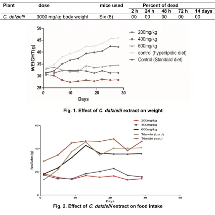
C. dalzielii extracts showed no mortality at 3000 mg/kg of body weight (bw) after 72 hours of observation following oral administration of the extract to the animals (Table 1). No sign of toxicity was observed in the wellness parameters during the 14-day observation period.
3.2 Effect of the Extract on Body Weight and Food Intake
Fig. 1 shows the weight variations of the animals during the experiment. Nutrition with hyperlipidic food (40% lipid) caused an increase in the weight of animals compared to control. Oral administration of C. dalzielii extract (200 mg / kg, 400 mg / kg, 600 mg / kg bw) showed a weight reduction in animals compared to control and those fed with a hyper lipid diet (P <0.05). The dose at 400 mg/kg body weight showed at the end of the experiment the greatest reduction in weight with an average weight of 2.2 ± 1.6 g (7.1%) against an increase of 12.85 ± 4.8 g
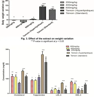
(38.16%) for the batch of animals that received only hyperlipidic food (Fig. 2). As for the variation of the food taken, the negative control lot, the batch that received only the hyperlipidic food and the one receiving the extract at 600 mg / kg of body weight showed a significant increase of the food taken with respectively a difference of consumption between the beginning and end of the experiment of 20.63 g, 27.3 g and 22.4 g. Animals treated at 200 and 400 mg / kg showed little variation in food intake. (1 to 6.9 g) (Fig. 3).
3.3 Effect of the extract on liver enzymes and lipid profile
Fig. 4 shows the effect of C. dalzielii extract on total cholesterol, HDL-cholesterol (HDLc), LDL-cholesterol (LDLc) triglycerides. Animals in the control group fed with hyper-lipid and standard food had a high triglyceride concentration compared to groups of animals treated with the plant extract. The group treated with a dose of 200 mg / kg had the lowest total cholesterol concentration. The extract has shown a reduction of triglyceride and LDL cholesterol (bad cholesterol) level, and they induced production of HDLc (Good Cholesterol) level in the animal. There was no significant difference between the different groups for ALT levels, Concerning the concentration of AST, the extracts caused a significant reduction compared to the control group (Fig. 5).
3.4 Effect of the Extract on the
Antioxidant Enzymes and MDA
Table 1. Oral acute toxicity study of the plant extract
Plant dose mice used Percent of dead
2 h 24 h 48 h 72 h 14 days C.dalzielii 3000 mg/kg body weight Six (6) 00 00 00 00 00
Fig. 1. Effect of C. dalzielii extract on weight
Fig. 2. Effect of C. dalzielii extract on food intake
3.5 In vitro Enzymatic Activities of the
Extract
The ethanolic extract of C. dalzielii showed an inhibition of 64.58 ± 0.86% on the pancreatic lipase at 100 µg / ml of the extract against 90.1 ± 2.27% for the orlistat used as a reference substance (Figure 8). For the activity on trypsin, the extract inhibited at 31.66 ± 2.88% for a concentration of 100 µg / ml of extract. Regarding the inflammatory enzyme inhibition test, Caralluma dalzielii inhibit lipoxygenase at 65.75 ± 0.05% at 1 mg/ml greater than that of gallic acid used as reference which gave an inhibition of 54.87 ± 0.04%; against xanthine oxidase at 100 µg / ml the extract showed an
activity of 80.55 ± 8.41% against 90.90 ± 00% for quercetin used as reference substances (Fig. 7).
3.6 Antioxidant Activity and
Phytochemical Composition of the Extract
The ethanolic extract of Caralluma dalzielii
Fig. 3. Effect of the extract on weight variation ***P-value is significant at p < 0.05
Fig. 4. Effects of the extract on lipid profiles of serum ***P-value is significant at p < 0.05
4. DISCUSSION
In this study, the anti-obesity activity of C. dalzielii extract was determined using hyperlipid food on animal models. Plants generally induce
weight loss through several mechanisms, they can increase satiety [32], increase
thermogenesis [33], prevent differentiation of preadipocytes [23] and lipase inhibition [34].
Table 2. Antioxidant activity and phytochemical composition of the extract
Activities C.dalzielii Gallic acid Quercétin
Antioxidant activity
DPPH 62.727 ± 2,4 ** 86,63±0,08 *** 82.17 ± 0.30***
FRAP 1.51 ± 0.03* 8.84± 0.45 *** 4.69± 0.05 **
Phytochemical composition
Total flavonoids content (mgEQ/1 g)
9.58 ± 0.35 ND ND
Total polyphenol content (mgEAG/1 g)
368,875 ± 16.2 ND ND
mgEAG/1 g = milligrams of gallic acid equivalent per 1 g of dry extract; mgEQ/1 g = milligrams equivalent quercetin for 1 g of dry extract; FRAP = Ferric Reducing Antioxidant Power, DPPH = (2,2-diphenyl-1-picrylhydrazyl. ***P-value is significant at p < 0.05; Mean ± S.E.M = Mean values ± Standard error of means of
Fig. 5. Hepatic biochemical parameters to liver enzymes AST = Aspartate Amino Transferase; ALT= Alanine aminotransferase
***P-value is significant at p < 0.05
Fig. 6. Effect of extracts on MDA in the liver
MDA= malondialdehyde, ***P-value is significant at p < 0.05; Mean ± S.E.M = Mean values ± Standard error of means of three experiments
Fig. 7. Effect of extracts on the antioxidant enzymes of the liver Cat= Catalase; SOD= Superoxide dismutase
Fig. 8. In vitro activity of Caralluma dalzielii extract
***P-value is significant at p < 0.05; Mean ± S.E.M = Mean values ± Standard error of means of three experiments
Oral administration of C. dalzielii extract resulted in weight reduction in the treated animals compared to untreated animals. The extract also has an anorectic effect at a dose of 400 and 200 mg/kg body weight. The ability of the plant extract to reduce food intake coupled with decreased weight gain in treated animals compared to their counterparts what received water (control groups), suggests that C. dalzielii
could be a good satiety inducer and weight management modulator. The Caralluma genus is characterised by the presence of many pregnane glycosides [11]., these molecules isolated in many plants like Caralluma fimbriata, Hoodia gordonii is known for their anorectic activity, anti-obesity [12-13]. The pregnant pregnans glycosides have indeed isolated from the whole plant of Caralluma dalzielii [15], it could, therefore, act on obesity by its anorectic effect. Obesity is a pro-inflammatory condition due to macrophages infiltrating in the adipose tissue, cytokines (produced by macrophages) and adipokines produced by fat cells alter lipid metabolism [35]. Proinflammatory cytokines, tumour necrosis factor α (TNF-α) and interleukin-1 (IL-interleukin-1) stimulate lipolysis in adipocytes by increasing circulating free fatty acid levels and the production of lipoproteins rich in triglyceride, which will contribute to the increase of serum triglycerides in obese patients. Dyslipidemia characterised by an increase in triglyceride levels with the growth of LDL, cholesterol [36]; which is a major risk factor for cardiovascular diseases.
C. dalzielii in the present study in addition to weight reduction by the three doses caused a significant decrease in triglyceride levels and a lower LDLc level than the control lot made obese with hyperlipidic food (Fig. 4). Our extracts could prevent cardiovascular disease by preventing the accumulation of triglycerides and LDLc in the body. This could explain the traditional use of this plant against heart problems in Mali [37].
induction of SOD and catalase synthesis in the presence of a hyperlipidic diet, but these decreased in obese animals. These two enzymes play an important role in the metabolism of reactive oxygen species. SOD is involved in the destruction of the superoxide anion (O2
-), and its production decreases with increasing body weight [41]. Catalase catalyses the degradation of hydrogen peroxide (H2O2) and
thus protects the body from the oxidative and harmful action of hydrogen peroxide. This result probably explains the protective effect of C. dalzielii extract against oxidative stress in the liver of the mice. The damage to the cell by the oxidant products in obesity is attenuated by the increase in antioxidant enzymes such as SOD, CAT and a reduction in MDA production.
The inhibition of digestive enzymes is a promising alternative for the treatment of obesity, mainly because they act on the small intestine, without acting on the central nervous system, where the usual anorectic agents act. Pancreatic lipase is a key enzyme in lipid digestion for the absorption of triglycerides in the small intestine. It is secreted by the pancreas and hydrolyses triglycerides into glycerol and free fatty acids [42]. This inhibition might be a good way to reduce lipid absorption. Trypsin is an enzyme of the pancreas. It is synthesised as a trypsinogen (inactive proenzyme) and stored in the enzymatic vesicles of acinar cells, from which it is excreted at the time of digestion. When trypsin inhibitors are present in a diet, they can lead to a reduction in the rate of growth in animals due to a decrease indigestible proteins, leading to weight loss and endogenous catabolism of proteins [43].
The extract of C. dalzielii showed a good inhibition of the digestive enzymes but lower than the reference molecules used. This activity observed could be due to the presence of polyphenols and flavonoids found in our extract. Indeed, a good number of natural antihyperlipidemic molecules have already been isolated from some plants; Epigallocatechin-3-gallate, kaempferol and quercetin have been shown to inhibit pancreatic lipase [44]. Trypsin inhibitors isolated from Tamarindus indica L. seeds showed a reduction in weight and an increase in Cholecystokinin (CCK) levels in the treated animals [45]. CCK is an anorectic hormone that sends satiety signals to the brain. Obesity is associated with an increase in the reactive oxygen species (ROS), this is probably due to the presence of excess adipose tissue itself since adipocytes and preadipocytes have
been identified as a source of pro-cytokines. therefore, an increase of cytokine concentration could be responsible for an increase in ROS [7]. The extract of C. dalzielii showed good antioxidant activity by inhibiting the DPPH radical and reducing the ferric ion to ferrous ion. It has also shown good potential to inhibit inflammatory enzymes such as xanthine oxidase and lipoxygenase (LOX). A glycoside isolated in the extract Conyza bonariensis have shown an inhibitory activity on xanthine oxidase [46]. Glycosides inhibitory of LOX are isolated in
Calendula officinalis L. [47]. The inhibitory potentials observed in the extract could be due to the glycosides contained in these species [48-49]. The extract contains polyphenols and flavonoids. These molecules indeed have antiobesity activities and would help reduce weight by their anti-lipase activity [44]. Rutin, quercetin, narregenine inhibit the activity of glycerol-3-phosphate dehydrogenase (GPDH) and the expression of PPARγ, C / EBPR genes involved in the differentiation of adipocytes. Adipocytes are the cells that store lipids in the body [50]. The presence of flavonoids in the extract might have acted on the fat cells for weight reduction.
5. CONCLUSION
In conclusion, this study indicates that C. dalzielii
extract appears to be a good and promising agent for the search for molecules capable of modulating obesity because of its potential in inhibiting digestive enzymes and its anorectic effect. They also showed a good reduction of triglycerides and LDLc levels in treated animals indicating its potential as a weight reducing agent.
CONSENT
It is not applicable.
ETHICAL APPROVAL
COMPETING INTERESTS
Authors have declared that no competing interests exist.
REFERENCES
1. Seidell JC, Flegal KM. Assessing obesity: classification and epidemiology. Br Med Bull. 1997;53:238–252
2. Anne-Laure W. Hoodia gordonii (Masson) Sweet ex Decne: Une plante d’Afrique du Sud, de son utilisation traditionnelle vers un éventuel avenir thérapeutique. Mémoire de thèse, Université de Lorraine; 2012. In French.
3. Skaal L, Pengpid S. Obesity and health problems among South African healthcare workers: Do healthcare workers take care of themselves? S Afr Fam Pract. 2011; 53(6):563-567.
4. Bationo AF. Obésité: Nous en sommes tous menacés. Ecodufaso/ Groupe Ecodafrik; 2018.
Available:http://www.ecodufaso.com/obesit
e-nous-en-sommes-tous-menaces/.Consulté le 30/08/2018. French 5. Bartolomucci A, Cabassi A, Govoni P,
Ceresini G, Cero C, Berra D. Metabolic consequences and vulnerability to diet-induced obesity in male mice under chronic social stress. Plos one. 2009;4:43 31.
6. Terra X, Montagut G, Bustos M, Llopiz N, Ardèvol A, Bladé C, et al. Grape-seed procyanidins prevent low-grade inflamma-tion by modulating cytokine expression in rats fed a high-fat diet. J. Nutr. Biochem. 2009;20:210–218.
7. Alba F, Eduardo M, Mirandeli B, Jaime E, Ángel M, Cesar E, et al. Inflammation, oxidative stress, and obesity. Int. J. Mol. Sci. 2011;12:3117-3132.
8. Priyanka M, Pragyanshu K, Sneha J, Mahendra B, Kanthi KK, Kamlesh KB. Screening of six ayurvedic medicinal plants for anti-obesity potential: An investigation on bioactive constituents from Oroxylum indicum (L.) Kurz bark. Journal of Ethnopharmacology. 2017;197:138-146. 9. Carette C, Muzard L, Radu A, Barsamian
C, Bretault M, Czernichow S. Traitement pharmacologique de l’obésité, Médecine. Clin. Endocrinol. Diabète. 2012;61:1-12. 10. Pare D, Hilou A, Ouedraogo N, Guenne S.
Ethnobotanical study of medicinal plants used as anti-obesity remedies in the
nomad and hunter communities of Burkina Faso. Medicines. 2016;3:1-9.
11. Van-Heerden FR, Marthinus HR, Maharaj VJ, Vleggaar R, Senabe JV, Gunning PJ. An appetite suppressant from Hoodia species. Phytochemistry. 2007;68(20): 2545-2553.
12. Gooda SN, Saari N, Ismail A, Khatib A, Mahomoodally F, Abdul Hamid A. Plants’ metabolites as potential antiobesity agents. Sci. World J; 2012.
DOI: 10.1100/2012/436039
13. Maclean D, Luo L. Increased ATP content/ production in the hypothalamus may be a signal for energy-sensing of satiety: Studies of the anorectic mechanism of a plant steroidal glycoside. Brain Research. 2014;10-20(1-2):1-11.
14. Nadembega P, Boussim JI, Nikiema JB, Poli F, Antognoni F. Medicinal plants in Boskouré, Kourittenga province, Burkina Faso: An ethnobotanical study. Journal of Ethnopharmagical. 2011;133:378-395. 15. De Leo M, De Tommasi N, Sanogo R,
Autore G, Marzocco S, Pizza C, et al. A. new pregnane glycosides from Caralluma dalzielii. Steroids. 2005;70:573-585.
16. Nishida C, Ko GT, Kumanyika S. Body fat distribution and noncommunicable diseases in populations: Overview of the 2008 WHO expert consultation on waist circumference and waist-hip ratio. Eur. J. Clin. Nutr. 2010;64:2–5.
17. Tanko Y, Halidu A, Mohammed A, Ahmed MK, Musa KY. Effect of ethanol extract of Caralluma dalzielii N.E.Br. (Asclepiadaceae) on blood glucose levels of fructose -induced insulin resistance in laboratory animals. Annals of Biological Research. 2012;3(11):4980-4984.
18. Ligne directrice 423 de l’OCDE pour les essais de produits chimiques, Toxicité orale aiguë - Méthode par classe de toxicité aiguë. 2001;1-14. In French 19. Mei-Yin C, Yu-Hua K, Jin-Ming C,
Chih-Min Y, Chao-Hsiang C. Effects of herbal mixture extracts on obesity in rats fed a high-fat diet. Journal of Food and Drug Analysis. 2016;24(3):594-601.
20. Changhyun R, Uhee J. Screening of crude plant extracts with antiobesity activity. International Journal of Molecular Sciences. 2012;13:1710-1719.
composition in soybean [Glycine max (L.) Merrill]. International Journal of Agronomy and Plant Production. 2013;4(12):2877-2884.
22. Ohkawa H, Ohishi N, Yagi K. Assay for lipid peroxidation in animal tissues by thiobarbituric acid reaction. Ann. Biochem.
1979;95:351–358.
23. Misra HP, Fridovich I. the role of superoxide anion in the epinephrine and a simple assay for superoxide dismutase autoxidation. J. Biol. Chem. 1972;247: 3170–3175.
24. Beers F, Sizer JR. Cambridge, a spectrophotometric method for measuring the breakdown of hydrogen peroxide by catalase. J. Biol. Chem. 1951;195:133– 140.
25. Velazquez E, Tournier HA, Mordiyavich-Buschiazza P, Saavedra G, Schinnella GR. Antioxydant activity of Paraguayan plants extracts. Fitoterapia. 2003;74:91-97. 26. Koleva II, Beck VT, Linssen PH, Groot A, Evstatieva NL. Screening of plants extracts for antioxidant activity: Comparative study on three testing methods. Phytochemistry Analysis. 2002;13:8-17.
27. Hinneburg I, Damien-Dordan HJ, Hiltunen R. Antioxidant activities of extracts from selected culinary herbs and spices. Food Chemistry. 2006;97(1):122-129.
28. Malterud KE, Rydland KM. Inhibitors of 15-lipoxygenase from orange peel. Journal of Agricultural and Food Chemistry. 2000;48: 5576-5580.
29. Filha FZSIF, Vitolo LG, Fietto JAL, Saude-Guimaraes DA. Xanthine oxidase inhibitory activity of Lychnophora species from Brazil ("Arnica"). J. Ethnopharmacol. 2006;107: 79-82.
30. Singleton LV, Orthofer R, Lamuela-Raventos RR. Analysis of total phenol and other oxydation substractes and anti-oxidants by mean of Folin- Ciocalteu reagent. Methode in Enzymology. 1999; 299:152-178.
31. Arvouet-Grand A, Vennat B, Pourrat A, Legret P. Standardisation d’un extrait de Propolis et identification des principaux constituants. Journal de Pharmacie de Belgique. 1994;49:462-468.
32. Walsh DE, Yaghoubian V, Behforooz A. Effect of glucomannan on obese patients: A clinical study. Int J Obes. 1984;8(4):289-93.
33. Haaz S, Fontaine KR, Cutter G, Limdi N, Perumean-Chaney S, Allison DB. Citrus
aurantium and synephrine alkaloids in the treatment of overweight and obesity: an update. Obes Rev. 2006;7(1):79-88. 34. Ozkan D, Ayse O, Damla A, Basak YD,
Serap D, Umit S. Inhibition of pancreatic lipase by culinary plant extracts. Int J Plant Biol Res. 2015;3(2):1038.
35. Lara-Castro C, Fu Y, Chung BH, Garvey WT. Adiponectin and the metabolic syndrome: Mechanisms mediating risk for metabolic and cardiovascular disease. Curr Opin Lipidol. 2007;18(3):263-70. 36. Klop B, Elte JWF, Cabezas MC.
Dyslipidemia in obesity: Mechanisms and potential targets. Nutrients. 2013;5(4): 1218–1240.
37. Sreelatha R, Pullaiah J. Induction of somatic embryogenesis and plant regeneration from intermodal explants of
Caralluma stalagnifera. Botany Res Internatinal. 2010;3(1):17-20.
38. Clark JM, Brancati FL, Dielh AM. The prevalence and etiology of elevated aminotransferase levels in the United States. Am J Gastroenterol. 2003;98:960-967.
39. Misra DS, Maiti R, Ghosh D. Protection of swimming-induced oxidative stress in some vital organs by the treatment of composite extract of Withania somnifera,
Ocimum sanctum and Zingiber officinalis
in male rat. Afr J Tradit Complement Altern Med. 2009;6(4):534–43.
40. Al-Dalaeen A, Al-Domi HA. Evaluation of oxidant-antioxidant status in obese children and adolescents. Pakistan Journal of Nutrition. 2016;15(10):942-947.
41. Fridovich I. Superoxide dismutase: Regularities and irregularities. Harvey Lect. 1985;79:51-75.
42. Nitin AL, Neeraj KP, Sneha CJ, Kamlesh KB. Inhibitors of pancreatic lipase: State of the art and clinical perspectives. EXCLI Journal. 2014;13:897-921.
43. Mcdougall GJ, Shpiro F, Dobson P, Smith P, Blake A, Stewart D. Different polyphenolic components of soft fruits inhibit α-amylase and α-glucosidase. J Agric Food Chem. 2005;53:2760-2766. 44. Sergent T, Vanderstraeten J, Winand J,
Beguin P, Schneider Y. Phenolic com-pounds and plant extracts as potential natural anti-obesity substances. Food Chemistry. 2012;135:68–73.
from Tamarindus indica L. seeds reduces weight gain and food consumption and increases plasmatic cholecystokinin levels. Clinics (Sao Paulo). 2015;70(2):136– 143.
DOI: 10.6061/clinics/2015(02)11
46. Kong LD, Abliz Z, Zhou CX, Li LJ, Cheng CH, Tan RX. Glycosides and xanthine oxidase inhibitors from Conyza bonariensis. Phytochemistry. 2001;58(4): 645-51.
47. Bezáková L, Grančai D, Obložinský M, Vanko M, Holková I, Paulikova I, et al. Effect of flavonoids and cynarine from
Cynara cardunculus L. on lipoxygenase activity. Acta Facultatis Pharmaceuticae
Universitatis Comenianae. 2007;54:48- 53.
48. Mohammed A, Essam A, Eric Gt. Pregnane Glycosides from Caralluma russeliana. J. Nat. Prod. 2000;63:1451-1453.
49. Nacoulma O. Plantes médicinales et pratiques médicales traditionnelles au Burkina Faso. Cas du plateau central; TOME I & II. Thèse d’Etat, Univ. Ouaga; 1996.
50. Chin-Lin H, Gow-Chin Y. Effects of flavonoids and phenolic acids on the inhibition of adipogenesis in 3T3-L1 adipocytes. J. Agric. Food Chem. 2007; 55(21):1-8.
_________________________________________________________________________________
© 2018 Pare et al.; This is an Open Access article distributed under the terms of the Creative Commons Attribution License
(http://creativecommons.org/licenses/by/4.0), which permits unrestricted use, distribution, and reproduction in any medium,
provided the original work is properly cited.
Peer-review history: