(UDC: 602.9)
Tissue engineering
G. Vunjak-Novakovic1
1Professor of Biomedical Engineering and Medicine Columbia University, New York NY
10032, USA
http://www.bme.columbia.edu/fac-bios/vunjak-novakovic/faculty.html
Abstract
Today, we live longer and better than ever before. The aging population and increasing expectations for an improved quality of life are driving a need for developing treatment modalities for the repair or replacement of tissues lost to injury, congenital abnormality or disease. Tissue engineering is responding to this need by developing methods for the use of stem cells from a variety of sources for full regeneration of tissue structure and function. At the same time, engineered tissues are serving as models for biological research, study of disease, testing of drugs, and development of personalized therapeutic modalities that are tailored to a specific patient and medical condition. We review here the general concept of tissue engineering, and the potential and challenges of this rapidly developing interdisciplinary field.
Key words: tissue engineering, artificial tissue, stem cells, personalized medicine
1. Historical perspective
While the state of the art of regenerative medicine is far from having ready-to-use “replacement organs”, we are getting increasingly close to the development of cell-based treatment modalities that will re-establish the structure and function of damaged or diseased tissues. The true beginning of the field is hard to pinpoint. As early as the 1870’s Julius Petri, a bacteriologist working with the famous Robert Koch, invented glass dishes that he named after himself, and the technique of cell cloning – both of which remain extensively used even to this day. At the beginning of the 20th century, Ross Harrison reported what can be considered the first example
of tissue culture, using a notoriously difficult system: neuronal outgrowth from embryonic tissue [Harrison 1907].
Tissue engineering was officially established in 1988, when the field was defined as “the application of principles and methods of engineering and life sciences toward fundamental understanding of structure-function relationships in normal and pathological mammalian tissues and the development of biological substitutes to restore, maintain, or improve tissue function”. This description still reflects the unifying concept of the field.
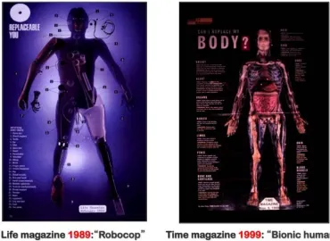
The 1990s mark a radical change in our capability to grow biological substitutes of native tissues [Lysaght, 2001]. An illustrative example is shown in Figure 1.
Fig. 1. Tissue engineering is an emerging field. Left: cover page of the Life magazine in 1989, depicting the envisioned options (all based on artificial materials) for replacing our failing organs. Right: cover page of the Time magazine in 1999, with the vision of tissue-engineered
“replacement parts” for human body.
In 1989, the scientific community envisioned that the options for “replacement parts” for our failing organs will be entirely based on the use of plastic, ceramic and metal-based implants. Just ten years later, a new vision emerged of fully biological tissue replacements engineered using cells, biomaterials and culture environments. The pace of the field may be best exemplified by the rapidly growing stream of publications – from about a dozen in 1991, and the first review in 1993 [Langer and Vacanti, 1993] to over 30,000 at the end of 2009.
2. Design of tissue engineering systems
is a growing recognition that the regeneration of an adult tissue, by stem cells or an engineered tissue construct, is regulated by the same principles that regulate native development. The fields of stem cells biology, bioengineering and regenerative medicine now realize that the cells respond to the entire context of their environment: soluble factors (oxygen, nutrients, growth factors), other cells (in direct contact or through secretion of paracrine factors), extracellular matrix (through its composition, structural and mechanical features), and physical signals (electrical, mechanical, hydrodynamic). The premise is that tissue-engineering systems should capture the dynamics of the native cellular environments, in order to mobilize cells into forming native-like tissue structures.
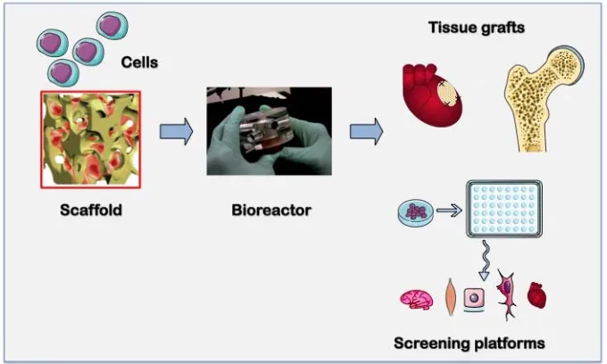
In general, the designs of tissue engineered systems are inspired by biology, in an attempt to create in vivo like (biomimetic) environments providing the cells with appropriate environmental cues - molecular, structural, and physical – that will result in tissue regeneration [Vunjak- Novakovic and Scadden, 2011], [Zimmerm,ann et al. 2006]. To direct cells to differentiate at the right time, in the right place, and into the right phenotype, one needs to recreate the right environment, with biology and engineering interacting at multiple levels. The control of the cellular environment is provided by an integrated use of two components: a biomaterial scaffold (a structural and logistic template for cell attachment and tissue formation) and a bioreactor (a culture system providing control over environmental factors, though facilitated mass transport to and from the cells and application of physical signals).
Fig. 2. Tissue engineering system. Stem or progenitor cells are placed into the biomaterial scaffold (providing a template for tissue formation) and cultured in a bioreactor (providing environmental control and the necessary biophysical signals). The resulting engineered tissues
can be used as implants for replacement or regeneration of native tissues lost to injury, abnormality or disease, or as test beds for biological research, study of disease or drug
screening.
3. Scaffold design criteria
Depending on what tissue is to be replaced, the properties of scaffolds will vary along several parameters, including biological substances used, porosity, elasticity, stiffness, and specific anatomical shapes. Extracellular matrix components have been made into various matrices, including hydrogels and porous scaffolds, using a variety of methods. Synthetic matrices made from polymers have been explored as scaffolds for tissue engineering, because of their easily controlled and reproducible properties. Their use, however, is often accompanied by surface modifications to enhance cell adhesion. Additionally, these materials do not have the advantage of providing biological signaling. In contrast, biologically derived matrices provide the cells with microenvironmental signals similar to those in intact native tissues [Godier-Fournemont et al. 2011].
Several studies have used scaffolds as a structure to improve cell adhesion and alter their behavior. RGD-peptides have been widely used as molecules tethered to scaffolds, to increase cell adhesion [Burdick and Vunjak-Novakovic, 2009]. It is possible to microencapsulate growth factors within a hydrogel, and depending on degradation rate of the hydrogel, growth factor release kinetics may be finely controlled. Thus, these scaffolds not only provide spatial control, but also control over biochemical signaling, in the temporal domain. Such technology brings us a step closer to mimicking the physiological setting. Control over biochemical signaling to cells is important for both in vitro studies of cell differentiation and in vivo therapies, where sustained drug release over a bolus injection of a drug is necessary to mediate repair.
New “cell-instructive” materials are now being utilized to mimic the native matrix and actively interact with the cells. These new scaffolds are functional at multiple length- and time-scales: molecular (by incorporation of integrin-binding ligands and regulation of availability of growth factors), cellular (directed migration, mediation of cell-cell contacts and stiffness as a differentiation factor), and tissue levels (establishment of interfaces, structural and mechanical anisotropy). The enormous variation of cell/tissue properties has led to the development of “designer scaffolds” [Freytes et al, 2009].
4. Bioreactor design criteria
Cells are central to any of our efforts to grow tissue grafts, to construct models of disease, or to develop in vitro platforms for therapeutic screening. In order to mobilize their full biological potential, the scaffold-bioreactor system should serve as an in vitro mimic of the in vivo milieu of the development, regeneration or disease.
Today, bioreactor designs are guided by a “biomimetic” approach, which attempts to recapitulate in vitro some of the important aspects of the native cellular milieu associated with tissue development and regeneration [Freytes et al. 2009]. Bioreactors can be designed to control cell environment (through enhanced mass transport to and from the cells), provide physical signals (hydrodynamic, mechanical, electrical), and enable insight into cellular behavior (through on-line imaging). Design of a tissue engineering bioreactor should ideally support cell viability and 3D organization by mechanisms similar to those present in the native cell environment. Overall, bioreactors provide an opportunity to manipulate and control only certain aspects of a given niche, but do allow for quantitative studies of cellular interactions with their environment.
signals. These conditions are far from the in vivo situation, where cells reside in a precisely controlled environment, and are subjected to spatial and temporal gradients of multiple factors.
To overcome these limitations, bioreactors are designed to provide tightly controlled, dynamic culture settings. With their capability to generate spatial gradients of regulatory signals, subject cells to dynamic changes in their environment, and offer insight into cellular responses in real time, these new technologies are providing physiologically meaningful conditions. This new generation of tissue engineering bioreactors is finding applications in fundamental biological research, engineering of functional tissue grafts, and studies of disease. We provide here one example of an advanced approach to tissue engineering currently studied for eventual translation into clinical application.
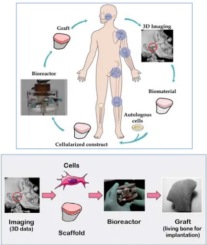
5. Engineering of anatomically shaped bone grafts
Damage or malformation of bone in head and face due to trauma, cancer surgery or birth defects not only leave the patient with the loss of tissue and its function, but also render them psychologically scarred. The burden of craniofacial injuries extends far beyond direct medical expenses as these injuries often impair the patient’s social integration and the ability to re-engage in economic activity at full capacity. Due to the complexity of bone reconstruction in this region, currently available treatment options (grafting of bone harvested from another area in the body after reshaping, or implantation of biomaterial spacers) fall short of providing adequate care. The availability of living bone grafts engineered in vitro would revolutionize the way we currently treat these defects.
Fig. 3. Personalized bone grafts. To precisely reconstruct the shape and structure of native bones (head and face, or body skeleton) the fabrication of the biomaterial scaffold and the bioreactor chamber are guided by imaging. The patient’s own cells are used to culture a living
tissue grafts for implantation.
Clinical scans of the affected area are used to make correctly shaped biodynamic scaffolds. The scaffolds are seeded with adult human stem cells (for example, derived from fat aspirates) and cultured in appropriately designed bioreactors, to grow personalized living human bone grafts.
6. Summary
Извод
Инжењерингткива
G. Vunjak-Novakovic1
1Professor of Biomedical Engineering and Medicine Columbia University, New York NY
10032, USA
http://www.bme.columbia.edu/fac-bios/vunjak-novakovic/faculty.html
Резиме
Данас живимо дуже и боље него икада. Старење становништва и повећана очекивања
побољшања квалитета живота воде ка развоју модалитета третмана за поправку или
замену ткива изгубљеног повредом, урођеном маном или услед болести. Ткивно
инжењерствоодговаранаовупотребуразвојемметодазакоришћењематичнихћелијаиз
различитих извора запотпуну регенерацијуструктуреи функције ткива. Уисто време,
инжењерска ткива служе као модели за биолошка истраживања, изучавање болести,
тестирање лекова, и развој персонали терапеутских модалитета који су подешени
специфичном пацијенту и медицинском стању. Овде дајемо преглед општег концепта
инжењерингаткива, каоипотенцијалаиизазоваовеинтердисциплинарнеобластикојасе
брзоразвија.
Кључне речи: инжењеринг ткива, вештачко ткиво, матичне ћелије, персонализована медицина
References
Burdick, J.A., and Vunjak-Novakovic, G. (2009) Engineered microenvironments for controlled stem cell differentiation. Tissue Eng Part A 15, 205
Freytes D, Wan L and Vunjak-Novakovic G. Geometry and Force Control of Cell Function.
Journal of Cellular Biochemistry 108(5): 1047-58, 2009 (cover article)
Godier-Fournemont A, Martens T, Koeckert M, Wan LQ, Parks J, Zhang G, Hudson J, and Vunjak-Novakovic G. Composite scaffold provides a cell delivery platform for cardiovascular repair PNAS 108(19): 7974-7979, 2011.
Grayson WG, Fröhlich M,,Yeager K, Bhumiratana S, Cannizzaro C, WanLQ, ChanME, Liu
ME, X. Edward GuoEX and Vunjak-NovakovicGV. (2009) Engineering anatomically shaped human bone grafts. Proceedings of the National Academy of Sciences USA 107(8):3299-3304
Harrison, R.G. (1907) Observations on the living developing nerve fiber. Proc Soc Exp Biol Med 4, 140
Langer R, Vacanti JP. Tissue engineering. Science. 260:920-6. 1993.
Lysaght MJ, Reyes J. (2001) The growth of tissue engineering. Tissue Eng. 7(5):485-93. Vunjak-Novakovic G and Scadden D.T. Biomimetic platforms for human stem cell research.