Pharmacophore 2016, Vol. 7 (5), 236-245
USA CODEN: PHARM7 ISSN 2229-5402
Pharmacophore
(An International Research Journal)
Available online at http://www.pharmacophorejournal.com/
Review Article
HYDROGEL DRUG DELIVERY SYSTEM
Hrushikesh Anantrao Joshi
*, Omkar Ashok Doiphode, Rahul Vinayak Jadhav and
Rajendra N. Patil
Shivnagar Vidya Prasrak Mandal's College of Pharmacy, Malegaon (BK), Baramati, India
ABSTRACT
The dissolution of a hydrophilic polymer in water can be prevented by adding cross-links via either a physical or a chemical process. A cross-linked hydrosol is called a hydrogel and swells in the surrounding liquid to a certain swelling ratio, depending on the number of cross-links, i.e., the cross-linking density. These hydrogels have many advantageous features including, stimuli responsive, good diffusion properties, low toxicity and good compatibility, Because of their chemical structures are similar to those of the bioactive Glycosoaminoglycan (GAG) molecules present in the extra-cellular matrix. Hydrogel preparation by freeze- thaw method involves physical cross-linking due to crystallite formation. This method does not require the presence of a cross-linking agent such physically cross-linked materials also exhibit higher mechanical strength than chemical or irradiative techniques. In Physical hydrogels, mechanical load can be distributed along the crystallites of the three dimensional structures. Extent crosslinking hydrogel analyzed by FTIR and DSC. The molecular transport phenomenon, as studied by the dynamic swelling experiments.
Keywords:
Hydrogel, Freeze-Thaw method, Crystallites, Swelling, Drug delivery system, FTIR, DSC.INTRODUCTION
Every drug molecule needs a delivery system to carry the drug to site of action upon administration to the patient. Delivery of the drugs can be achieved using various types of dosage forms including tablets, capsules etc. Most of these conventional drug delivery systems are known to provide release of the drug with little or no control over delivery rate. Hydrogel drug delivery system in a simple binary system of a polymer and a liquid, a sol is formed when the polymer- liquid interaction are more favored than both polymer- polymer and liquid – liquid interactions. If the polymer is hydrophilic and liquid is water, the product of the polymer- liquid interaction is called a Hydrosol. The dissolution of a hydrophilic polymer in water can be prevented by adding cross-links. Hydrogel can only swell in surrounding liquid to a certain swelling ratio, depending on the number of cross- links, i.e. the cross-linking density. Hydrogel
have many advantageous features including, low toxicity and good biocompatibility, because their chemical structures are similar to those of the bioactive glycosoaminoglycan (GAG) molecules (e.g heparin sulfate, chondrotin sulfate and hyaluonan) present in the native extracellular matrix.8,17,21,20
Physical and Chemical Gels
Hrushikesh Anantrao Joshi et al. / Pharmacophore 2016, Vol. 7 (5), 364-373 chemical hydrogels are permanent and
irreversible as a result of configuration. The hydrogel will be available as a drug reservoir; loaded drugs will be released by diffusion from the hydrogels or by erosion of the hydrogel. Water inside the hydrogel allows free diffusion of the some molecules, while the polymer serves as a matrix to hold water together. The network structure of hydrogel can be characterized by a number of parameters. One parameter of particular concerns to this work is the mesh size. The mesh size is the term used to define the distance between cross-links in hydrogel network. The Change in mesh size alters the diffusion of a therapeutic moiety (proteins) from a hydrogel carrier. The releasing mechanism can be controlled by swelling or erosion of the hydrogels. The major mechanism in the erosion is very complex because it depends on the degradation, swelling, dissolution, or diffusion of oligomer and monomer residues. When a in cross-linked, amorphous, glassy polymer is brought into contact with thermodynamically compatible solvent, the latter dissociates into polymer, and when the solvent concentration in the swollen polymer reaches a critical value, chain disentanglement begins to dominate and the polymer is eventually dissolved. On the basis of this idea, polymer swelling due, to solvent penetration, and relaxation- controlled polymer dissolution kinetics had been proposed. The dissolution flux was expressed as the difference between the polymer stress gradient and the solvent osmotic pressure gradient.The dissolution process can be understood as the transformation undergone by the polymer from on entangled gel- like phase to a disentangled liquid solution. The dynamics of these polymer chains have been discussed by means of the reputation idea and the release is executed during this process and the release behavior would be affected by the property of the hydrogel because there can be diverse interactions between the hydrogel and drug. Hydrogel show minimal tendency to adsorb proteins from body fluids because of their low interfacial tension. Many hydrogels provide inert surfaces that prevent nonspecific adsorption of
proteins. Further, the ability of molecules of different sizes to diffuse into (drug loading) and out (hydrogel).Hydrogels allows to possible use in drug delivery systems. Many, polypeptide drugs example insulin, are difficult to dissolve in aqueous medium under physiological condition, due to their hydrophobic character. Numerous designs and materials for the successful delivery of hydrophobic polypeptide drug had been reported. Hydrogel posses a hydrophobic domain, which is suitable for adopting hydrophobic drug such as insulin. This hydrogel showed unique erosion behavior depending on the pH condition the hydrogel remains stable under acidic pH condition but to formation of the hydrogen bonds. On the other hand, the hydrogel showed spontaneous erosion behavior under a neutral pH condition, due to breakage of the hydrogen bonds by the ionization procedure of carboxyl group. The pH dependence would be available for oral polypeptide drug carrier, protecting under acidic pH conditions (stomach) and neutral pH conditions (small intestine).1,3,5,14,15,16,
Hydrogel Oral Drug Delivery System
Eudragit, the most prominent acrylic acid based enteric coating and numerous types have been developed for specific applications. Variation in the small intestine can be exploited to target breakdown of the enteric coating to a specific region of the GI tract as discussed above. Eudragit as aqueous, anionic polymer composed of methacrylic acid and methacrylates. The exact composition can be varied target breakdown of the coatings at a specific pH. pH sensitive drug delivery system developed for protein- peptide drug delivery, colon targeted drug delivery, for foul tasted drugs. At low pH ( ~2) protection of drug in at low pH environment Complexation
and increase in mesh size occurs due to ionic repulsion and swelling of the polymer at high pH.
Properties of pH Sensitive Hydrogels
Hrushikesh Anantrao Joshi et al. / Pharmacophore 2016, Vol. 7 (5), 364-373 properties depending on the ph of the
environment. The pendant acidic or basic groups on polyelectrolytes undergo ionization like acidic or basic groups of monoacids or monobases, ionization of polyelectrolytes, however, is more difficult due to electrostatic effects exerted by other adjacent ionized groups. This tends to make the apparent dissociation constant (Ka) Different from that of the corresponding monoacid or monobase. The presence of ionizable groups on polymer chains in swelling of the hydrogels much beyond that can achievable by non- electrolytes polymer hydrogels, the presence of ionizable groups on polymer chain results in swelling of the hydrogels. Since the swelling of polyelectrolyte hydrogels is mainly due to electrostatic repulsion such as pH, ionic strength, type of counter ions. The swelling and pH responsiveness of polyelectrolyte hydrogels can be adjusted by using neutral co-monomers, such 2-hydroethyl methacrylate, methyl methacrylate and maleic anhydride.6,12,18
Applications of the Ph Sensitive Hydrogels pH sensitive hydrogels have been most frequently used to develop controlled release formulations for oral administration. The pH in the stomach (<3) is quite different from the neutral pH in intestine and such a difference is large enough to elicit pH dependent behavior of polyelectrolyte hydrogels. For polycationic hydrogels, the swelling is minimal at neutral pH. This property has been used to prevent release of foul tasting drug in the neutral pH environment of the mouth. For Polyanionic hydrogels were developed for colon specific drug delivery system. Swelling of such hydrogels in the stomach is minimal and thus drug release is also minimal. The extent of swelling increase as the hydrogel passes down, the intestinal tract due to the increase in pH leading to ionization of the carboxylic groups.18,19 Freeze/Thaw Method of Preparation
Hydrogel preparation involves physical cross- links due to crystallite formation. These method does not require the presence of a cross-linking agent such physically crosslinked materials also exhibit higher mechanical strength than chemical
Hrushikesh Anantrao Joshi et al. / Pharmacophore 2016, Vol. 7 (5), 364-373 different areas such as intelligent polymers,
medicine, drug release, sensors, and cell encapsulation material. Etc. One important feature of PVA hydrogels (PVA HG) is the ability to change their mass, volume, and density in contact with electrolyte solution by a certain quantity of water which was firstly retained by the hydrogel. These modifications could be explained by water elimination from the hydrogels that initially reached the equilibrium of swelling. Polyvinyl alcohol, henceforth referred to as PVA, has become a prime candidate for improved biomaterials and drug delivery systems. PVA is a relatively inert polymer which is easily processable. PVA is hydrophilic and therefore swells in the presence of water or biological fluids to form hydrogels. This properly is particularly useful because it can allow for the release of drugs incorporated into these hydrogels. Other polymers such as polyacrylic acid (PAA) and polyethylene glycol (PEG) can be blend with PVA to impart additional properties such as pH- sensitivity or improved blood response. One problem with preparation of such biomaterials is the use of crosslinking agents and other reacting to form networks needed to product stable materials. This agent may include glutaraldehyde and formaldehyde, among others. Any residual material left after the formation of the polymer networks may reach out and cause harm to the body, as these agents are generally toxic. Thus, alternative methods of forming polymer networks or gels have been studied. In PVA films, crystallites that stabilize the material can be formed either by freezing and thawing cycles or by annealing using heating and cooling. These techniques form useful stable biomaterials without addition of toxic adjuvants. Physical mixtures and secondary bonding: In addition, hydrogels are formed by polymer blends between chitosan and other water- soluble nonionic polymers, such as poly vinyl alcohol (PVA). After a lyophilization or a series of freeze- thaw cycles. These polymer mixture forms junction points in the form of crystallites and inter- polymer complexation. The chain-chain interactions perform as cross-linking sites of the
hydrogel formation. In the case of Chitosan- PVA polymer blends, increasing the chitosan content negatively affects the formation of PVA crystallites leading to the formation of poor hydrogel structures.
Cryogenic Gelation
Hrushikesh Anantrao Joshi et al. / Pharmacophore 2016, Vol. 7 (5), 364-373
Figure 1: Multimembrane onion like physical hydrogel
Figure 2: Illustration of a freeze- thaw treatment yielding highly porous PVA physical hydrogels, 1:
Macromolecules in solution, 2: Liquid microphase, 3: Frozen solvent, 4: Macropores 5: Polymer network.
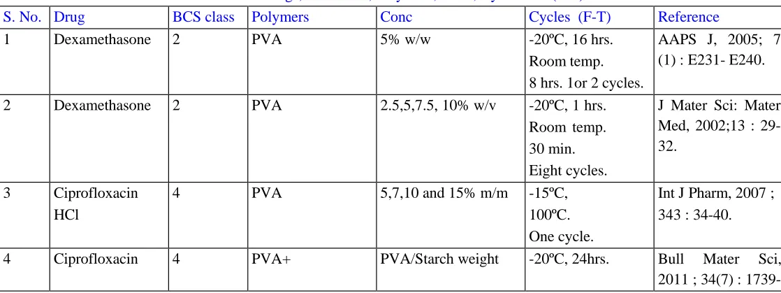
Table1: Drugs, BCS class, Polymers, Conc, Cycles and (F-T)
S. No. Drug BCS class Polymers Conc Cycles (F-T) Reference
1 Dexamethasone 2 PVA 5% w/w -20ºC, 16 hrs.
Room temp. 8 hrs. 1or 2 cycles.
AAPS J, 2005; 7 (1) : E231- E240.
2 Dexamethasone 2 PVA 2.5,5,7.5, 10% w/v -20ºC, 1 hrs.
Room temp. 30 min. Eight cycles.
J Mater Sci: Mater Med, 2002;13 : 29- 32.
3 Ciprofloxacin
HCl
4 PVA 5,7,10 and 15% m/m -15ºC,
100ºC. One cycle.
Int J Pharm, 2007 ; 343 : 34-40.
4 Ciprofloxacin 4 PVA+ PVA/Starch weight -20ºC, 24hrs. Bull Mater Sci,
2011 ; 34(7) : 1739-
Freezing Thawing
1 2 3 4 5
First membrane space
Hrushikesh Anantrao Joshi et al. / Pharmacophore 2016, Vol. 7 (5), 364-373
HCl Starch fraction
20/80,33.3/66.7 and 42.8/57.2
Room Temp. 1 hrs.
At least Three
cycles.
1748.
5 Sulfadizine
silver
2 PVA and
Chitosan
PVA- 7.5% Chitosan- 0.5, 0.75 and 1.0%
-20ºC, 18hrs. Room Temp. 6 hrs.
Minimum Three
cycles
J Pharm Bioallied Sci, 2012 : S53- S56.
6 Theophylline 1 PVA 15 and 20% wt -20ºC, 6 or 12 hrs.
25ºC, 2hrs. Two- three cycle.
Eur J Pharm
Biopharm, 1997; 43 : 51-58.
7 Theophylline 1 PVA -20ºC,
2 to 24 hrs. 25±1ºC, 2 to 24 hrs.
Two to nine cycles.
J Memb Sci, 1995; 107 : 229-237.
8 Naproxen
sodium
2 Acrylamide
+ MBAAm
0.765gm in 20 ml water
-18ºC, 16 hrs. room temp.
J Pharm Pharmcol, 2014 ; 2 : 527-533
9 Aspirin 1 PVA and
PAA
-80ºC, Room temp. Up toTen cycles
Afr J Pharm
Pharmcol, 2014 ; 8 (24) : 674-684.
10 Ketanserin 2 PVA -20ºC, 12hrs.
25ºC, 2 hrs. Two cycles.
J Biomater Sci
Polym Ed, 1996 ; 7(12) : 1055-1064.
11 Atenolol 3 PVA 10% wt -20ºC, 16 hrs.
Room temp. 8 hrs. Five cycles.
J Saudi Chem Soc, 2010 ;14 : 237-240.
12 Clindamycin 1 PVA +
Sodium alginate
PVA- 10% w/v SA -3% w/v
-20ºC, 18 hrs. Room temp. 6 hrs. Three cycles
Biol Pharm Bull, 2008 ;31(12) :2277- 2282.
13 Propranolol
HCl
1 PVA 10,15 and 20% w/w -20ºC, 15 hrs.
5ºC, 24 hrs.
Chem Pharm Bull, 1989 ; 37(9) : 2491- 2495.
Atenolol 3
14 Indomethacin 2 PVA
MP Sorbitol
PVA - 10,15,20,25 and 30 %w/v MP- 0,1,2,3
and 4 % w/v Sorbitol- 0,10,20,30, and 40 % w/v
-20ºC, 25ºC. Four Cycles.
Arch Pharm Res, 1993 ;16(1) :43-49.
15 Indomethacin 2 PVA
PAA
PVA- 8,10,15,20,25%
PAA/PVA% wt
fraction- 0/100,
-10ºC, 15 hrs.
4-5ºC, 24 hrs.
Hrushikesh Anantrao Joshi et al. / Pharmacophore 2016, Vol. 7 (5), 364-373
10/90,20/80,
30/70,40/60 and 100/0
16 Econazole
nitrate
2 PVA 10,15and 20% w/v -12ºC, 16 hrs.
Room temp. 8 hrs.
Four, six and eight cycles.
J Drug Del, 2014 : 1-14.
17 Captopril 3 PVA 2%w/w -30ºC, 48 hrs.
4-5ºC, 24 hrs.
Acta Pharmacol
Sin, 2000,21 (7) : 591-595.
18 Ephedrine
HCl
1 PVA 10,12,14, 16 and 18%
w/v
-20ºC, 12 hrs. 15ºC, 12 hrs.
Repeated Three
cycles.
Colloid Polym Sci, 2014 ;
292 : 1665 -1673.
19 Pentamidine 4 PVA
PLGA
0,3 and 6% w/v 5,10, and 15% w/v
-20ºC, 16 hrs. Room temp. 8 hrs.
One full cycle.
Pharm Res, 2002 ; 19(11) : 1713-1714.
20 Rosiglitazone
maleate
2 PVA+ Chitosan
+ Glyoxal
PVA 4% w/w Chitosan 2,4,6 ,and 8% w/w Glyoxal 2,4,6 and 8% w/w
-60ºC, 12 hrs. Room temp. 4 hrs.
Daru , 2010 ; 18 (3) : 200-210.
21 Ampicillin
sodium
3 PVA+
Hydroxy ethyl starch
not given -20ºC, 18hrs.
25ºC, 6 hrs. Three cycles.
Arabian J Chem , 2014 ; 7 : 372-380.
22 Oxprenolol 1 PVA 15 and 20% wt -20ºC, 6 or 12 hrs.
25ºC, 2hrs. Two- five cycles.
Eur J Pharm
Biopharm, 1997; 43;51-58
23 Gentamicin 3 PVA+
Dextran
PVA -2.5, 3.75, 5.0, 6.25,7.5,and 10 %w/v
Dextran- 0.38,0.56, 0.75, 0.94 and 1.13 %w/v
-20ºC, 18 hrs. 25ºC, 18 hrs. Three consecutive cycles.
AAPS Pharm Sci Tech, 2010 ; 11(3) : 1092-1103.
24 Insulin 4 PVA 5 and 10% w/v -20ºC, 20hrs.
Room temp. 4 hrs.
One- seven cycles.
J Mater Sci:Mater
Med, 2007 ;18:
2205-2210.
25 Enrofloxacin 1 PVA and
Pectin
PVA 15% w/v Pectin 2.0-4.0% w/v
-18ºC for 20 hrs 25ºC for 8 hrs 3-5 cycles
Bioresour Technol, 2013;145:280-284
26 Savlon
(Chlorohexidine gluconate, Cetrimede)
PVA Chitosan
PVA/ CS fraction
2.9,3.0,4.0,5. 6,6.0,8.2 and 12.1
Three cycle
Hrushikesh Anantrao Joshi et al. / Pharmacophore 2016, Vol. 7 (5), 364-373
27 Aminophylline
Theophylline, Ampicillin.
1 3
PVA, Chitosan, Sorbiton Sequinoleate (Castor oil) Sesame oil
PVA- 15% w/v Chitosan - 1% w/v
-20ºC for 20 hrs. 4ºC for 4 hrs. One cycle
Biol Pharm Bull, 1998 ;21(11): 1202- 1206.
28 Theophylline 1 PVA, PVA- 7%
NaCl - 11%
-20ºC for 24hrs. Int j Pharmcol,
2006 ; 2(3) :286- 292.
29 Chondroitin
Sulphate
PVA, chitosan
PVA- 9% w/v Chitosan- 1,2 and 5% w/v
-20ºC for 24 hrs. Room temperature 4 hrs.
Four cycles
J Biotechnol
Biomater, 2012 ; 2(4) .
30 Clindamycin 1 PVA,
Sodium alginate
PVA- 10% w/v SA- 3%
-20ºC for 18 hrs. Room temperature for 6 hrs.
Three cycles.
Biol Pharm Bull, 2008 ; 31(12):2277- 82
31 Nill 0 PVA
Amorphus Sulfonated Polyesters
PVA- 10% w/v PES- 5% w/v
-22ºC for 17 hrs Room Temperature
Mater Res, 2007 ; 10(1):43-46
32 Nill 0 PVA
Sodium decylsulfate
PVA 11wt% -22ºC for 22 hrs.
25ºC for 4hrs. One - nine cycles
J Phy Chem B, 2007; 111: 2166- 2173
33 Nill 0 PVA not given -20ºC for
12 hrs.
25ºCFor 12 hrs. One - three Cycles
Proc Estonian Acad Sci, 2009 ,58 (1) :63-66
34 Nill 0 PVA PVA - 10 to 16 % w/v 0ºC ± 2ºC
37ºC ± 2ºC 15-45 cycles
Biomed Mater,
2012; 7.
35 Nill 0 PVA not given -25ºC for
24 hrs.
25ºC for 24 hrs. One cycle
.
J Polym Sci: Polym Phy , 1997 ; 35 ; 2421-2427.
36 Nill 0 PVA,
Water Soluble chitosan
PVA -7 % Wt WS chitosan -2% wt Glycerol - 1% wt
-20ºC for 24 hrs.
25ºC for 24 hrs. One cycle
Hrushikesh Anantrao Joshi et al. / Pharmacophore 2016, Vol. 7 (5), 364-373
Glycerol,.
37 Niil 0 PVA,
Glycidyl acrylate, Glutaradehyde HCl
20 % PVA- Acrylate -15ºC for 24hrs.
Room temperature 24 hrs.
Two cycles
Polym, 2004 ; 41 :7715-7722.
38 Nill 0 PVA,
Collagen
2.5,5,8,10 and 15% w/v
-30ºC for 12 hrs. Room temperature. 1-16 cycles
J Mater Sci: Mater Med,1993 ; 4 :538- 542
39 Nill 0 PVA, PAA,
DMSO,water.
PAA/PVA-0/100, 10/90,20/80,30/70, 40/60 and 100/0
J Mater Sci, 2006 ; 41 : 2393-2404.
40 Nill 0 PVA, KCl,
NaCl
PVA- 11.14 % w/v. -15ºC for 12 hrs.
room temperature
12 hrs. Three
cycles.
Eur Polym J, 2007 ; 43 : 460-467.
REFERENCES
1. Alves, M and Jensen, B (2001), “Poly(vinyl hydrogels Produced By Conventional alcohol) Physical Hydrogels : New Vista On Crosslinking or By Freezing/Thawing A long Serving Biomaterial”, Methods”, Adv Polym Sci, Vol.153, 37-65.
Macromolecular Biosci, Vol.11, 1293-1313. 9. Kutz, M (2004), “Standard Handbook of
2. Berger, J and Reist, M (2004), “Structure and Biomedical Engineering and Design”, Interactions in Chitosan Hydrogels Formed by Mcgraw- Hill Professional, 22.1-22.14. Complexation or Aggregation for Biomedical 10. Nam, K and Watanabe, J (2004), “The Application”, European J Pharm Biopharm, Characteristics of Spontaneously Forming
Vol. 57, 35-52. Physically Crosslinked Hydrogels composed
3. Chauhan, S and Harikumar, S (2012), of two Water Soluble Phospholipid Polymers “Hydrogels: A Smart Drug Delivery for Oral Drug Delivery Carrier - I : Hydrogel Systems”, Int J Res Pharma Chem, Vol. Dissolution and Insulin Release Under
2(3), 603-614. Neutral pH condition”, European J Pharma
4. Dash, M and Chillini, F (2011), “Chitosan- A Sci, Vol. 23, 261-270.
Versatile Semi-Synthetic polymer in 11. Ottenbrite, R and Park, K (2010), Biomedical Applications”, Progress Polym “Biomedical Applications of Hydrogels
Sci, Vol. 36, 981-1014. Handbook”, Springer Science,1-15.
5. Ebara, M and Narain, R (2014), “Smart 12. Parrdossi, G and Cavalieri, F(2002),
Biomaterials”, Springer Business Media, 9- “Tailoring of Phsyical and Chemical
65. Properties of Macro- microhydrogels Based
6. Gerlach, G and Arndt, K (2010), “Hydrogels on Telechelic PVA”, Biomacromolecule, Vol. Sensors and Actuaters”, Springer Business 3,1255-1262.
Media, 1-14. 13. Patachia, S and Valente, A (2007), “Effect of
7. Gupta, S and Wenster, T (2011), “Evaluation Non- Associated Electrolyte Solution on The of PVA Gels Prepared Without Crosslinking behavior of Polyvinyl alcohol Based Agents As A Cell Adhesive Surface”, J Mater Hydrogels”, European Polym J, 2007, Vol.
Sci: Mater Medica, Vol. 22, 1763-1772. 43, 460-467.
Hrushikesh Anantrao Joshi et al. / Pharmacophore 2016, Vol. 7 (5), 364-373 and their blends with poly(acrylic acid) and
poly(ethylene glycol) for drug delivery applications”, J Drug Deliv Sci Tech, 2004, 14(4), 294-297.
15.Peppas, N and Wood, K (2004), “Hydrogels For Oral Delivery of Therapeutic Proteins”,
Expert Opinion Biological Therapeutics,
Vol. 4(6), 1-7.
16.Qiu, Y and Park, K (2001), “Environment Sensitive Hydrogels for Drug Delivery”, Adv
Drug Deliv Rev, Vol.53, 321-339.
17.Singh, A and Sharma, P (2010), “Hydrogels: A Review”, Int J Pharma Sci Rev Res, Vol. 4(20), 97-105.
18.Siepmann, J and Siegel, R and Rathore, M (2012), “Fundamentals and applications of Controlled Release Drug Delivery”, Springer
Science and Business Media, 75-106.
19.Vlijn, R and Bibi, N (2007), “Bioresponsive hydrogels”, Material Today, Vol. 10(4), 40- 48.
Correspondence Author:
Hrushikesh Anantrao Joshi
*
Shivnagar Vidya Prasrak Mandal's College of Pharmacy, Malegaon (BK), Baramati, India
Cite This Article:
Hrushikesh, Anantrao Joshi; Omkar, Ashok Doiphode; Rahul, Vinayak Jadhav andRajendra, N Patil (2016), “Hydrogel drug delivery system”, Pharmacophore, Vol. 7 (5), 364-373.