IJSRSET185339 | Published : 28 April 2018 | March-April-2018 [(5) 3 : 244-260]
Themed Section : Engineering and Technology © 2018 IJSRSET | Volume 5 | Issue 3 | Print ISSN: 2395-1990 | Online ISSN: 2394-4099
244
Image Segmentation and Bias Correction by Using Maximum
Likelihood Algorithm
Dr. P. D. Sathya1, M.Thenmozhi2
1Assistant Professor, Annamalai University, Tamil Nadu, India 2ME-Communication Systems, Annamalai University, Tamil Nadu, India ABSTRACT
This paper presents a novel optimization approach for de-noising and bias correction of MR image with intensity in-homogeneity. Intensity of inhomogeneous objects to be Gaussian distributed with different means and variances are modeled, and then a sliding window is introduced to map the original image intensity onto another domain, where the intensity distribution of each object is still Gaussian but can be better separated. The means of the Gaussian distributions in the transformed domain can be adaptively estimated by multiplying the bias field with a piecewise constant signal within the sliding window. A maximum likelihood energy functional is then defined on each of the local regional, which combines the bias field, the membership function of the object region, and the constant approximating the true signal from its corresponding object. The energy functional is then extended to the whole image domain by the Bayesian learning approach. Furthermore, the smoothness of the obtained optimal bias field is ensured by normalized convolutions without extra cost. Experiments on the real image demonstrate the superiority of the proposed algorithm to other state-of-the-art representative methods.
Keywords: Intensity in Homogeneity, Bias Correction, Brain Segmentation, Bias Field Estimation
I. INTRODUCTION
Intensity in homogeneity caused by imperfection of imaging devices and subject-induced susceptibility effects can lead to serious misclassifications by intensity-based segmentation algorithms. Medical image acquisition devices and protocols that have hugely evolved over the last decades provide a vast amount of data out of which the information essential for diagnosis, therapy planning and execution, and monitoring the progress of disease or results of treatment has to be extracted. Preprocessing step is required automatically extracting clinical useful information usually requires a preprocessing step by which various image artifacts, which may degrade the results of subsequent image analysis algorithms, are removed. Method that deal with aof preprocessing with spurious smoothly varying image intensities, i.e.,
Figure 1. Intensity in homogeneity in MRI brain
image
Intensity In homogeneity: Smooth spatially varying function that alters image intensities that otherwise would be constant for the same tissue type regardless of its position in an image It is the most easy form, the method assumes that intensity in homogeneity is perform multiplicative or additive, i.e., the intensity in homogeneity field multiplies or adds to the image intensities. Most frequently, the multiplicative model has been used as it is consistent with the inhomogeneous sensitivity of the reception coil. Due to induced currents in homogeneities for modeled as non uniform excitation, the multiplicative model is less appropriate. In addition to intensity in homogeneity, the MR image formation model should incorporate high-frequency noise. This noise is known to have a Rician distribution. However, as long as the signal-to-noise ratio (SNR) is not too low, noise can be approximated by a quasi-Gaussian distribution. This approximation is appropriate for image areas corresponding to tissues but not for no-signal areas, such as air.
Classification of correction methods: Intensity in homogeneity correction number of methods have been proposed in the last two decades. In the following, we propose a classification scheme, accordingly classify correction methods, and discuss the advantages and disadvantages of different correction strategies. From the very beginning, two main approaches have been applied to minimize the intensity in homogeneity in MR images, namely the
prospective and retrospective approach. The first aims at calibration and improvement of the image acquisition process, while the latter relies exclusively on the information of the acquired image and sometimes also on some a priori knowledge. Based on the classification proposed below, we have further divided into the prospective methods into those that are based on phantoms, multi-coils, and special sequences. The retrospective methods are further classified into filtering, surface fitting, segmentation, and histogram based.
Prospective • Phantom
• Multi-coil
• Special sequences
Reprospective • Filtering
• Homomorphic
• Homomorphic unsharp masking
• Surface fitting
• Intensity
• Gradient
• Segmentation
• ML, MAP
• Fuzzy c-means
• Nonparametric
• Histogram
• High frequency maximization
• Information minimization
• Histogram matching
A.Prospective Methods
acquired phantom image or water is usually used for phantoms and median filtering is applied for image smoothing. The phantom based method, which is a major drawback of this approach because it cannot correct for patient-induced in homogeneity. The remaining intensity in homogeneity can be as high as 30%. Another limitation of this approach is the temporal and spatial variation of the coil profile that calls for frequent acquisitions of the phantom image. To reduce the number of phantom acquisitions, the authors in proposed to carefully record the orientation and position of the phantom and the coils to be able to geometrically transform the estimated in homogeneity field to any acquired image. Attempts have been made to first mathematically model the in homogeneity field by polynomials, curves [or by integration based on the Biot Savart law, and then fit the obtained model to the phantom image. A method that minimizes the dependency of the in homogeneity field on the scanner and object has been proposed , deriving a mathematical model from the equation for T1 signal generation and using a phantom image, flip angle mapping, and reference objects. However, the usefulness of this method is limited by the specific imaging conditions and sensitivity to input parameters.
Multi-coil: There are two types of coils, surface and volume are most frequently used in MRI. Surface coils usually have good SNR but induce severe intensity in homogeneity, while volume coils exhibit less in homogeneity but have poor SNR. The intensity in homogeneity field is obtained by dividing the filtered surface coil image with the body coil image and smoothing the resulting image. The main disadvantage of these methods is the prolonged acquisition time. The final in homogeneity field was modeled by a spline surface that had been fitted to a set of in homogeneity field estimates. This method could also handle multispectral images (T1, T2) if acquired by the same coils. In general, the usability of the multi-coil methods is limited by prolonged
acquisition times, special coil settings, and incomplete in homogeneity correction.
Special Sequences: This group of techniques is predominantly related to specific acquisition (hardware) designs and is thus only briefly described. For certain pulse sequences, the spatial distribution of the flip angle can be estimated and used to calculate the intensity in homogeneity. The mathematical model behind this approach requires the acquisition of two images, the second one with doubled nominal flip angle of the first. Other techniques, such as sensitivity encoding by multiple receiver coils, also reduce the in homogeneity artifact but they were mainly developed to speed up the scanning process. In, echo planar imaging (EPI) phased modulation maps were estimated to remove the in homogeneity field in the original image. Another method, proposed for the in homogeneity field minimization of 4.7T images, modified certain pulses in the modified driven equilibrium Fourier transform (MDEFT) imaging. Besides, for MRI contrasts, such as diffusion weighting, magnetization transfer ratios, two point quantitative T1, and two point quantitative T2, in which the final image is a ratio, most of the multiplicative intensity in homogeneity is cancelled and the images are relatively homogeneous. This is because the sensitivity variation of the receive coil remains unchanged even if the sequence parameters are varied.
B. Retrospective Methods
an MR scanner, retrospective methods can also remove patient dependant in homogeneity.
Filtering Methods: Assume filtering methods that can be separated from the high-frequency signal of the imaged anatomical structures by low-pass filtering. However, this assumption is valid only if the imaged anatomical structures are relatively small and thus contain no low frequencies that might be mistakenly removed by low-pass filtering. For most of the anatomical structures imaged by MR this assumption does not hold, which results in overlap of anatomy and in homogeneity frequency spectra. This limits the feasibility of filtering methods. Besides, high image contrasts generate filtering artifacts known as edge effects, manifesting themselves as a distortion of homogeneous tissues near the edges. The strongest edges that are usually at the object/background transitions can be removed by either masking out the background, replacing background pixels by average intensity values or by extrapolating tissue intensities over the background. Nevertheless, substantial intensity in homogeneity usually remains in an image after correction by these methods. Two main filtering approaches, Homomorphic filtering and Holomorphic un-sharp masking (HUM), have been proposed. Morphological filtering and simple high-pass filtering also belong to this group of methods but, in contrast to some successful applications to microscopic images, they have not been found useful for MRI.
Surface Fitting Methods: These methods fit a parametric surface to a set of image features that contain information on intensity in homogeneity. The resulting surface, which is usually polynomial or spline based, represents the multiplicative in homogeneity field that is used to correct the input image. Based on the image features used for surface fitting, this methods are further sorted into intensity and gradient based methods.
Intensity Based: A parametric surface in the form of thin plate splines was least squares fitted to intensities of a set of pixels, which were assumed to belong to
the same tissue and were distributed over the entire image. Manual selection of pixels corresponding to a dominant tissue and automatic selection, which was based on neural network classification, were investigated. Although subjective and time-consuming, manual selection was shown to give better results. In, an automated iterative method was proposed, incorporating segmentation of homogeneous areas of the major tissue, followed by fitting a second order polynomial to intensities of the segmented tissue. Gaussian main tissue model, was applied to estimate the in homogeneity fields and merge several surface coil (phased array) images. The major drawback of these methods is that the in homogeneity field is estimated only from intensities of one major tissue and then blindly extrapolated over the whole image. A combination of B-spline surface fitting and a histogram based method was proposed in.
homogeneous image areas are large and distinctive, such as the white matter in brain images.
Segmentation Based Methods: On the other hand, correct segmentation makes intensity in homogeneity correction rather trivial. Intensity in homogeneity correction and segmentation can thus be viewed upon as two intertwined procedures. In intensity in homogeneity correction methods segmentation based these two procedures are merged so that they benefit from each other, simultaneously yielding better segmentation and in homogeneity correction. These intensity in homogeneity correction methods are further classified according to the image segmentation method utilized.
ML, MAP Based: The maximum-likelihood (ML) or the maximum a posteriori probability (MAP) criterion may be used to estimate the image intensity probability distribution by parametric models. Finite mixture and more frequently finite Gaussian mixture models are used and modified to incorporate intensity in homogeneity. The models' parameters can be estimated by the expectation-maximization (EM) algorithm, iterating between classification and intensity in homogeneity correction. An additional class, named “other,” which had a uniform intensity probability distribution was introduced in to model the intensities not belonging to any of the main tissues. Another similar approach uses additional mixed tissue classes to model the partial volume effect and background by Rayleigh distribution. Because the Gaussian model is only an approximation of a single tissue probability density, several Gaussians can be used per one main tissue, for example, 3 for white matter and 2 for grey matter in the brain, and much more for minor, less significant tissues. A unique approach, first published in and refined later, upgraded the EM iterative scheme by adding a special step in which the resulting in homogeneity field was scaled to minimize a new criterion, namely, the classification error rate (CER). In this way, the whole algorithm was primarily guided by the results that would be produced by a coarse segmentation. The
criterion of minimum image entropy was used in to estimate the in homogeneity field, while the classical EM procedure was implemented to optimize ML in search for model parameters. To deal with the high number of searched parameters, e.g. classification and in homogeneity correction steps, or to speed up the algorithm, optimization schemes such as generalized EM (GEM) , iterative conditional modes (ICM) or expectation conditional minimization (ECM) have been proposed.
One of the main requirements of the EM based approaches is the initialization of explicitly modeled classes and spatial distribution of tissues. Initialization can be obtained by manual selection of representative points for each class, which is subjective and irreproducible. A self-adaptive vector quantization and tree-structure -means classification were implemented to estimate the initial class means, standard deviations or brain mask. Other automatic approaches used a statistical probability atlas, initially registered to the processed image, to estimate the required parameters.
Model of intensity probability do not exploit the information about spatial connectedness of neighboring pixels belonging to the same class, (hidden) Markov random fields [(H) MRF] were frequently incorporated. This resulted in improved segmentation, which was less sensitive to noise and had smoother tissue borders. Even though, to reduce over smoothing of tissue borders, spatial connectedness should not be too strong. The suggestion of authors to limit HMRF smoothing by penalizing only the neighboring intensity combinations that were implausible due to the specific topology of the imaged object.
probability maps. The ICM scheme estimated different groups of parameters, using EM to find the mixture-classification parameters and Levenberg– Marquardt optimization for in homogeneity field and registration steps. This method of algorithm, although rather tedious and time-consuming, seems promising due to the integration of segmentation, registration and intensity in homogeneity correction, which had been treated separately in the past.
Fuzzy C-Means: These methods use the standard fuzzy c-means (FCM) segmentation and modify the objective function to adapt to intensity in homogeneity. The main property of FCM methods is the soft classification model, which assumes that image voxels belong to more than one class. This is consistent with the partial volume effect observed in MR images and thus eliminates the explicit modeling of mixed classes, which is required by the abovementioned FGM models. The authors in proposed an adaptive fuzzy c-means method, which multiplies the class centroid values by a function of location, estimating the intensity in homogeneity. By using a spatial regularization terms, penalizing first and second derivatives of the in homogeneity field, were added to the objective function to preserve its smoothness. Because the weights of these regularization terms are difficult to set, they have to be tuned manually. An automatic procedure based on histogram mode searching was used to set the initial values of class centroids, while the objective function was minimized by the Jacobi iterative scheme applied on a multi-grid algorithm to speed up the process. The method was later generalized to 3-D multispectral images and accelerated by the same authors. In and refined later in, spatial information was incorporated by adding a spatial regularization term that enabled the class membership of a voxel to be influenced by its neighbors. This approach proved tolerant to salt and pepper noise, resulting in smoother segmentation. By tuning repeatedly a regularization parameter determining the smoothness of segmentation and, implicitly, also the in homogeneity field. Another approach was proposed
in where the in homogeneity field was modeled by a B-spline surface, while the spatial voxel connectivity was implemented by a dissimilarity index, which enforced the connectivity constraint only in the homogeneous areas. In this way the tissue boundaries were better preserved. Yet another method that performed local fuzzy c-means classification and thereby completely avoided the need for modeling the intensity in homogeneity function was presented in.
Nonparametric: Nonparametric segmentation based in homogeneity correction methods were proposed in and, using nonparametric max shift or mean shift clustering. The methods did not require any a priori knowledge on the intensity probability distribution, e.g., tissue class means and variances, but blindly classified the voxels according to the main modes of the feature space that combined voxel intensities and corresponding second derivatives. The latter were incorporated to exploit the spatial voxel connectivity, i.e., to incorporate spatial information into classification. In homogeneity correction by a parametric polynomial model is based on iterative minimization of class square error, i.e., within class scatter, of the intensity distribution that is due to intensity in homogeneity.
Histogram Based Methods: The method of histogram based on operate directly on image intensity histograms and need little or no initialization and/or a priori knowledge on the intensity probability distribution of the imaged structures. This makes these methods fully automatic and highly general so that they can usually be applied to various images with or without pathology. A numerous segmentation based methods also operate on image intensity histograms, the distinction between the segmentation based and histogram based methods is that the latter provide no segmentation results.
uniformity normalization), was proposed in. The method is iterative and seeks the smooth multiplicative field that maximizes the high frequency content of the distribution of tissue intensity. The method is fully automatic, requires no
a priori knowledge and can be applied to almost any MR image. Interestingly, no improvements have been suggested for this highly popular and successful method.
Information Minimization: These methods are based on the assumption that intensity in homogeneity corruption introduces additional information to the in homogeneity-free image. The removal of intensity in homogeneity is, therefore, based on constrained minimization of image information, which is estimated by image entropy. Image entropy can be computed from the original intensity distributions or from the log-transformed distributions. In the first case, multiplicative correction model has to be constrained so as to avoid uniform scaling of image intensities (contrast changing), which would otherwise result in completely uniform image with no anatomical information.By the use of other method, in log-transformed intensity domain, the multiplicative correction model becomes additive and thereby requires no scaling constraints. However, numerical computation of entropy becomes far more difficult due to the nonlinear log-transformation of image intensities and associated problems with histogram binning. Nevertheless, the information minimization methods can be widely applied to different types of MR images because they use solely the information that is present in an image, without making assumptions on spatial and intensity distributions.
An information minimization based intensity in homogeneity correction method was first considered in 1995. Refined applications on microscopic and MR images followed in year 2000 and latter. The method in utilized fast annealing to minimize a three part energy function, consisting of image entropy, field smoothness constraint and a mean preserving regularization term. In, image correction was
performed by a linear model consisting of multiplicative and additive components, which were modeled by a combination of smoothly varying basis functions that were constrained to preserve the global intensity statistics. An interesting attempt to extent the information minimization method was proposed in, optimizing the first-order conditional entropy. Inter-volume in homogeneity correction, removing only the differences between two in fields to allow inter-volume comparisons, was solved by minimization of joint volume entropy. Proposed an iterative correction strategy in which intensity in homogeneity correction forces, reducing the global entropy of the feature space, were estimated for each voxel. Besides intensities, spatial information in the form of second order derivatives was incorporated in the feature space. This method was further extended in with the aim to integrate spatial and intensity information from multispectral MR images, i.e., from T1, T2, and proton density (PD) weighted images. The advantage of this method was its ability to incorporate complementary information of the multispectral images and their derived features for better in homogeneity correction.
spatial and in intensity correspondence. This suggests that simultaneous information theoretic registration and in homogeneity correction are well worth further exploration.
Histogram Matching: The image is divided into a small sub-volumes in proposed histogram matching method which intensity in homogeneity was supposed to be relatively constant. Local intensity in homogeneity was estimated by least square fitting of the intensity histogram model to the actual histogram of a sub-volume. The model of histogram was a finite Gaussian mixture with seven parameters, initialized from the global histogram of the image. No segmentation was required as only intensity in homogeneity estimation was needed for each sub-volume. These local estimates were then tested for outliers and interpolated by a B-spline surface to produce the final in homogeneity field. Another method found the Legendre polynomial in homogeneity model by nonlinear optimization based on a special valley function, which was shaped by the a priori given mean intensities and standard deviations of the main tissue classes. As histogram matching methods require several input parameters and a tissue model, they are far less general as, for example, the information minimization methods. Other Methods: Three methods that cannot be easily classified into any of the above categories are briefly described. By the registration based method proposed in, an image undergoing intensity in homogeneity correction was registered to a reference image. Normalized mutual information and a B-spline deformation model were used to perform multi-scale rigid and non rigid registrations. The in homogeneity field was extracted by smoothing and dividing the two registered images. The major drawback of this approach is the requirement for an application-specific reference image. The second method relies on singularity function analysis. The main idea behind this method was to correct an image in such a way that its high frequency spectrum remained unchanged while the intensity in homogeneity corrupted low frequency part was removed and later reconstructed
by a model that enforced piecewise intensity constancy in the image domain. The algorithm works on one dimensional image profiles, alternating between columns and rows. The final in homogeneity field is obtained by smoothing the quotient of the original and the reconstructed image. The method requires no a priori knowledge or background removal but may be sensitive to a number of input parameters. The third method combines a set of techniques for embedding the physics of the imaging process that generates a class of MR images into segmentation or registration algorithm.
This project presents a novel vibration approach to simultaneous bias correction and segmentation. By exploiting local image redundant information, we define a mapping from original image domain to another domain so that the intensity probability model is more robust to noise. We then define an ML energy functional based on the intensity distributions in each local region in the transformed domain, which combines the bias field, the membership function of each object region, and the constant approximating the true signal from its corresponding object. Finally, the ML energy functional is extended to the whole image domain, which we call the criterion of maximum likelihood in transformed domain (MLTD). The MLTD criterion achieves a global minimum with respect to each of its variables. Moreover, analysis of the MLTD criterion shows that it is a soft classification model, which assumes that each pixel intensity belongs to more than one class, while the hard classification assigns the intensity of each pixel to only one class. Therefore, the MLTD criterion obtains a better corrected bias field.
II.LITERATURE REVIEW
• It automatically determines the curve to shrink or expand by utilizing the Bayesian rule to involve the regional features of images.
• It drives the curve evolve with an appropriate speed to avoid the leakage at weak boundaries.
• It reduces the influence of false boundaries, i.e., edges far away from objects of interest.
[1] A. Anderson et al.described a new energy minimization method for simultaneous tissue classification and bias field estimation of magnetic resonance (MR) images. We first derive an important characteristic of local image intensities--the intensities of different tissues within a neighborhood form separable clusters, and the center of each cluster can be well approximated by the product of the bias within the neighborhood and a tissue-dependent constant. We then introduce a coherent local intensity clustering (CLIC) criterion function as a metric to evaluate tissue classification and bias field estimation. An integration of this metric defines an energy on a bias field, membership functions of the tissues, and the parameters that approximate the true signal from the corresponding tissues. Thus, tissue classification and bias field estimation are simultaneously achieved by minimizing this energy. The smoothness of the derived optimal bias field is ensured by the spatially coherent nature of the CLIC criterion function. As a result, no extra effort is needed to smooth the bias field in our method. Moreover, the proposed algorithm is robust to the choice of initial conditions, thereby allowing fully automatic applications. Our algorithm has been applied to high field and ultra high field MR images with promising results.
[2] L. Wang et al. described a new energy minimization framework for simultaneous estimation of the bias field and segmentation of tissues for magnetic resonance images. The bias field is modeled as a linear combination of a set of basis functions, and thereby parameterized by the coefficients of the basis
functions. We define an energy that depends on the coefficients of the basis functions, the membership functions of the tissues in the image, and the constants approximating the true signal from the corresponding tissues. This energy is convex in each of its variables. Bias field estimation and image segmentation are simultaneously achieved as the result of minimizing this energy. We provide an efficient iterative algorithm for energy minimization, which converges to the optimal solution at a fast rate. A salient advantage of our method is that its result is independent of initialization, which allows robust and fully automated application. The proposed method has been successfully applied to 3-Tesla MR images with desirable results. Comparisons with other approaches demonstrate the superior performance of this algorithm.
[3] B. Wang et al. presented a novel level set method (LSM) for image segmentation. By utilizing the Bayesian rule, we design a nonlinear adaptive velocity and a probability-weighted stopping force to implement a robust segmentation for objects with weak boundaries. The proposed method is featured by the following three properties: 1) it automatically determines the curve to shrink or expand by utilizing the Bayesian rule to involve the regional features of images; 2) it drives the curve evolve with an appropriate speed to avoid the leakage at weak boundaries; and 3) it reduces the influence of false boundaries, i.e., edges far away from objects of interest. We applied the proposed segmentation method to artificial images, medical images and the BSD-300 image dataset for qualitative and quantitative evaluations. The comparison results show the proposed method performs competitively, compared with the LSM and its representative variants.
curves in their previous subregions. The final result is obtained by combining all boundaries detected in these subregions. The proposed method has the following three advantages: 1) It can be automatically executed without human-computer interactions; 2) it applies the edge-based level set method with relay fashion to detect all boundaries; and 3) it automatically obtains a full segmentation without specifying the number of relays in advance. The comparison experiments illustrate that the proposed method performs better than the representative level set methods, and it can obtain similar or better results compared with other popular segmentation algorithms.
[5] U. Vovk et al.presented a medical image acquisition devices provide a vast amount of anatomical and functional information, which facilitate and improve diagnosis and patient treatment, especially when supported by modern quantitative image analysis methods. However, modality specific image artifacts, such as the phenomena of intensity in homogeneity in magnetic resonance images (MRI), are still prominent and can adversely affect quantitative image analysis. In this paper, numerous methods that have been developed to reduce or eliminate intensity in homogeneities in MRI are reviewed. First, the methods are classified according to the in homogeneity correction strategy. Next, different qualitative and quantitative evaluation approaches are reviewed. Third, 60 relevant publications are categorized according to several features and analyzed so as to reveal major trends, popularity, evaluation strategies and applications. Finally, key evaluation issues and future development of the in homogeneity correction field, supported by the results of the analysis, are discussed.
[6] E. McVeigh et al. described a receiver coil response is a major cause of no uniformities in magnetic resonance images. The spatial dependence of the sensitivity and phase of single-saddle receiver coils has been investigated quantitatively by calculating the H1 field and comparing the results
with measurements of a uniform phantom. Agreement between the measurements and calculations is excellent. A method is developed which corrects for both the non-uniform sensitivity and the phase shifts introduced by receiver coils.
[9] M. Ahmed et al.We present a novel algorithm for fuzzy segmentation of magnetic resonance imaging (MRI) data and estimation of intensity in homogeneities using fuzzy logic. MRI intensity in homogeneities can be attributed to imperfections in the radio-frequency coils or to problems associated with the acquisition sequences. The result is a slowly varying shading artifact over the image that can produce errors with conventional intensity-based classification. Our algorithm is formulated by modifying the objective function of the standard fuzzy c-means (FCM) algorithm to compensate for such in homogeneities and to allow the labeling of a pixel (voxel) to be influenced by the labels in its immediate neighborhood. The neighborhood effect acts as a regularization and biases the solution toward piecewise-homogeneous labeling. Such regularization is useful in segmenting scans corrupted by salt and pepper noise. Experimental results on both synthetic images and MR data are given to demonstrate the effectiveness and efficiency of the proposed algorithm.
[10] B. Brinkmann et al.presented a grayscale in homogeneities in magnetic resonance (MR) images confound quantitative analysis of these images. Homomorphic un-sharp masking and its variations have been commonly used as a post-processing method to remove in homogeneities in MR images However, little data is available in the literature assessing the relative effectiveness of these algorithms to remove in homogeneities, or describing how these algorithms can affect image data. In this study, the author address these questions quantitatively using simulated images with artificially constructed and empirically measured bias fields. The authors' results show that mean-based filtering is consistently more effective than median-based algorithms for removing in homogeneities in MR images, and that artifacts are frequently introduced into images at the most commonly used window sizes. The authors' results demonstrate dramatic improvement in the effectiveness of the algorithms with significantly larger windows than are commonly used.
[11] P. Vemuriet al. presented a magnetic resonance (MR) images can be acquired by multiple receiver coil systems to improve signal-to-noise ratio (SNR) and to decrease acquisition time. The optimal SNR images can be reconstructed from the coil data when the coil sensitivities are known. In typical MR imaging studies, the information about coil sensitivity profiles is not available. In such cases the sum-of-squares (SOS) reconstruction algorithm is usually applied. The intensity of the SOS reconstructed image is modulated by a spatially variable function due to the non-uniformity of coil sensitivities. Additionally, the SOS images also have sub-optimal SNR and bias in image intensity. All these effects might introduce errors when quantitative analysis and/or tissue segmentation are performed on the SOS reconstructed images. In this paper, we present an iterative algorithm for coil sensitivity estimation and demonstrate its applicability for optimal SNR reconstruction and intensity in homogeneity correction in phased array MR imaging.
exploration to choose automatically the number of classes and the associated parameters that give the best output. This requires the authors to define what is meant by "best output" and for this they propose the application of minimum entropy. The methods developed have been implemented and are illustrated throughout on simulated and real data (brain and breast MR).
[13] K. Leemput et al. proposed a model-based method for fully automated bias field correction of MR brain images. The MR signal is modeled as a realization of a random process with a parametric probability distribution that is corrupted by a smooth polynomial in homogeneity or bias field. The method the authors propose applies an iterative expectation-maximization (EM) strategy that interleaves pixel classification with estimation of class distribution and bias field parameters, improving the likelihood of the model parameters at each iteration. The algorithm, which can handle multichannel data and slice-by-slice constant intensity offsets, is initialized with information from a digital brain atlas about the a priori expected location of tissue classes. This allows full automation of the method without need for user interaction, yielding more objective and reproducible results. The authors have validated the bias correction algorithm on simulated data and they illustrate its performance on various MR images with important field in homogeneities. They also relate the proposed algorithm to other bias correction algorithms.
[14] D. Pham et al. presented the fuzzy segmentation of two-dimensional (2-D) and three-dimensional (3-D) multispectral magnetic resonance (MR) images that have been corrupted by intensity in homogeneities, also known as shading artifacts. The algorithm is an extension of the 2-D adaptive fuzzy C-means algorithm (2-D AFCM) presented in previous work by the authors. This algorithm models the intensity in homogeneities as a gain field that causes image intensities to smoothly and slowly vary through the image space. It iteratively adapts to the
intensity in homogeneities and is completely automated. In this paper, the authors fully generalize 2-D AFCM to three-dimensional (3-D) multispectral images. Because of the potential size of 3-D image data, they also describe a new faster multi grid-based algorithm for its implementation. They show, using simulated MR data, that 3-D AFCM yields lower error rates than both the standard fuzzy C-means (FCM) algorithm and two other competing methods, when segmenting corrupted images. Its efficacy is further demonstrated using real 3-D scalar and multispectral MR brain images.
[15] B. Likar et al. describes the problem of retrospective correction of intensity in homogeneity in magnetic resonance (MR) images is addressed. A novel model-based correction method is proposed, based on the assumption that an image corrupted by intensity in homogeneity contains more information than the corresponding uncorrupted image. The image degradation process is described by a linear model, consisting of a multiplicative and an additive component which are modeled by a combination of smoothly varying basis functions. The degraded image is corrected by the inverse of the image degradation model. The parameters of this model are optimized such that the information of the corrected image is minimized while the global intensity statistic is preserved. The method was quantitatively evaluated and compared to other methods on a number of simulated and real MR images and proved to be effective, reliable, and computationally attractive. The method can be widely applied to different types of MR images because it solely uses the information that is naturally present in an image, without making assumptions on its spatial and intensity distribution. Besides, the method requires no preprocessing, parameter setting, nor user interaction. Consequently, the proposed method may be a valuable tool in MR image analysis.
Intensity in homogeneity is usually ascribed to a smooth and spatially varying field multiplying the true signal of the same object in the measured image. This spatially varying smooth field is named as bias field. Existing method Bias correction is a procedure to estimate the bias field from the measured image to reduce its side effect .In the existing bias correction approaches can be categorized into two categories, namely prospective and retrospective approaches. Prospective methods aim at calibrating and improving image acquisition processing by applying specific hardware or devising special imaging sequences. However, these methods cannot correct patient-induced in homogeneity. Comparatively, retrospective methods only rely on the acquired images and sometimes some prior knowledge. Thus, they are relatively more general, and can be used to correct patient-induced in homogeneity from different sources. The retrospective methods can be further categorized into several categories based on filtering, surface fitting, histogram and segmentation . Among various retrospective methods, segmentation based ones are most attractive, since they unify segmentation and bias correction under a single framework to benefit from each other, simultaneously yielding better segmentation and bias correction results. In these methods, parameter model based on the maximum-likelihood (ML) or maximum a posterior (MAP) probability criterion is often used, in which the corresponding parameters are often estimated by the expectation maximization (EM) algorithm. However, an appropriate initialization of the EM algorithm is critical to such algorithms, which requires either a close estimate of the bias field or a coarse segmentation. Manual selections of seed points for each class are often used, but it is subjective and irreproducible.Neighboring pixels information belonging to the same class, the segmentation results are often sensitive to noise and the tissue borders may not be smooth. Markov random fields (MRF) model can yield improved segmentation results that are less sensitive to noise.
Recently, Li et al. proposed a parametric method for simultaneous bias field correction and segmentation by minimizing a least square energy functional. The bias field is modeled as a linear combination of a set of orthogonal polynomial basis function. In 7T MRI even though this leads to a very smooth bias field, some bias fields cannot be well fitted by polynomials, such as the bias field. Moreover, each pixel is assigned to one tissue class. However, intensities of the partial volume voxels are composed of multiple class intensities in images, and the proportion of the partial volume voxels in low-resolution datasets can be up to 30%. Thus, the calculated bias field may be partially wrong. Li et al. proposed a variation level set (VLS) approach to simultaneous segmentation and bias correction. However, this method needs to alternatively iterate two partial differential equations, which is very time-consuming. Furthermore, the energy functional in the VLS method is not convex in the set of characteristic functions, making it easy to be trapped into local minima.
A. Overview of Existing Approach
There are two categories in bias correction, namely prospective and retrospective approaches.
Prospective methods
• Calibrating and improving image acquisition processing by applying specific hardware or devising special imaging sequences.
• These methods cannot correct patient-induced in homogeneity.
Retrospective methods
• Used to correct patient-induced in homogeneity.
• But it can’t separate patient induced in homogeneity and device caused intensity in homogeneity.
This project presents a novel variation approach to simultaneous bias correction and segmentation. By exploiting local image redundant information, we define a mapping from original image domain to another domain so that the intensity probability model is more robust to noise. We then define an ML energy functional based on the intensity distributions in each local region in the transformed domain, which combines the bias field, the membership function of each object region, and the constant approximating the true signal from its corresponding object. Finally, the ML energy functional is extended to the whole image domain, which we call the criterion of maximum likelihood in transformed domain (MLTD). The MLTD criterion achieves a global minimum with respect to each of its variables. A soft classification model, shows that each pixel intensity belongs to more than one class, while the hard classification assigns the intensity of each pixel to only one class in MLTD criterion. Therefore, the MLTD criterion obtains a better corrected bias field. Moreover in MLTD criterion the recently proposed CLIC can be specially viewed. while the MLTD is more accurate to model in homogeneous image intensity.
A. Proposed and Related Works
Let us revisit the probability density function (PDF) of the intensity I(y) represented by (2). For the clustering center point x , we define a mapping T: I(x|αi) → I(x|αi) from original image intensity domain D(T) to another domain R(T) as follow.
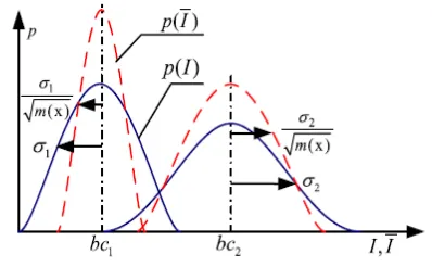
Refer to the red dashed curves in Fig 3.1the overlapping tails of the distributions are suppressed to some extent. Therefore, the misclassification caused by the overlapping intensity can be alleviated to some extent in the transformed domain R(T). In the following, we will design our energy functional on the domain R(T) by means of the well-defined relationship between domain D(T) and R(T).
Figure 1. Original image intensity domain (blue solid curves) and the transformed domain (red dashed
curves)
B. Energy Minimization
The whole minimization procedure consists of the following three steps, which are implemented iteratively.
• Keep u fixed, optimize and update the variable sets c, b, and σ.
• Keep c, b, and σ fixed, optimize and update u.
• Check whether the convergence has been reached. If not, return to 1).
Everything is detailed in Matlab Program(c,b,σ).
C. Advantages Of The MLTD Model
If we set to be, the membership function of region to be a truncated Gaussian window will be the same as the CLIC energy functional with except for some trivial constant factors. In addition our MLTD model the CLIC model can be specially viewed and the MLTD will be more accurate to model the image with intensity in homogeneity than the CLIC model. It is claimed that the CLIC model will yield a hard classification (the image intensity in a fixed position only belongs to one tissue). Differently, we will discuss that the proposed MLTD model leads to a soft classification (image intensity in a fixed position belongs to all tissues with a corresponding probability).
In medical imaging, partial volume voxels often have
an intensity composed of multiple class
partial volume voxels in low resolution datasets. The partial volume effect severely influences the accuracy of estimated bias field if a hard classification method is adopted because the hard classification method assumes the image intensity in a fixed position only belongs to one tissue. Our proposed methods are soft classification methods which assume that intensity of each tissue is composed of multiple class intensities, thereby alleviating partial volume effect to some extent.
D. Maximum Likelihood Algorithm
The MLTD method is considered to be one of best estimation methods in statistics. For example, in adult female penguins but be unable to measure the height of every single penguin in a population due to cost or time constraints.
The unknown mean and variance of heights are normally distributed as we assumed, the mean and variance can be estimated with MLE while only knowing the heights of some sample of the overall population. The mean and variance would accomplish by MLE as parameters and finding particular parametric values that make the observed results the most probable given the model.
V.RESULTS AND DISCUSSION
Original image
Bias field
Segmented result
Histogram of original Image
Histogram of Bias Corrected Image
However, there still exist some small dot regions that are misclassified due to the strong noise.
In the sagittal slice fig. compares results by MLTD and RMLTD, CLIC and VLS on a 3-tesla MRI. The estimated bias fields, bias corrected images, and segmentation results are shown 6.7, 6.8, 6.9 respectively. The histogram of the original MRI image and the histograms of the bias corrected images by our methods, CLIC and VLS are shown. From the histograms of the bias corrected images recovered by MLTD, CLIC, and VLS, we see that the histograms of specific tissues approximately satisfy Gaussian distribution but with significantly different variances. These results validate that MLTD model is better consistent with the intensity distribution of the image with intensity in homogeneity than CLIC and VLS which do not consider the variance of intensities belonging to different tissues. Testing competing methods on Brain normal subjects (1 mm isotropic spacing, no added noise, discrete anatomical labeling) with stimulated bias fields with 0%, 20%,and 40% intensity in homogeneity, respectively. From Mc Gill Brain. The 5%, 10%, and 20% levels of Gaussian noises are then added to these images. Moreover ,the segmentation results of MLTD are much closer to the brain anatomy than CLIC and VLS.
Application to Simultaneous Segmentation and Bias Correction
Finally, show the segmentation results of MLTD method on a 7T MRI image. The original images, estimated bias fields, and the bias corrected images are shown in fig 6.1, 6.2, 6.3 respectively. Obviously, the image qualities are significantly improved and some regions whose intensity contrast is too low to be identified are able to be distinguished easily after correction.
VI. REFERENCES
[1] C. Li, C. Xu, A. Anderson, and J. Gore, “MRI tissue classification and bias field estimation based on coherent local intensity clustering A unified energy minimization framework,” in Proc. Inf.
Process. Med.Image, Williamsburg, VA, USA, 2009, pp. 288–299.
[2] C. Li, C. Gatenby, L. Wang, and J. Gore, “A robust parametric method for bias field estimation and segmentation of MR images,” in Proc.IEEE Conf. Comput. Vis. Pattern Recognit., Miami, FL, USA, 2009,pp. 218–223.
[3] B. Wang, X. Gao, D. Tao, and X. Li, “A nonlinear adaptive level set for image segmentation,” IEEE Trans. Cybern., vol. 44, no. 3, pp. 418–428,Mar. 2014.
[4] X. Gao, B.Wang, D.Tao, and X. Li, “A relay level set method for automatic image segmentation,” IEEE Trans. Syst., Man, Cybern. B,Cybern., vol. 41, no. 2, pp. 518–525, Apr. 2011.
[5] U.Vovk, F. Pernus, and B. Likar, “A review of methods for correction of intensity in homogeneity in MRI,” IEEE Trans. Med. Imag., vol. 26,no. 3, pp. 405–421, Mar. 2007.
[6] E. McVeigh, M. Bronskill, and R .Henkelman, “Phase and sensitivity of receiver coils in magnetic resonance imaging,”Med. Phys., vol. 13,no. 6, pp. 806–814, Nov./Dec. 1986.
[7] A.Simmons, P.Tofts, G. Barker, and S. Arridge, “Sources of intensity non-uniformity in spin echo images at 1.5 t,” Magn. Reson. Med., vol. 32, no. 1, pp. 121–128, Jul. 1994.
[8] A. Fan et al., “A unified variational approach to de-noising and bias correction in MR,” in Proc. Inf. Process. Med. Imag., Ambleside, U.K.,Jul. 2003, pp. 148–159.
[10] B. Brinkmann, A. Manduca, and R. Robb, “Optimized homomorphic unsharp masking for MR grayscale in homogeneity correction,” IEEE Trans. Med. Imag., vol. 17, no. 2, pp. 161–171, Apr. 1998.
[11] P. Vemuri, E. Kholmovski, D. Parker, and B. Chapman, “Coil sensitivity estimation for optimal SNR reconstruction and intensity in homogeneity correction in phased array MR images,” in Proc. Inf. Process. Med.Imag, Glenwood Springs, CO, USA, 2005, pp. 603–614.
[12] R. Guillemaud and M. Brady, “ Estimating the bias field of MR images,” IEEE Trans. Med. Imag., vol. 16, no. 3, pp. 238–251, Jun. 1997.
[13] K. Leemput, F. Maes, D. Vandermeulen, and P. Suetens, “Automated model-based bias field correction of MR images of the brain,” IEEE Trans. Med. Imag., vol. 18, no. 10, pp. 885–896, Oct. 1999.
[14] D. Pham and J. Prince, “Adaptive fuzzy segmentation of magnetic resonance images,” IEEE Trans. Med. Imag., vol. 18, no. 9, pp. 737– 752, Sep. 1999.