Abebaw Bitew, Birhanemeskel Tegene, Biruk Yeshitila, Rawleigh Howe, Ebba Abate,Mulat Dagnew. Ethiop Med J, 2019, Supp. 1
ORIGINAL ARTICLE
BACTERIAL PROFILE, ANTIBACTERIAL SUSCEPTIBILITY PATTERN AND
AS-SOCIATED FACTORS AMONG WOMEN ATTENDING ANTENATAL AND
POST-NATAL HEALTH SERVICES AT THE UNIVERSITY OF GONDAR TEACHING
HOSPITAL, NORTHWEST ETHIOPIA.
Abebaw Bitew, MSc1*, Birhanemeskel Tegene, MSc2, Biruk Yeshitila, MSc3, Rawleigh Howe, MD, PhD3, Ebba Abate, PhD4, Mulat Dagnew, PhD 2
ABSTRACT
Background: Bacterial infections are an important cause of maternal morbidity and mortality especially in re-source limited countries such as Ethiopia. The major bacterial infections include urinary tract infections, septice-mia and endometritis. Antibiotic resistant bacterial pathogens have become a growing problem worldwide and pose a serious threat to vulnerable populations, including mothers. However, studies which address the problem in the Ethiopian setting are scarce.
Objective: To assess the bacterial profile, antibacterial susceptibility pattern and associated factors among moth-ers attending antenatal and postnatal care health services.
Methods: A cross-sectional study was conducted on 222 study participants at the University of Gondar Teaching Hospital from January 1 to May 31, 2016. Clinical specimens such as urine, blood and cervical discharge speci-mens were collected from patients and antimicrobial susceptibility tests conducted following standard procedures. Data were entered and analyzed with SPSS version 20. Bivariate and multivariate logistic regression models were applied in data analysis.
Results: Out of 222 specimens collected, 57(25.7%) bacterial species were isolated. The predominant bacterial isolates from urine culture were Escherichia coli (24/47; 51.1%) and Staphylococcus aureus (16/47;34%). From blood cultures, Staphylococcus aureus (2/8; 25%), Coagulase negative staphylococci (2/8;25%), Klebsiella pneu-moniae (2/8;25%) and Streptococcus pyogenes 2/8(25%) were isolated. Neisseria gonorrheae (2/27;7.4%) was isolated from cervical discharge cultures. The majority of the isolates were resistant to amoxacillin and ampicillin but susceptible to ceftriaxone. Many multidrug resistant bacterial species were isolated. Being in the first trimes-ter of pregnancy and having a history of diabetes mellitus were strongly associated with the presence of bactrimes-terial infections.
Conclusion: The overall prevalence of bacterial infections was high with many being resistant to commonly pre-scribed antimicrobial agents. This calls for an urgent need to conduct screening of bacterial infections in both antenatal as well as postnatal women.
Keywords: antibiotic susceptibility pattern, bacterial profile, associated factors
INTRODUCTION
Bacterial infection is an important cause of maternal morbidity and mortality. The most common infec-tions during pregnancy and following delivery in-clude urinary tract infections (UTI), septicemia and endometritis (1-4). UTI is a common urinary tract problem among pregnant women. Its complication usually begins at about the sixth week of pregnancy and peaks between the second to twenty-fourth week due to urethral dilatation, increased bladder volume, decreased bladder and urethral tone, all of which contribute to increased urinary stasis.
Up to 70% of pregnant women develop glucosuria which encourages bacterial growth in the urine. The highest incidence of UTI occurs in women of child bearing age and this has been linked to sexual activ-ity and aging (5). The predominant organisms that cause UTI are Escherichia coli (E. coli), Staphylo-coccus aureus (S. aureus), Klebsiella pneumoniae (K. pneumoniae), Pseudomonas aeruginosa (P. aeruginosa), Proteus spp, Streptococcus faecalis and Enterobacter spps (6). E. coli and S. aureus are ma-jor pathogens that have been associated with UTI and they underpin the empiric choice of antibiotics (7).
1 Debre Markos University, Department of Medical Microbiology.
2 Department of Medical Microbiology, School of Biomedical and Laboratory Sciences, College of Medicine and
Health Sciences, University of Gondar. 3Armaur Hanson Research Institute, Ethiopia. 4Ethiopian Public Health
Institute.
Sepsis is the consequence of the body’s inflamma-tory response to bacterial endotoxins and exotoxins. Pregnant and postpartum women are particularly vulnerable to developing severe sepsis because of their high susceptibility to bacterial infection due to changing physiology, biochemistry and immune re-sponse during pregnancy (8). This leads to an as-cending postnatal infection of the genital tract, due to onset of the rupture of membranes or prolonged la-bour (9, 10). It was a major cause of peripartum morbidity and mortality throughout the pre-antibiotic era (11). Still, it is an important cause of maternal mortality in developing countries, and younger moth-ers have a higher risk, because of their higher rates of contamination with bacteria (12, 13). The major pathogens that cause sepsis are Group A Streptococ-cus (GAS), E. coli, S. aureus, S. pneumoniae, methi-cillin-resistant S. aureus (MRSA), Clostridium septi-cum (C. septisepti-cum)and Morganella morganii (M. morganii) (14).
Endometritis an infection of the lining of the uterus, due to the rise of vaginal flora through the cervix and into the uterus. In the vagina, there is an overgrowth of pathogenic bacteria, which results in the reduction of hydrogen peroxide-producing lactobacilli. This condition aggravates preterm delivery, low birth weight and late onset postnatal endometritis in moth-ers. The most commonly isolated organisms include N. gonorrheae, Ureaplasma urealyticum (U. urea-lyticum), Peptostreptococcus, Gardnerella vaginalis (G. vaginalis), Bacteroides, Group B Streptococcus (GBS), and Chlamydia (15). Currently drug resistant pathogens are an increasing problem worldwide (16). The rates of MRSA and Gram negative cepha-losporin-resistant (extended-spectrum beta-lactamase) infections have increased globally by 2.1% and 12% for UTI, respectively (17, 18, 19). Generally, around the time of childbirth, bacterial infections are the leading causes of maternal mortal-ity, accounting for about one tenth of the global bur-den of deaths (20). Annually, it leads to an estimated 358,000 deaths in the world of which 99% occur in developing countries. Sub Saharan Africa alone ac-counts for half of the deaths in developing countries. Apart from death, women who experience peripar-tum infections are prone to severe morbidity and long-term disabilities such as chronic pelvic pain, fallopian tube blockage and secondary infertility (21, 22, 23, 24). In the literature, pre and postnatal mater-nal infections have been associated with several risk factors, such as malnutrition, diabetes, obesity, se-vere anemia, premature rupture of membranes, mul-tiple per vaginal examinations, manual removal of the placenta, caesarean section, use of steroids and the lack of pre-incision antimicrobial care (25).
Screening and treatment of bacterial infections dur-ing the pre and postnatal periods have improved ma-ternal health outcomes and reduced the prevalence of drug resistant pathogens. However, there is a scar-city of studies in the medical literature from devel-oping countries such as Ethiopia which report on the bacterial profile and antibiotic susceptibility patterns of bacteria isolated from prenatal and post-natal women.
MATERIALS AND METHODS
A cross sectional study was conducted from January 1 to May 31, 2016 at the University of Gondar Teaching Hospital of Amhara region. The study population consisted of all mothers who presented with different clinical complaints in either the ante-natal or postante-natal periods. A total of 222 individuals suspected for urinary tract infection, septicemia or endometritis who self-reported to the hospital during the study period were examined. Convenient sam-pling technique was used to select the study partici-pants from the University of Gondar Teaching Hos-pital antenatal and postnatal clinic. All mothers who presented consecutively during the study period were included in the study.
A minimum of 1 to 10 blood-to-broth ratio was maintained. The blood culture broths were incubated at 37oC and checked for signs of bacterial growth daily up to 7 days. Bottles were examined for the visible evidence of bacterial growth such as turbidity, hemolysis, and presence of gas bubbles. Bottles which showed signs of growth were subcultured onto BAP, MAC, and MSA at 37oC for 24-48 hours aero-bically while chocolate agar plates (CAP) were incu-bated at 37oC for 24-48 hours with 10% carbon diox-ide (CO2). Blood culture broths with no bacterial growth were subcultured onto BAP, CAP, MSA and MAC plates, before being reported as negative (26). Gram staining was performed to differentiate Gram positive and Gram-negative bacteria.
Biochemical tests were conducted to specifically identify different pathogenic bacterial species from positive culture media. The collected cervical swabs were inoculated onto non-selective CAP and selec-tive agar modified Thayer-Martin medium (MTMM) since some fastidious bacterial species are more sus-ceptible to concentrations of vancomycin, colistin, nystatin and trimethoprim lactate (VCNT) which are used in the selective media. The inoculated plates were incubated at 37°C for 72 hours with 10% CO2 by using a candle jar (26). Subsequently oxidase test and Gram stains were performed. N. gonorrheae was differentiated from other Neisseria species by its characteristic of producing acid only from glucose. Identification of bacteria was based on colony mor-phology and Gram reactions. Biochemical character-istics such as Catalase, Coagulase, Novobiocin, Ba-citracin and Optochin sensitivity test results were used to identify Gram positive pathogenic bacteria. Triple sugar iron agar TSI, indole test, motility test, urea test, hydrogen sulphide production (H2S), citrate test, Lysine decarboxylase (LDC) and oxidase tests were used for identification of Gram negative bacte-ria. All identified clinical strains were subjected to in vitro susceptibility testing using the Kirby Bauer disk diffusion method as described in Clinical Laboratory Standard Institute (CLSI) (2016) guidelines and in-terpreted accordingly (26).
The antibiotic discs used for UTI were norfloxacillin (10µg), nitrofurantoin (300µg), ciprofloxacillin (5µg), trimethoprim-sulfamethoxazle (1.25/23.75µg), tetracycline (30µg), amoxacillin (10µg), ampicillin (10µg), ceftriaxone (30µg), gentamycin (10µg) and amikacin (30µg). Antibiotic discs used for septice-mia caused by Gram-positive bacteria were penicillin (10IU), cloxacillin (30μg) and clindamycin (2µg).
Gentamycin (10μg), nalidixic acid (30μg), ciproflox-acillin (5µg) and amikacin (30µg) were used for Gram negative organisms. Certain antibiotics such as amoxicillin (10μg), ampicillin (10μg),cefoxitin (30µg), cefixime (5µg), ceftriaxone (30 µg), trimethoprim-sulfamethoxazole (1.25/23.75 µg), ceftazidime (30 µg) were used to test the antibiotic susceptibility profile of both Gram positive and Gram negative organisms. Antibacterial susceptibil-ity tests for gonococci were conducted on Muller Hinton Agar (MHA) containing 5% sheep blood for the following antimicrobial agents: tetracycline (30μg), penicillin (10 IU), ciprofloxacin (5μg), ceftri-axone (30μg), spectinomycin(100μg). Plates were incubated at 37oC for 18- 24hours with 10% CO2. Data were entered and statistically analyzed by SPSS version 20 software. Bivariate and multivariate logis-tic regressions models were applied to determine possible associated factors with bacterial infections. Odds ratio was used as a measure of the strength of association and reported with 95% confidence inter-vals (95%CI) to determine statistically significant factors associated with presence of bacterial infec-tion. P value ≤ 0.05 was considered to be statistically significant.
In order to maintain the quality of the data and labo-ratory tests, data collectors and labolabo-ratory assistants were trained. Daily collected data was recorded and compiled. All steps in data collection and recording were monitored. The reagents were checked for ex-piry date and appropriate storage under recom-mended temperature and humidity conditions. Stan-dard operating procedures were prepared and strictly followed. The quality of culture media and antim-icrobial susceptibility testing were checked by using quality control standard strains of E. coli (ATCC
25922), S. aureus (ATCC 25923), E. faecalis (ATCC
29212) and K. pneumoniae (ATCC@BAA1705).
McFarland standards (0.5% BaSO4) were used to standardize the inoculum density of bacterial suspen-sions for susceptibility testing. The acceptance range of optical density of the 0.5% McFarland standards was 0.08-0.1 (CLSI, 2016).
RESULTS
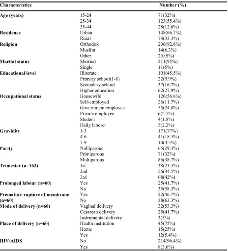
The majority of the study participants were urban dwellers (66.7%), Orthodox Christian (92.8%), mar-ried (95%) and housewives (56.8%). In the present study 162 (73%) of the women were on antenatal care (ANC) follow up; of these 68 (42%)
were in their third trimester of pregnancy. Sixty (27%) of the participants were on postnatal care follow up and were admitted after delivery (Table 1).
Table 1: Sociodemographic, obstetric and clinical variables from women attending antenatal and postnatal services at the University of Gondar Teaching Hospital January 1, 2016–May 31, 2016. (N=222*).
Characteristics Number (%)
Age (years) 15-24 71(32%)
25-34 123(55.4%)
35-44 28(12.6%)
Residence Urban 148(66.7%)
Rural 74(33.3%)
Religion Orthodox 206(92.8%)
Muslim 14(6.3%)
Other 2(0.9%)
Marital status Married 211(95%)
Single 11(5%)
Educational level Illiterate 101(45.5%)
Primary school(1-8) 22(9.9%)
Secondary school 37(16.7%)
Higher education 62(27.9%)
Occupational status Housewife 126(56.8%)
Self-employed 26(11.7%)
Government employee 55(24.8%)
Private employee 6(2.7%)
Student 4(1.8%)
Daily laborer 5(2.2%)
Gravidity 1-3 171(77%)
4-6 41(18.5%)
7-9 10(4.5%)
Parity Nulliparous 65(29.3%)
Primiparous 71(32%)
Multiparous 86(38.7%)
Trimester (n=162) 1st 38(23.5%)
2nd 56(34.5%)
3rd 68(42%)
Prolonged labour (n=60) Yes 25(41.7%)
No 35(58.3%)
Premature rupture of membrane (n=60)
Yes 22(36.7%)
No 38(63.3%)
Mode of delivery (n=60) Vaginal delivery 32(53.3%)
Cesarean delivery 25(41.7%)
Instrumental delivery 3(5%)
Place of delivery (n=60) Health institution 45(75%)
Home 15(25%)
Yes 12(5.4%)
HIV/AIDS No 214(96.4%)
Yes 8(3.6%)
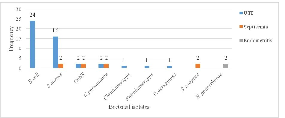
Figure 1. Profile and frequency of bacterial isolates from mothers during antenatal and postnatal period at Univer-sity of Gondar teaching Hospital from January 1, 2016 –May 30, 2016.
Isolation of pathogenic bacteria from different in-fection sites
A total of 134 urine, 61 blood and 27 cervical dis-charge samples were collected from patients in our study. The choice of which specimen to collect was determined by the chief complaint of the presenting women at the University of Gondar Teaching Hospi-tal during the study period. From these suspected clinical specimens, bacterial infection was present in urine samples from 47(35.1%) individuals. Likewise, the presence of bacterial infection was confirmed in 8(13.1%) blood samples and in 2(7.4%) cervical dis-charge samples. Overall, pathogenic bacteria were isolated in 57(25.7%) of the women. Of these, 47 (35.1%) were isolated from UTI suspected partici-pants, 8(13.1%) and 2(7.4%) were isolated from blood and from cervical discharge samples respec-tively.
The prevalence of bacteria differed based on the sample type; for instance a high proportion (61.7%) of Gram negative bacteria were isolated from urine. On the other hand, a higher rate of Gram positive bacteria was isolated from blood infections (75%). The predominant bacterial isolates from urine culture were E. coli (24/47; 51.1%), S. aureus (16/47;34%), K. pneumoniae (2/47;4.3%) and CoNS (2/47;4.3%) . On the other hand, the most common bacterial iso-lates identified from blood culture were S. aureus (2/8;25%), CoNS (2/8;25%), K. pneumoniae (2/8;25%), and S. pyogenes (2/8;25%). N. gonor-rheae was the only pathogenic bacteria isolated from cervical discharge samples (2/2; 100%) (Figure 1).
Antibacterial susceptibility pattern for bacterial iso-lates from urine samples
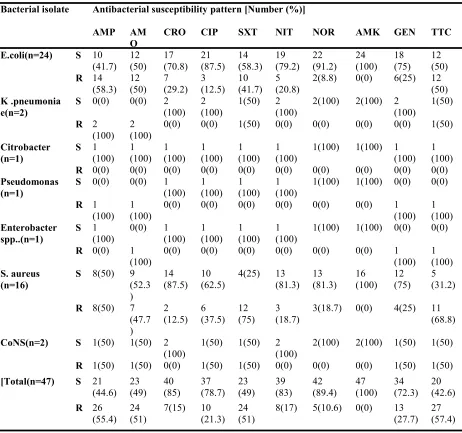
Antibacterial susceptibility testing was done for pure isolates obtained from urine samples. All pathogens isolated from urine were susceptible to amikacin (100%) and most to norfloxacillin (86.5%), nitrofu-rantoin (80.8%), and ceftriaxone (69.2%). Among Gram negative bacteria, all K. pneumoniae isolates were resistant to ampicillin and amoxicillin (100%). E. coli isolates were resistant to amoxicillin (58.3%), and trimethoprim-sulphamethoxazole (41.7%) (Table 2).
Table 2: Antibacterial susceptibility pattern of Gram negative and Gram positive UTI causing bacterial pathogens from women attending antenatal and postnatal services at the University of
Gondar Teaching Hospital from January 1 – May 31, 2016.
Bacterial isolate Antibacterial susceptibility pattern [Number (%)]
AMP AM O
CRO CIP SXT NIT NOR AMK GEN TTC
E.coli(n=24) S 10 (41.7)
12 (50)
17 (70.8)
21 (87.5)
14 (58.3)
19 (79.2)
22 (91.2)
24 (100)
18 (75)
12 (50) R 14
(58.3) 12(50) 7(29.2) 3(12.5) 10(41.7) 5(20.8) 2(8.8) 0(0) 6(25) 12(50) K .pneumonia
e(n=2) S
0(0) 0(0) 2 (100)
2 (100)
1(50) 2 (100)
2(100) 2(100) 2 (100)
1(50) R 2
(100) 2(100) 0(0) 0(0) 1(50) 0(0) 0(0) 0(0) 0(0) 1(50) Citrobacter
(n=1) S
1 (100)
1 (100)
1 (100)
1 (100)
1 (100)
1 (100)
1(100) 1(100) 1 (100)
1 (100) R 0(0) 0(0) 0(0) 0(0) 0(0) 0(0) 0(0) 0(0) 0(0) 0(0) Pseudomonas
(n=1)
S 0(0) 0(0) 1
(100) 1(100) 1(100) 1(100) 1(100) 1(100) 0(0) 0(0) R 1
(100) 1(100) 0(0) 0(0) 0(0) 0(0) 0(0) 0(0) 1(100) 1(100) Enterobacter
spp..(n=1)
S 1
(100) 0(0) 1(100) 1(100) 1(100) 1(100) 1(100) 1(100) 0(0) 0(0) R 0(0) 1
(100) 0(0) 0(0) 0(0) 0(0) 0(0) 0(0) 1(100) 1(100) S. aureus
(n=16)
S 8(50) 9 (52.3 )
14
(87.5) 10(62.5) 4(25) 13(81.3) 13(81.3) 16(100) 12(75) 5(31.2) R 8(50) 7
(47.7 )
2
(12.5) 6(37.5) 12(75) 3(18.7) 3(18.7) 0(0) 4(25) 11(68.8) CoNS(n=2) S 1(50) 1(50) 2
(100) 1(50) 1(50) 2(100) 2(100) 2(100) 1(50) 1(50) R 1(50) 1(50) 0(0) 1(50) 1(50) 0(0) 0(0) 0(0) 1(50) 1(50) [Total(n=47) S 21
(44.6) 23(49) 40(85) 37(78.7) 23(49) 39(83) 42(89.4) 47(100) 34(72.3) 20(42.6) R 26
(55.4) 24 (51)
7(15) 10 (21.3)
24 (51)
8(17) 5(10.6) 0(0) 13 (27.7)
27 (57.4)
AMP=Ampicillin; AMO=Amoxicillin; NIT=Nitrofurantoin; NOR=Norfloxacin; CIP=Ciprofloxacin; SXT=Trimethoprim-sulphamethoxazole; CRO=Ceftriaxone; GEN= Gentamicin; AMK=Amikacin; TTC= tetracy-cline
CoNS= Coagulase -negative staphylococcus; S= Susceptible; R= Resistant Antibacterial susceptibility pattern for bacterial
isolates from blood samples
The majority of Gram positive bacterial isolates from blood were resistant to ceftazidime (83.3%) and trimethoprim-sulphamethoxazole (66.7%).
Table 3: Antibacterial susceptibility pattern of Gram positive bacterial pathogens causing septicemia
in women attending antenatal and postnatal services at the University of Gondar Teaching Hospital, from January 1, 2016 – May 31, 2016.
AMO=Amoxacillin; AMP=Ampicillin; P=Penicillin; CAZ= Ceftazidime;SXT=Trimethoprim-sulphamethoxazole; CXM=Cefixime; CXC=Cloxacillin; DA=Clindamycin; CXT=Cefoxitin; CoNS= Coagulase-negative staphylococ-cus; S=Susceptible; R= Resistant.
Bacterial isolate Antibacterial susceptibility pattern
AMO AMP CRO P CAZ SXT CXM CXC DA CXT
S. aureus (n=2)
S 1(50) 1(50) 1(50) 1(50) 1(50) 1(50) 1(50) 1(50) 1(50) 1(50) R 1(50) 1(50) 1(50) 1(50) 1(50) 1(50) 1(50) 1(50) 1(50) 1(50) CoNS
(n=2)
S 1(50) 1(50) 1(50) 2 (100)
0(0) 0(0) 0(0) 1(50) 2 (100)
2(100) R 1(50) 1(50) 1(50) 0(0) 2
(100) 2(100) 2(100) 1(50) 0(0) 0(0) S.
pyo-genes (n=2)
S 2(100) 2
(100) 2 (100)
2 (100)
0(0) 1(50) 2 (100)
2 (100)
2 (100)
1(50)
R 0(0) 0(0) 0(0) 0(0) 2
(100) 1(50) 0(0) 0(0) 0(0) 1(50) Total
(n=6) S 4(66.7) 4(66.7) 4(66.7) 5(83.3) 1(16.7) 2(33.3) 3(50) 4(66.7) 5(83.3) 4(66.7) R 2(33.3) 2
(33.3) 2(33.3) 1(16.7) 5(83.3) 4(66.7) 3(50) 2(33.3) 1(16.7) 2(33.3)
Antibacterial susceptibility pattern for bacterial iso-lates from cervical discharge samples
N. gonorrheae was the only pathogenic bacterial iso-late from cervical discharge samples. All isoiso-lates of N. gonorrheae were resistant to tetracycline (100%) and partially resistant to penicillin (50%), ceftriaxone (50%) and spectinomycin (50%). However all the N. gonorrheae isolates were sensitive to ciprofloxacin (100%).
Multidrug resistance of isolated bacterial pathogens Taking all the bacterial isolates from the different bio-logical samples into account, multidrug resistance (defined as resistance to ≥2 drugs of different classes) was observed in 54.5% and 95.8% of Gram negative and Gram positive bacteria respectively (Table 4). Risk factors associated with presence of bacterial infections
In the bivariate logistic analysis, demographic and clinical factors such as age, occupation, marital status, religion, level of education, gravidity and parity were not significantly associated with having bacterial in-fection.
Table 4: Multidrug resistance (MDR) of bacterial isolates from women attending antenatal and postnatal services at the University of Gondar Teaching Hospital from January 1, 2016 – May 31, 2016.
Antibiograms Bacterial isolates E.coli
(=24) K. pneu-moniae (n=4) Citro-bacter (n=1) Entero-bacter (n=1) P.
aerugi-nosa(n=1) N. gonor-rheae (n=2)
AMX,TTC 1(4.2%) - - - - -
AMX,CRO 1(4.2%) - - - - -
AMX,SXT,GEN,TTC 1(4.2%) - - - - -
AMP,CRO,SXT,NOR,GEN ,TTC
1(4.2%) - - - - -
AMX,CRO,SXT,TTC 2(8.4%) - - - - -
AMP,SXT,NOR,GEN,TTC 1(4.2%) - - - - -
AMX,SXT.TTC AMX,CPR,SXT,NIT,TTC SXT,GEN AMX,CRO,SXT,GEN,TTC ,NIT AMX,NIT
CPR,SXT,NIT, GEN, TTC CPR, CRO, NIT
AMX, GEN, TTC AMX, CRO, GEN, TTC P, CRO, TTC
Total =18(54.5%) 1(4.2%) 1(4.2%) 1(4.2%) 1(4.2%) 1(4.2%) 1(4.2%) 1(4.2%) - - - 13(54.2%) 2(50%) - - 2(50%) - - - - - - - - - - - - - - - - - - 1(100%) - - 1(100%) - - - - - - - - 1(100%) - 1(100%) - - - - - - - - - 1(50%) 1(50%) Antibiograms for Gram
positive bacteria
S. aureus
(n=18) CoNS (n=4) S. pyo-genes (n=2)
AMX, SXT,GEN, TTC 1(5.5%) - -
AMX,CPR,SXT,NOR,NIT,
GEN,TTC 1(5.5%) 1(25%) -
AMX,CRO,CPR,SXT,GEN ,TTC
1(5.5%) - -
CPR,SXT,NOR,NIT 1(5.5%) - -
AMP,CPR,SXT,NOR,TTC 1(5.5%) - -
AMP,CPR,SXT,NOR,GEN ,TTC
1(5.5%) - -
AMX, TTC 1(5.5%) - -
AMX, CPR, SXT, TTC 2(11.1%) - -
CPR, SXT, TTC 1(5.5%) - -
CRO, SXT, TTC 1(5.5%) - -
SXT, TTC 1(5.5%) - -
CPR, TTC 1(5.5%) - -
AMX, CPR, SXT, NIT, GEN
1(5.5%) - -
AMX, CPR, GEN, TTC 1(5.5%) - -
AMX, CRO, SXT 2(11.1% - -
AMP, SXT, CAZ, DA - 1(25%) -
AMP, CAZ - 1(25%) -
AMP, CAZ, CXC, SXT - 1(25%) -
CRO, SXT, CXC - - 2(100%)
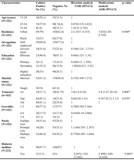
Table 5: Bivariate and multivariate analysis to assess factors associated with bacterial infections in
women attending antenatal and postnatal services at the University of Gondar Teaching Hospital, January 1, 2016 – May 31, 2016.
AOR=adjusted odds ratio; COR=crude odds ratio; 1=reference group; 95% CI=95% confidence interval.
Characteristics Culture Bivariate analysis
COR (95%CI) Multivariate analysis AOR (95%CI)
p-value Positive
No (%) Negative No (%)
Age (years) (N=222)
15-24 16(28.1) 55(33.3) 1
25-34 33(57.9) 90( 54.6) 0.87(0.315-2.422)
35-44 8(14) 20(12.1) 1 (0.41-2.716)
Residence (N=222)
Urban 45(79) 103(62.4) 2 (1.037- 4.315) 3.032(1.05-
8.729) 0.040*
Rural 12(21) 62(37.6) 1
Occupation
(N=222) Self-employed 39(68.4) 128(77.6) 1
Govern-ment
18(31.6) 37(22.4) 0.54(0.124 - 2.315)
Education
(N=222) Illiterate 23(40.4) 78(47.3) 0.66(0.323- 1.35)
Primary 3(5.3) 17(10.3) 0.42(0.11- 1.595)
Secondary 11(19.3) 26(15.8) 1.002(0.413- 2.43)
Higher
education 20(35) 44(26.7) 1
Marital status (N=222)
Married 52(91.2) 159(96.4) 0.37(0.109-1.273)
Single 5(8.8) 6(3.6) 1
Trimester (n=162)
1st 14(13.7) 24(43.75) 3.6(1.6-8.24) 5.8 (1.67-20.16) 0.006*
2nd 42(45.1) 14(21.9) 0.6(0.28-1.41) 0.367 (0.12-1.12) 0.078*
3rd 46(41.1) 22(34.4) 1 1
Gravidity (N=222)
1-3 44(77.2) 127(77) 0.78(0.194-3.166)
4-6 10(17.5) 31(17.8) 0.656(0.14-3.066)
7-9 3(5.3) 7(4.2) 1
Parity (N=222)
Nullipa-rous 18(31.6) 47(28.5) 1
Primipa-rous 16(28) 55(33.3) 1.114(0.538- 2.307)
Multipa-rous 23(40.4) 63(38.2) 0.779(0.369- 1.646)
Diabetes mellitus (N-222)
No
50(87.7)
160(97)
1
1
Yes 7(12.3) 5(3)
Multivariate analysis was carried out on covariates which showed significant association with bacterial infection in the bivariate analysis at a p-value of ≤ 0.2. The backward stepwise regression was em-ployed for controlling the confounding effect. Being an urban dweller, in the first trimester of pregnancy and having a history of diabetes mellitus were highly associated with the presence of bacterial infections (p ≤ 0.05). Moreover, women who were in their first trimester were 4.8 times more likely to develop bac-terial infections as compared to those in their second trimester (AOR= 4.8;95% CI:1.841-12.761, p=0.001). Likewise, women with diabetes mellitus (DM) were 5 times more likely to develop a bacterial infection as compared to those without DM (AOR= 5;95%CI:1.621-16.620, p=0.003) (Table 5).
DISCUSSION
The present study has identified the bacteria believed to be responsible for causing a range of clinical manifestations that are indicative of bacterial infec-tions in mothers attending antenatal and post-natal health services. In this study, the overall prevalence of bacterial infection during the antenatal and postna-tal period was 25.7%, indicating that pregnant women, as well as women who have delivered, con-tinue to be vulnerable to contracting bacterial infec-tions. This could have adverse repercussions on the health of neonates. Thus the current findings have important public health significance. The findings in the present study mirror findings reported from other studies from Ethiopia and elsewhere (27, 28, 29, 30, 31).
In this study, the prevalence of symptomatic UTI was 35.1% which is comparable to studies reported from Addis Ababa (31.6%) and also India (36.1%) (27, 31) but higher than in reports from Gondar (17.9%), Bahir Dar (18.9%), Dire Dawa (17%), Tanzania (17.9%) and another study from India (20.1%) (33, 34.35,36, 37). However a study from Nigeria has reported a much higher prevalence of symptomatic UTI (55%) (38).
The various estimates could reflect differences in sample size, laboratory methods employed and geo-graphical location. E. coli, S. aureus, CoNS, K. pneu-moniae, Citrobacter spp, Enterobacter spp, and P. aeruginosa were the most common bacterial patho-gens causing UTI in pregnant women in this study which agrees with reports by other investigators (5, 6, 33, 34, 35).
The most common bacterial isolates from urine sam-ples were Gram negative bacterial pathogens, which is also consistent with what is reported in the litera-ture (5, 30, 35). In our study, the second most pre-dominant isolate was S. aureus which is similar to what has been observed in other studies conducted in Ethiopia (5, 34, 35).
In this study, 13.1% of pathogenic bacteria were identified from blood samples. This finding is lower than reports from other studies conducted in Ethiopia (19.7%, 8.8%, 28%, 24%) (39- 42), Sudan (72.6%) (28) and the Netherlands (46%) (29). The possible explanation for the differences in bacterial preva-lence between the different studies could be the sam-ple size used, blood culture system, duration of study time, seasonal variation, nature of patient popula-tions, and differences in infection control practices of the different hospitals (41,42 ).
In the current study, 75% and 25% of the blood in-fections were due to Gram positive and Gram nega-tive bacteria respecnega-tively. The preponderance of in-fections from blood cultures were also due to Gram positive bacteria in other studies from different parts of Ethiopia which included Jimma, Mekele and Gon-dar (39,40,41,42). In this study, S. aureus, CoNS, S. pyogenes and K. pneumoniae were identified from blood samples. More or less similar observations have been recorded in different studies and countries (39, 40, 41, 42). In the present study, S. aureus was isolated in similar proportions as other bacteria (25%). In contrast, S. aureus was the predominant isolate in other studies from Ethiopia (39, 40). Out of the 27 cervical discharge samples, N. gonor-rhea was isolated from 7.4% of the women, which concurs with data from Sub Saharan African coun-tries (43, 44, 45). However, our result was lower than the findings from Gambella and another study from Gondar which reported 11.3% and 38.4% prevalence of N. gonorrhea respectively (46). This variation may be due to the difference in the diagnostic meth-ods used to detect N. gonorrhea infection. The study cited previously from Gondar used syndrome-based diagnosis and not laboratory confirmation of the etio-logic agent, possibly resulting in an over estimation (47).
On a positive note, in the present study, S. aureus isolates were wholly susceptible to amikacin. Three other antibiotics (ceftriaxone, nitrofirantoin and nor-floxacillin) were reasonably effective to treat S. aureus infections (88.2%, 76.5%, and 76.5% respec-tively). This finding was in line with studies from Dire Dawa (35) and Tanzania (51). A study from Iran reported a lower efficacy of these antibiotics to treat S. aureus infections (52) while an Indian study found these antibiotics to be very effective (31). In the present study, E. coli showed significant resis-tance to amoxacillin (58.3%), comparable to findings from Bahir Dar (34) and Nigeria (38). Likewise most Gram positive isolates such as S. aureus showed sign i f i c a sign t r e s i s t a sign c e t o t r i m e t h o p r i m -sulphamethoxazole (76.5%) and tetracycline (70.6%). CoNS showed a significant level of resis-tance to tetracycline (66.7%), amoxacillin (66.7%), ampicillin (66.7%), trimethoprim-sulphamethoxazole (66.7%) and gentamicin (66.7%), which was more or less comparable to findings from Nigeria (38) and India (48).
Different antibacterial susceptibility patterns were observed for bacterial isolates from blood. S. aureus was resistant to methicillin in the present study. In this study, we screened for MRSA using Cefoxitin which has been reported to be a surrogate marker for MRSA. The presence of MRSA in pregnant women may cause sepsis that could be transmitted to the fetus (14). K. pneumoniae isolates from blood were resistant to ceftazidime and trimethoprim-sulphamethoxazole, amoxacillin and ampicillin. The increased resistance of blood isolates in this study may be a signal of indiscriminate and continuous usage of sub-therapeutic doses of commonly avail-able drugs in the public health sector (39, 40). This could very well challenge the management and treat-ment of patients.
N. gonorrheae was highly resistant to tetracycline and penicillin. This finding is similar to previous reports in Ethiopia and other countries 929, 44, 46, 53, 54, 55, 56). In our study, N. gonorrheae was sen-sitive to ciprofloxacin, contrary to findings from studies in Bahir Dar and Gambella, Ethiopia (44, 46). However, a low level of susceptibility to ceftriaxone was reported in our study. The introduction of syn-dromic management for sexually transmitted infec-tions (STIs), and the use of fluoroquinolones and cephalosporin to treat such infections is fairly rou-tine, although there is always the risk that this could create a selective advantage for bacteria which are resistant to these antimicrobial agents (44).
The findings from this study are alarming because they point to the resistance of bacteria to commonly used antimicrobial agents in a resource limited coun-try such as Ethiopia where there are few alternatives for the management of gonorrhea. The occurrence of untreated and complicated gonorrhea may fuel the transmission of HIV and other STIs.
In the current study, a significant level of multidrug resistance (resistance to ≥2 classes of drugs) was noted. From the total number of isolates, 54.5% and 95.8% of Gram negative and Gram positive bacteria, respectively, exhibited multidrug resistance. This situation raises serious concerns and suggests the existence of a high resistance gene pool, perhaps due to gross misuse and inappropriate usage of antimicro-bial agents (57). Taken together, these findings clearly show how resistant strains are expanding at an alarming rate in the area. With this trend an antibi-otic which was effective only one year ago, might be rendered useless in a year’s time. This creates a huge burden, especially for people living in resource lim-ited countries where many are not assured of getting their daily food, let alone afford expensive medica-tions such as new antibiotics. All this places an un-bearable strain on health care provision in poor coun-tries.
Conclusion
The overall prevalence of bacterial infections was high among women who attended antenatal and post-natal care in the study area. Most of the bacterial isolates were resistant to the most commonly pre-scribed antibiotics. This finding highlights the sig-nificant threat posed by the rise in antibiotic resis-tance, especially for such vulnerable population groups. Therefore, treatment of bacterial infections in the study area needs to be guided by antimicrobial susceptibility testing. An effective surveillance sys-tem needs to be instituted to monitor trends in antim-icrobial resistance. At the same time prudent use of existing antibiotics is warranted to avoid further es-calation of the problem. Longitudinal studies are recommended to track development of bacterial re-sistance resulting from usage of antimicrobials and also to determine further risk factors which predis-pose women in antenatal or postnatal care, to acquire bacterial infections that could be detrimental for them and their infants.
ACKNOWLEDGEMENTS
The study was funded by the Ministry of Health through the Clinical Research Capacity Building program at the Armauer Hansen Research Institute (AHRI). We would like to acknowledge study par-ticipants for their participation in the study. We are grateful to Dr. Dawit Kassahun (Gynecologist) for his support during specimen collection and manage-ment of patients. We would also like to acknowl-edge the Department of Medical Microbiology for availing necessary materials to conduct this study. Finally, we want to thank the Armauer Hansen Re-search Institute, the Federal Ministry of Health and the Amhara Regional Health Bureau for allowing us to conduct the study as well as for their technical, material and financial support.REFERENCES
1. Gravett CA, Gravett MG, Martin ET, Bernson JD, Khan S, Boyle DS et al Serious and life-threatening preg-nancy-related infections: Opportunities to reduce the global burden. PLoS Med. 2012; 9(10): doi:10.1371/ journal.pmed.1001324.
2. Dhar H, Al-Busaidi I, Rathi B, Nimre EA, Sachdeva V, Hamdi I. A study of post-caesarean section wound infections in a regional referral hospital. Clinical and Basic Res. 2014; 14(2): 211-217.
3. Lorenzo M. The journal of nutrition, postnatal development of intestinal microflora as influenced by infant nutrition. J Nutrition Italy. 2008; 138( 9):1791S-5S.
4. Neu J, Rushing J. Cesarean versus vaginal delivery: Long term infant outcomes and the hygiene hypothesis. Clinical Perinatal. 2011; 38(2):321–31.
5. Alemu A, Moges F, Shiferaw Y, Tafess K, Kassu A, Anagaw B et al. Bacterial profile and drug susceptibil-ity pattern of urinary tract infection in pregnant women at Universsusceptibil-ity of Gondar Teaching Hospital, North-west Ethiopia. BMC Res Notes. 2012, 5:197.
6. Obiogbolu CH, Okonko IO, Anyamere CO, Adedeji AO, Akanbi AO, Ogun AA, et al. Incidence of urinary tract infections among pregnant women in Akwa metropolis, southeastern Nigeria. Scientific Research and Essay. 2009; 4 (8):820-824.
7. Sriskandan S.In infectious diseases, national centre for infection prevention and management, department of infectious diseases & immunity, Imperial College London, UK. J R Coll Physicians Edinb.2011; 41: 339– 46.
8. Seale AC, Mwaniki M, Newton CR, Berkley JA. Maternal and early onset neonatal bacterial sepsis: burden and strategies for prevention in sub-Saharan Africa. Lancet Infect Dis. 2009; 9(7): 428–438.
9. Van Dillen J, Zwart J, Schutte J, van Roosmalen J. Maternal sepsis: epidemiology, etiology and outcome; Curr Opin Infect Dis. 2010; 23(3):249-54.
10. Mary T, Busowski LM, John D, Busowski, Akhter M. Puerperal Group A Streptococcal infection. Case Reports in Medicine 2013; doi.org/10.1155/2013/751329USA.
11. Glazener MA, Macarthure C.; Postnatal morbidity, puerperal sepsis; 2001;3(4):179-83.
13. Rimawi BH. Infectious Comorbidities Encountered in Obstetrics and Neonatology: Intraamniotic infections; OMICS Group eBooks. 2014: www.esciencecentral.org/ebooks.
14. Trabert B, Misra DP. Risk factors for bacterial vaginosis during pregnancy among African-American women; Am J Obstet Gynecol. 2007; 197(5): 477.
15. Gelband H, Delahoy M. Policies to address antibiotic resistance in low- and middle-income countries. Dis-cussion Paper. Center for Disease Dynamics, Economics & Policy. 2014:1-36. https://cddep.org/wp-content/ uploads/2017/06/abrinlmics_cddep_gelband_and_ delahoy_9-14. Pdf (Accessed Dec 9, 2018).
16. Mach KE, Baron EJ, Shih M, Gau V, Wong PK, Liao JC et al. A biosensor platform for rapid antimicro-bial susceptibility testing directly from clinical samples, university of arizona. Tucson, Arizona. J Urol. 2011; 185(1):148-53.
17. Royal College of Obstetricians and Gynecologists (RCOG): Bacterial sepsis following pregnancy. London (UK). Green-Top Guideline. No. 64b April 2012; 21(64): 1-21.
18. Kasolo F, Yahaya AA, Ndihokubwayo JB, Impouma B, Oxenford CJ, Cognat S. Guide for establishing laboratory-based surveillance for antimicrobial resistance; World Health Organization Regional Office for Africa. 2013:1-25.
19. Gilbert NM, O’Brien VP, Hultgren S, Macones G, Lewis WG, Lewis AL. Urinary Tract Infection as a preventable cause of pregnancy complications: opportunities, challenges, and a global call to action; Global Adv Health Med. 2013; 2(5):59-69.
20. Yudin MH, Money DM. Screening and management of bacterial vaginosis in pregnancy of Canada obstet’ gynaecol can; Ann Intern Med. 2008; 149(1):W20-4.
21. Conde-Agudelo A, Belizan JM, Lammers C. Maternal-perinatal morbidity and mortality associated with adolescent pregnancy in Latin America: Cross-sectional study; Am J Obstet Gynecol. 2004; 192:342–349. 22. Mitt P, Lang K, Peri A, Maimets M. Surgical-Site infections following cesarean section in an Estonian
uni-versity hospital: Post discharge surveillance and analysis of risk factors; Infect Control Hosp Epidemiol 2005; 26(5):449-454.
23. Dolea C, Stein C. Global burden of maternal sepsis in the year, World Health Organization, Geneva; Br Med Bull. 2003; 67 (1): 1-11.
24. Mason DM, Aronof KL. Postpartum Group A Streptococcus Sepsis and maternal immunology. Am J Re-prod Immunol. 2012; 67(2):91–100.
25. Rivlin ME, Alderman E. Postpartum endometritis and disseminated infection in both mother and neonate. Obstet Gynecol. 2012; 120(2):471-3.
26. Clinical and Laboratory Standards Institute. Performance standards for antimicrobial susceptibility testing: twenty-fourth informational supplement M100-S24. CLSI, Wayne, PA, USA, 2016.
27. Mengistie Z, Woldeamanuel Y, Asrat D, Adera A. Prevalence of bacterial vaginosis among pregnant women attending antenatal care in TikurAnbessa University Hospital, Addis Ababa, Ethiopia. BMC Res Notes. 2014; 7:822.
28. Oud L. Pregnancy-Associated Severe Sepsis: Contemporary State and Future Challenges. Infect Dis Ther-apy. 2014; 3 (2):175-189.
29. Ahmed MI, Babiker AR. Puerperal sepsis in a rural hospital in Sudan, susceptibility to antibiotics. Mat Soc Med. 2013; 25(1): 19-22.
30. Imade PE, Izekor PE, Eghafona NO, Enabulele OI, Ophori E. Asymptomatic bacteriuria among pregnant women. N Am J Med Sci. 2010; 2(6): 263.
31. SamagaMP. Bacteriological Profile of Urinary Tract Infections in Pregnant Women; Indian J Microbiol Res. 2016; 3(1):17-21.
32.Al-Badr A, Al-Shaikh A.Recurrent Urinary Tract Infections Management in Women. Sultan Qaboos Unive
Med J,2013; 13(3): 359–367. 33. Ferede G, Gizachew Y, Wondimeneh Y, Sisay Z. The Prevalence and antimicrobial susceptibility pattern of
bacterial uropathogens isolated from pregnant women, of Gondar, Ethiopia. Europ J Exp Bio 2012;2
(5):1497-502. 34. Demilie T, Beyene G, Melaku S, Tsegaye W. Urinary bacterial profile and antibiotic susceptibility pattern
among pregnant women in northwest Ethiopia; Ethiop J Health Sci. 2012; 22(2):121-128. 35. Derese B, Kedir H, Teklemariam Z, Weldegebreal F, Balakrishnan S. Bacterial profile of urinary tract in
36. Masinde A, Gumodoka B, Kilonzo A, Mshana SE. Prevalence of urinary tract infection among pregnant women at Bugando Medical Centre, Mwanza, Tanzania. Tanzan J Health Res. 2009; 11(3): 154-161. 37. Gupta A, Garg P, Nigam S. Bacterial Vaginosis in Pregnancy (<28 weeks and its effect on pregnancy
out-come: A study from a western up city. Indian J Clinical Practice. 2013; 23 (11):740-744.ed
38. Parveen K, Momen A, Ara Begum A, Begum M. Prevalence Of Urinary Tract Infection During Pregnancy. J Dhaka National Med Coll Hos. 2011; 17 (02): 8-12.
39. Asrat D, Amanuel YW. Prevalence and antibiotic susceptibility pattern of bacterial isolates from blood cul-ture in Tikur Anbassa Hospital, Addis Ababa, Ethiopia. Ethiop Med J. 2001, 39(2):97-104.
40. Tizazu Z, Kannan S, Yilma D, Beyene G. Invasive bacterial pathogens and their antibiotic susceptibility patterns in Jimma University Specialized Hospital, Jimma, and Southwest Ethiopia. Ethiop J Health Sci.
2011;21(1): 1-8. 41. Gebreyesus A, Negash L, Aregawi S et al. Bacteriological profile and drug susceptibility pattern of blood
culture isolates among febrile patients in Mekele hospital, Northern Ethiopia. SpringerPlus 2015;4:314 42. Ali J, Kebede Y. Frequency of isolation and antimicrobial susceptibility pattern of bacterial isolation from
blood culture in Gondar University Hospital. Ethiop. Med J. 2008; 46(2):155–161.
43. Taffa N, Bjune G, Sundby J, Gaustad P, Alsterom A. Prevalence of Gonococcal and Chlamydial Infections and Sexual Risk Behavior Among Youth in Addis Ababa, Ethiopia. Sex Transm Dis. 2002;29:828–33 44. Tibebu M,Shibabaw A, Medhin G, Kassu A.Neisseria gonorrhoeae non-susceptible to cephalosporins and
quinolones in Northwest Ethiopia. BMC Infect Dis. 2013;13:415.
45. Hailemariam M, Abebe T, Mihret A, Lambiyo T. Prevalence of Neisseria Gonorrhea and Their Antimicro-bial Susceptibility Patterns Among Symptomatic Women Attending Gynecology Outpatient Department in Hawassa Referral Hospital, Hawassa, Ethiopia. Ethiop J Health Sci. 2013; 23(1): 10–18.
46. Ali S, Sewunet T, Sahlemariam Z, Kibru G. Neisseria gonorrhea among suspects of sexually transmitted infection in Gambella hospital Ethiopia: Risk factors and drug resistance. BMC Res Notes 2016;9:439 47. Moges B, Yismaw G, Kassu A, Megabiaw B, Alemu S,Amare B et al. Sexually transmitted infections based
on the syndromic approach in Gondar town, northwest Ethiopia: a retrospective study. BMC Public Health. 2013; 13: 143.
48. Vaijanathrao CY, Nalini YL, Reddy CM. Antibiotic Sensitivity Pattern of Uropathogens: A Comparative Study between Symptomatic and Asymptomatic Bacteriuria in Pregnant Women. Int J Curr Microbiol App Sci. 2015; 4(6): 689-695.
49. Bager P, Simonsen J, Ethelberg S, Frisch M. Cesarean Delivery and Risk of Intestinal Bacterial infection, C-Section and Intestinal Infection, Denmark; JID. 2010; 201 (15):898-902.
50. Dagnew M, Yismaw G, Gizachew M, Gadisa A, Abebe T, Tadesse T et al. Bacterial profile and antimicro-bial susceptibility pattern in septicemia suspected patients attending Gondar University Hospital, Northwest Ethiopia. BMC Res Notes 2013; 6:283
51. Mpogoro FJ, Mshana SE, Mirambo MM, Kidenya BR, Gumodoka B, Imirzalioglu C. Incidence and pre-dictors of surgical site infections following caesarean sections at Bugando Medical Centre, Mwanza, Tanzania. Antimicrob Res Infect Control. 2014; 3:25.
52. Jalali M, Shamsi M, Roozbehani N, KabirK. Prevalence of Urinary Tract Infection and Some Factors Af-fected in Pregnant Women in Iran Karaj City Middle East. J Scientific Research. 2014; 20 (7): 781-785. 53. Shamna MS, KalaichelvanVK, FazilMarickar YM, Deepu S.Cesarean Section and Prophylactic Antibiotics;
IOSR Journal of Pharmacy and Biological Sciences.2014; 9 (2): 51-54.
54. Pokharel MS. Study on antibiotics sensitivity pattern of bacterial flora in cases of pre-labour rupture of mem-brane: Dissertation Kathmandu, Nepal: Tribhuvan University; 2004: 108.
55. Kankuriesko E, Kurkitatio T, Carison T, Esmaa HM. Incidence, treatment and outcome of peripartum sepsis. Acta Obstet Gynecol Scand. 2003; 82(8); 730-735.
56. Walker CK, Sweet RL.Gonorrhea infection in women: prevalence, effects, screening, and management. Int J Womens Health. 2011; 3: 197–206.
57. Komolafe AO, Adegoke AA. Incidence of bacterial septicemia in life Metropolis, Nigeria. Malaysian J Mi-crobiol 2008; 4(2): 51-61.