R E S E A R C H
Open Access
Control of post-translational modifications in
antithrombin during murine post-natal
development by miR-200a
Raúl Teruel, Irene Martínez-Martínez, José A Guerrero, Rocío González-Conejero, María E de la Morena-Barrio,
Salam Salloum-Asfar, Ana B Arroyo, Sonia Águila, Nuria García-Barberá, Antonia Miñano, Vicente Vicente,
Javier Corral and Constantino Martínez
*Abstract
Background:Developmental haemostatic studies may help identifying new elements involved in the control of key haemostatic proteins like antithrombin, the most relevant endogenous anticoagulant.
Results:In this study, we showed a significant reduction of sialic acid content in neonatal antithrombin compared with adult antithrombin in mice. mRNA levels ofSt3gal3andSt3gal4, two sialyltransferases potentially involved in antithrombin sialylation, were 85% lower in neonates in comparison with adults.In silicoanalysis of miRNAs overexpressed in neonates revealed that mir-200a might target these sialyltransferases. Moreover,in vitrostudies in murine primary hepatocytes sustain this potential control.
Conclusions:These data suggest that in addition to the direct protein regulation, microRNAs may also modulate qualitative traits of selected proteins by an indirect control of post-translational processes.
Keywords:miRNAs, Sialytransferases, Antithrombin, Post-translational modifications, Microarray, Post-natal development
Background
MicroRNAs (miRNAs) are small non-coding RNAs impli-cated in the modulation of a large number of physiological and pathological processes [1,2] through a mechanism based on the repression of protein translation or degrad-ation of messenger RNAs [3]. MiRNAs have been recently involved in the modulation of several haemostatic factors such as fibrinogen, tissue factor, and proteins implicated in platelet function [4-6]. Actually, miRNAs can also be involved in the quantitative variations of elements of the haemostatic system observed during development [7]. In humans, levels of haemostatic factors go in constant in-crease after birth until reaching levels similar to those found in adults within the first year [8-10]. In particular, antithrombin, an anticoagulant serpin crucial in the con-trol of the hemostatic system [11], is significantly reduced (50%) in plasma of neonates in comparison with adults
[8]. Despite these differences, neonates maintain a perfect haemostatic equilibrium. Accordingly, a developmental model between neonatal and adult period is ideal to study the mechanisms that regulate haemostatic protein levels and the adaptation of this system to particular conditions. In addition to these quantitative changes, few works have shown that neonatal antithrombin has lower levels of si-alic acid than its adult counterpart but the molecular mechanism of this regulation is unknown [12,13]. Thus, the developmental model may also allow to investigating the regulation of N-glycosylation by miRNAs and thus to further enlarge the effects of miRNAs in gene regulation.
Methods Mouse samples
Non-inbred Swiss CD1 mice from different litters were sacrificed by cervical dislocation or decapitation at differ-ent ages, from day one after birth to adult age. Livers finely dissected were immediately snap-frozen in liquid nitrogen. Blood was anticoagulated with trisodium citrate,
* Correspondence:[email protected]
Centro Regional de Hemodonación, University of Murcia, IMIB, Spain, C/Ronda de Garay S/N, 30003, Murcia, Spain
centrifuged at 1,500 × g for 5 minutes to obtain platelet poor plasma and immediately stored in aliquots at−80°C. All experimental procedures strictly followed the Univer-sity of Murcia approved Institutional Animal Care guide-lines and were approved by the local ethical committee (#C131002043; 15/02/2010).
Antithrombin levels and activity
Antithrombin activity was determined by chromogenic methods, as previously described [14]. Anti-factor Xa (anti-FXa) assay was performed with pentasaccharide, bovine FXa, and S-2765 chromogenic substrate (Chromogenix, IZASA, Spain). Antithrombin levels were determined by enzyme-linked immunosorbent assay and electro-immunodiffusion (Laurell), as previously reported [15]. Values were expressed as a percentage relative to a pool of citrated plasma from 10 adult control mice (100%).
Electrophoretic analysis of antithrombin
Mouse plasma samples were run in polyacrylamide gel electrophoresis under denaturing and non-denaturing conditions, blotted onto PVDF membranes, and immuno-detected with goat anti-human antithrombin polyclonal antibody (Sigma-Aldrich, Madrid, Spain) and rabbit anti-goat IgG-horseradish peroxidase conjugate (Sigma-Aldrich, Madrid, Spain), with detection via an ECL kit (Amersham Biosciences, Little Chalfont, UK), essentially as described elsewhere [16].
Isoelectrofocusing
Plasma samples from adult and neonate mice were subjected to isoelectrofocusing (IEF) analysis and electroelution using an OFFGEL fractionator with strips of 12 cm with a pH gradient of 4–7 (Agilent 3100, Agilent Technologies, Madrid, Spain). Each fraction collected was run in SDS-PAGE gel and immunodetected as described above.
Glycosylation analysis
Plasma from adult and neonate (+1 day) mice (10 μL)
were treated with 2 Uα2-3,6,8,9 neuraminidase (sialidase) (N 3786, Sigma-Aldrich, Saint Louis, USA) at 37°C for 18 hours in 50 mM sodium phosphate buffer, pH 6.0. Sam-ples were resolved by SDS-PAGE and detected as previ-ously described.
RNA Isolation
Total RNA was isolated from frozen liver using Trizol® Reagent (Invitrogen, Carlsbad, CA) following
manufac-turer’s instructions. The RNA concentration and 260/
280 ratio were determined by using NanoDrop spectro-photometer (Thermo Scientific, Wilmington, DE) and RNA integrity was verified by lab-on-chip technology using the Experion automated electrophoresis system (Bio-Rad Laboratories, Madrid, Spain).
MicroRNA microarray
MicroRNAs microarray profiling was performed using total RNA extracted from the liver from one adult mouse (day +50) and one neonate mouse (day +1) using the LC Sciences technology (LC Sciences, Houston, TX). The arrays were designed to detect and quantify miRNA tran-scripts corresponding to 558 mature miRNAs contained in the Sanger mirBase Release 10.0 (miRMouse 10.0: ftp:// mirbase.org/pub/mirbase/10.0/). We used two chips (1 and 2) in which RNAs from each sample were labeled ei-ther with cy3 or with cy5. The signal values were derived by background subtraction and normalization. Additional details on the array are available elsewhere [7].
In silico studies
Several web databases and algorithms of miRNA target prediction were used for the search of miRNA targeting sialyltransferases. We essentially used TargetScan [17] (re-lease 5.1: http://www.targetscan.org), which provides the prediction results computed by the TargetScanS algo-rithm, PicTar (http://pictar.mdc-berlin.de) [18], and mi-Randa (http://www.microrna.org/microrna/home.do) [19].
Murine hepatocyte primary culture
Hepatocytes were isolated from livers of Swiss CD1 mice using a modified version protocol from Wu et al. [20]. Mice were anesthetized with an intraperitoneal injection of a ketamine/xylazine mixture. A 24G clear cannula was inserted into the posterior vena cava and secured with a ligature. A second ligature was placed around the anterior vena cava, between the liver and the heart, and the portal vein was severed, allowing outflow of solution. The liver was then perfused at 37°C with oxygenized HBSS (in mM: 137 NaCl, 5.4 KCl, 0.8 MgSO4.7H2O, 0.3 NaHPO4.2H2O, 0.44 KH2PO4, 26 NaHCO3, pH 7.4) 3 min at 5 mL/min and 5 min at 7 mL/min. The perfu-sion solution was then changed to HBSS supplemented with 4 mM CaCl2 and containing 0.12% collagenase (Sigma-Aldrich, Madrid, Spain) for 8 min at 5 mL/min. The liver was additionally incubated with HBSS with 0.12% collagenase for 15 min, filtered through a cell
strainer (100μm from Becton Dickinson, Madrid, Spain)
and hepatocytes were isolated by repeated 50 ×g centri-fugations. Viability was assessed using trypan blue to be >90% in all the cases. Six-well plates were pre-coated with
50μg/mL collagen from Stemcell (Grenoble, France) for
12 h at 4°C and cells were seeded at 250,000/well.
Hepatocyte transfection
nmol/L of precursor molecules for miR-17-3p, miR-200a, and negative scrambled control (Applied Biosystems, Madrid, Spain) by using siPORT™NeoFX™transfection agent (Applied Biosystems, Madrid, Spain). The cells were collected 48 hours after transfection and total RNA was extracted.
qRT-PCR and validation assays
Total RNA from mouse livers and from transfected hepato-cytes was isolated using Trizol® Reagent (Invitrogen, Madrid, Spain). RNA integrity was verified using bioanalyzer
(Bio-Rad, Madrid, Spain). RNA samples were stored at −80°C
until used in the experiments. The miRNA and mRNA quan-tification were carried out as previously described [5]. For St3gal3,St3gal4, andSt6gal1, as well as forserpinc1 tran-scripts relative quantification, retrotranscription reactions were performed using 100 ng of total RNA for each sample according to the manufacturer instructions (SuperScript First Strand, Invitrogen, Madrid, Spain). One set of primers and a probe were chosen from the Applied Biosystems list of TaqMan® Gene Expression Assays for these sialyltransferases (Hs00544033_m1, Hs00920871_m1, and Hs00949382_m1, respectively). For serpinc1 expression was measured using assay Hs00166654_m1 (Applied Biosystems). Sialyltransferase
mRNA expression analysis was performed in triplicate for
each sample. Expression ofβ-actin (Hs99999903_m1) was
used as endogenous reference control. The PCR reactions were performed using an LC480 Real Time PCR system (Roche Applied Science, Barcelona, Spain). We employed the 2-ΔCt method to calculate the relative abundance of miRNA and mRNA compared with endogenous control expression. Ct is the Threshold Cycle andΔCt = Ct sam-ple gene - Ct endogenous control.
MiRNA assay kits for miR-200a (Applied Biosystems, Madrid, Spain) were used to validate expression levels in mouse hepatocytes during post-natal development (neo-nates day+1, n=14; adults day+50, n=5). Expression of U6 snRNA (Applied Biosystems, Madrid, Spain) was used as endogenous reference control.
Results
Quantitative differences of antithrombin between neonate and adult mice
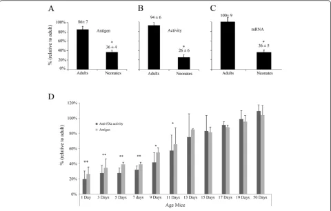
Antithrombin levels in plasma of neonates (day+1) were
60% lower than in adults (day+50) [36±4% (n=13) vs.
86±7% (n=6)] (Figure 1A). As expected, correlating values were observed in antithrombin activity [neonate (n=13):
26±6% vs. adult (n=6): 94±6%] (Figure 1B). We checked
the association between these values andserpinc1mRNA
levels in liver. As shown in Figure 1C,serpinc1mRNA levels
in neonates and adults [36±5% (n=13) vs. 100±9% (n=6)]
matched with previously published data [7], and correlated with antigen and functional levels in plasma. In order to bet-ter delineate the variations observed along the development, we determined the antigenic levels and activity of antithrom-bin of three different mice litters from day one after birth to adult age. Our results showed that antithrombin antigen and activity levels paralleled. At day+13 after birth, antithrombin levels were similar to those observed in adults (Figure 1D).
Qualitative differences of antithrombin in neonate and adult mice
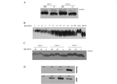
SDS-PAGE analysis of plasma antithrombin revealed that plasma antithrombin from neonate mice had a lower
molecular weight than its adult counterpart (Figure 2A). We next performed native gel electrophoresis with plasma samples extracted at different times during mouse post-natal development. Newborn mice had a plasma anti-thrombin with slower migration than the adult one (Figure 2B), and this result is compatible with a lower global negative charge in neonate’s antithrombin. Plasma antithrombin concentration was not responsible for the differences in electrophoretic mobility (Figure 2B, last lanes). Interestingly, we observed that at day+15, plasma antithrombin from neonate had the same electrophoretic characteristics (Figure 2B) and size (Figure 2C) than adult antithrombin.
To further evaluate the differences of neonatal and adult antithrombins, we performed IEF of plasma from neonate and adult mice. Our results showed that neo-nates expressed more antithrombin isoforms with higher
pI (5.19) and lacked of isoforms with lower pI (4.94) (Figure 2D). These results were in accord with those obtained in native electrophoresis.
Antithrombin glycosylation
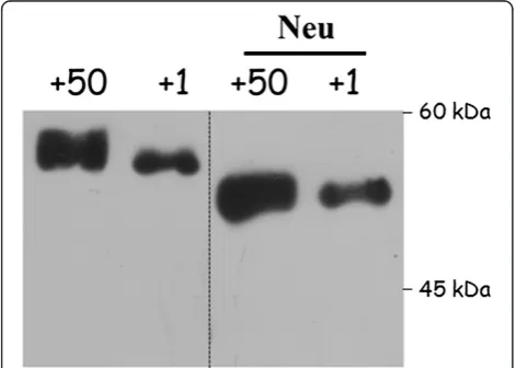
In order to evaluate the role of glycosylation in the qualitative changes of neonate’s antithrombin, we treated plasma from adult and neonate mice with neuramini-dase. This treatment rendered the same electrophoretic
mobility in SDS-PAGE gels for adult’s and neonate’s
antithrombins and sustained an incomplete content of sialic acid for neonate’s antithrombin (Figure 3) that may explain the different migration of neonate’s antithrombin observed in SDS, native electrophoresis, as well as the IEF results (Figure 2).
Expression of sialyltransferases potentially involved in glycosylation of antithrombin
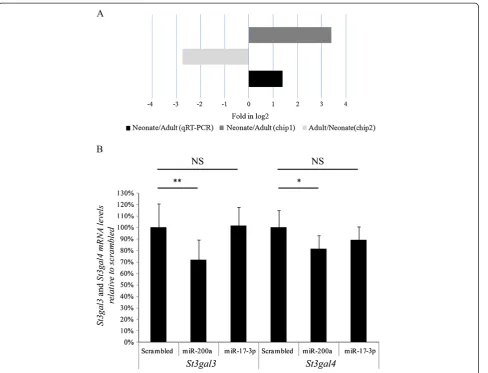
There are three different sialyltransferases able to sialylate N-linked glycoproteins like antithrombin, i.e. St3gal-III, St3gal-IV, and St6gal-I. In order to evaluate if any of these three sialyltransferases were down regulated in neonates, we measured their mRNA levels in liver from neonates and adults by qRT-PCR. As shown in Figure 4, our results indicated an ~85% reduction in neonates in comparison
with adults for St3gal3 and St3gal4 expression, whereas
levels ofSt6gal1 mRNA remained unchanged. As it
hap-pened for antithrombin, mRNA levels of St3gal3 and
St3gal4were similar to those observed in adults at day+13 after birth (Figure 4).
Regulation of St3gal3 and St3gal4 by miR-200a
One hypothesis to explain the variation of the levels of St3gal3and St3gal4 during post-natal development may reside in a miRNA-dependent regulation. Using target predicting algorithms, we found that miR-200a may
target St3gal3 and St3gal4 (Table 1) and thus it was a
valuable candidate to explain the lower sialylation of antithrombin in neonate mice. Interestingly, the analysis of the subtractive miRNA array revealed that miR-200a is over-expressed in neonates in comparison with adults in both chips. Moreover, validation studies in 5 adults and 14 neonates by qRT-PCR confirmed this result (Figure 5A). The next step to demonstrate the potential
regulation of St3gal3 and St3gal4 by miR-200a was to
perform transfection studies of primary hepatocytes from adult mouse with miR-200a. Interestingly, this
procedure provoked a significant reduction of St3gal3
and St3gal4 (31% and 20%, respectively), whereas no effect was observed when a scrambled oligonucleotide was employed or when cells were transfected with miR-17-3p, a miRNA expressed at high levels in neonates that, according to in silico predictions, does not modu-late these sialyltransferases (Figure 5B).
Discussion
Antithrombin is the main endogenous anticoagulant and, thus, its role in regulating haemostasis is absolutely essential. Indeed, complete antithrombin deficiency is in-compatible with life and partial deficiency is an import-ant risk factor for developing venous thrombosis [21]. Besides its role in haemostasis, antithrombin may also regulate other important physiological processes such as inflammation, angiogenesis or apoptosis [22-24]. Intri-guingly, the levels of antithrombin in neonates are severely reduced in comparison with adults without relevant physiological consequences [8,9,25]. This study confirms that the difference not only relies on protein expression levels but also in post-translational modifications. Here, we studied the expression, features, and functionality of antithrombin in a mouse model to deepen into the impact of post-translational modifications of this protein in devel-opmental haemostasis.
Our previous results suggested that the lower levels of antithrombin in neonate mice are mainly explained by a concomitant reduction of mRNA in hepatocytes [7]. In addition, electrophoretic data in the present study sug-gest that the lower molecular weight of antithrombin from neonates is due to a post-translational modifica-tion: an abnormalN-glycosylation (Figures 2 and 3). Our results show that the sialic acid content of antithrombin is smaller in neonates than adults. Unfortunately, we were unable to perform fine glycomic studies to calculate the exact sialic acid content of neonate antithrombin due Figure 3Glycosylation of antithrombin from neonate (day +1)
to the large amount of purified protein that is required for this procedure. Interestingly, a reduced sialylation of anti-thrombin has also been described for antianti-thrombin in chicken and sheep neonates [12,13]. These data strongly suggest that this has to be a process highly regulated in different species. Aiming to identify the mechanisms involved in such control, we evaluated the mRNA levels of three sialyltransferases potentially involved: St3gal3, St3gal4, andSt6gal1. Indeed, St6gal-I performsα2-6 sialic acid linkage as that present in antithrombin [26]. Accor-dingly, this enzyme seems to be the main responsible for the sialylation of antithrombin. However, in St6gal-I KO
mice, St3gal-IV, that performs α2-3 linkages, may also
achieve α2-6 linkages in von Willebrand factor [27]. In addition, a study by Fan et al. revealed that recombinant human antithrombin expressed in baby hamster kidney cells is fully sialylated containingα2-3 linkage [28]. Thus, it is worth suggesting that the lower levels of sialic acid in neonate’s antithrombin might be explained by the reduced
expression of St3gal3 and St3gal4. Further experiments
are necessary to clarify these issues.
The next step to understand the mechanism respon-sible for these differences was the identification of the element(s) controlling the levels of these sialyltransferases. In this framework, the recent report suggesting that some con-served genes implicated in glycosylation pathway may be reg-ulated by miRNAs during animal development [29], reveals miRNAs as potential candidates. n fact,in silicosearching identified miR-200a as an excellent regulator ofSt3gal3and St3gal4. Interestingly, the levels of this miRNA during deve-lopment show a fully compatible change (overexpressed in neonate mice, but reduced expression in adults). The final proof indicating the control of these sialyltransferases by miR-200a was obtained by transfecting this miRNA in adult primary hepatocytes. These experiments suggest that miR-200a may be in part implicated in the regulation ofSt3gal3 and, in a lesser degree, in the regulation ofSt3gal4, as pre-dicted by in silico studies (Table 1). Specificity of this
Table 1 miR-200a putative target site inSt3gal3andSt3gal4mRNA using different target prediction software
miRNA target prediction software Parameters St3gal3 St3gal4
TargetScan (release 5.2) [17] Seed match 7mer-m8 7mer-m8
Context score percentile 85 75
PCT 0.35 0.17
Pictar [18] Score 4.67
-Target site number 1
-Free energy (Kcal/mol) −21.4
-microRNA.org [19] mirSVR score −0.9262 −0.2421
PhastCons score 0.5877 0.5271
Figure 4Levels of selected sialyltransferases mRNA and of miR-200a in neonate and adult liver.Levels of mRNA from three
regulation is further suggested by the lack of effect of another miRNA overexpressed in neonate’s liver, miR-17-3p. How-ever, other mechanisms and additional miRNAs still to characterize may be involved in the reduced expression of these two sialyltransferases in neonate mice.
Finally, it would be of great interest to evaluate whether or not these qualitative modifications regulated indirectly by miRNAs could have functional significance apart of contributing to an increased clearance [30]. In our case, it is necessary to investigate the functional relevance of the lower sialylation in antithrombin, not only on the anti-coagulant function, which might contribute to explain the dramatic change of the haemostatic system after birth, but also on other functions of this molecule.
Conclusions
involved in transcriptional [32,33], translational or post-translational processes.
Competing interests
The authors declare that they have no competing interests.
Authors’contributions
RT, IMM, MEMB, SS-A, and SA performed biochemical assays (WB, IEF; qRT-PCR). JAG and NGB performed work with mice. ABA and RGC performed in vitroassays. AM measured protein levels and activities. RT, JC, and CM designed the research, analyzed the results, and wrote the paper. VV critically read the manuscript. All authors read and approved the final manuscript.
Acknowledgments
MEM-B is a holder of a predoctoral research grant from ISCIII. CM and IM-M are investigators from Fundación para la Formación e Investigación Sanitarias de la Región de Murcia (FFIS). This work was supported by research grants from ISCIII and FEDER (PI11/00566, PI12/00657, Red RECAVA RD12/0042/0050).
Received: 21 February 2013 Accepted: 8 May 2013 Published: 16 May 2013
References
1. Fabbri M, Croce CM, Calin GA:MicroRNAs.Cancer J2008,14:1–6. 2. Bartel DP:MicroRNAs: target recognition and regulatory functions.
Cell2009,136:215–233.
3. Guo H, Ingolia NT, Weissman JS, Bartel DP:Mammalian microRNAs
predominantly act to decrease target mRNA levels.Nature2010,466:835–840. 4. Fort A, Borel C, Migliavacca E, Antonarakis SE, Fish RJ, Neerman-Arbez M:
Regulation of fibrinogen production by microRNAs.Blood2010,
116:2608–2615.
5. Teruel R, Perez-Sanchez C, Corral J, Herranz MT, Perez-Andreu V, Saiz E, Garcia-Barbera N, Martinez-Martinez I, Roldan V, Vicente V, López-Pedrera C, Martinez C:Identification of miRNAs as Potential Modulators of Tissue Factor Expression in Patients with Systemic Lupus Erythematosus and Antiphospholipid Syndrome.J Thromb Haemost2011,9:1985–1992. 6. Nagalla S, Shaw C, Kong X, Kondkar AA, Edelstein LC, Ma L, Chen J,
McKnight GS, Lopez JA, Yang L, Jin Y, Bray MS, Leal SM, Dong JF, Bray PF:
Platelet microRNA-mRNA coexpression profiles correlate with platelet reactivity.Blood2011,117:5189–5197.
7. Teruel R, Corral J, Perez-Andreu V, Martinez-Martinez I, Vicente V, Martinez C:
Potential role of miRNAs in developmental haemostasis.PLoS One2011,
6:e17648.
8. Andrew M, Paes B, Milner R, Johnston M, Mitchell L, Tollefsen DM, Castle V, Powers P:Development of the human coagulation system in the healthy premature infant.Blood1988,72:1651–1657.
9. Andrew M, Paes B, Milner R, Johnston M, Mitchell L, Tollefsen DM, Powers P:
Development of the human coagulation system in the full-term infant.
Blood1987,70:165–172.
10. Andrew M, Vegh P, Johnston M, Bowker J, Ofosu F, Mitchell L:Maturation of the hemostatic system during childhood.Blood1992,80:1998–2005. 11. Rau JC, Beaulieu LM, Huntington JA, Church FC:Serpins in thrombosis,
hemostasis and fibrinolysis.J Thromb Haemost2007,5(Suppl 1):102–115. 12. Amrani DL, Mosesson MW, Koide T:Evidence that chicken antithrombin III
is a developmentally regulated glycoprotein synthesized by hepatocytes.
Biochim Biophys Acta1985,847:324–334.
13. Niessen RW, Lamping RJ, Peters M, Lamers WH, Sturk A:Fetal and neonatal development of antithrombin III plasma activity and liver messenger RNA levels in sheep.Pediatr Res1996,39:685–691.
14. Ishiguro K, Kojima T, Kadomatsu K, Nakayama Y, Takagi A, Suzuki M, Takeda N, Ito M, Yamamoto K, Matsushita T, Kusugami K, Muramatsu T, Saito H:
Complete antithrombin deficiency in mice results in embryonic lethality.
J Clin Invest2000,106:873–878.
15. Hagiwara S, Iwasaka H, Shingu C, Matsumoto S, Uchida T, Noguchi T:
High-dose antithrombin III prevents heat stroke by attenuating systemic inflammation in rats.Inflamm Res2010,59:511–518.
16. Larsson H, Akerud P, Nordling K, Raub-Segall E, Claesson-Welsh L, Bjork I:A novel anti-angiogenic form of antithrombin with retained proteinase binding ability and heparin affinity.J Biol Chem2001,276:11996–12002.
17. Guerrero JA, Teruel R, Martinez C, Arcas I, Martinez-Martinez I, de la Morena-Barrio ME, Vicente V, Corral J:Protective role of antithrombin in mouse models of liver injury.J Hepatol2012,57:980–986.
18. Manco-Johnson MJ, Jacobson LJ, Hacker MR, Townsend SF, Murphy J, Hay W Jr:Development of coagulation regulatory proteins in the fetal and neonatal lamb.Pediatr Res2002,52:580–588.
19. McCoy AJ, Pei XY, Skinner R, Abrahams JP, Carrell RW:Structure of beta-antithrombin and the effect of glycosylation on antithrombin's heparin affinity and activity.J Mol Biol2003,326:823–833.
20. Ellies LG, Ditto D, Levy GG, Wahrenbrock M, Ginsburg D, Varki A, Le DT, Marth JD:Sialyltransferase ST3Gal-IV operates as a dominant modifier of hemostasis by concealing asialoglycoprotein receptor ligands.Proc Natl Acad Sci USA2002,99:10042–10047.
21. Fan B, Crews BC, Turko IV, Choay J, Zettlmeissl G, Gettins P:Heterogeneity of recombinant human antithrombin III expressed in baby hamster kidney cells. Effect of glycosylation differences on heparin binding and structure.J Biol Chem1993,268:17588–17596.
22. Zhang L, Hammell M, Kudlow BA, Ambros V, Han M:Systematic analysis of dynamic miRNA-target interactions during C. elegans development.
Development2009,136:3043–3055.
23. Ashwell G, Harford J:Carbohydrate-specific receptors of the liver.
Annu Rev Biochem1982,51:531–554.
24. Kahai S, Lee SC, Lee DY, Yang J, Li M, Wang CH, Jiang Z, Zhang Y, Peng C, Yang BB:MicroRNA miR-378 regulates nephronectin expression modulating osteoblast differentiation by targeting GalNT-7.PLoS One2009,4:e7535. 25. Guttilla IK, White BA:Coordinate regulation of FOXO1 by miR-27a, miR-96,
and miR-182 in breast cancer cells.J Biol Chem2009,284:23204–23216. 26. Wan HY, Guo LM, Liu T, Liu M, Li X, Tang H:Regulation of the transcription
factor NF-kappaB1 by microRNA-9 in human gastric adenocarcinoma.
Mol Cancer2010,9:16.
27. Raja SM, Chhablani N, Swanson R, Thompson E, Laffan M, Lane DA, Olson ST:
Deletion of P1 arginine in a novel antithrombin variant (antithrombin London) abolishes inhibitory activity but enhances heparin affinity and is associated with early onset thrombosis.J Biol Chem2003,278:13688–13695. 28. Hernandez-Espinosa D, Minano A, Martinez C, Perez-Ceballos E, Heras I,
Fuster JL, Vicente V, Corral J:L-asparaginase-induced antithrombin type I deficiency: implications for conformational diseases.Am J Pathol2006,
169:142–153.
29. Martinez-Martinez I, Ordonez A, Navarro-Fernandez J, Perez-Lara A, Gutierrez-Gallego R, Giraldo R, Martinez C, Llop E, Vicente V, Corral J:
Antithrombin Murcia (K241E) causing antithrombin deficiency: a possible role for altered glycosylation.Haematologica2010,95:1358–1365. 30. Lewis BP, Burge CB, Bartel DP:Conserved seed pairing, often flanked by
adenosines, indicates that thousands of human genes are microRNA targets.Cell2005,120:15–20.
31. Krek A, Grun D, Poy MN, Wolf R, Rosenberg L, Epstein EJ, Macmenamin P, Da PI, Gunsalus KC, Stoffel M, Rajewsky N:Combinatorial microRNA target predictions.Nat Genet2005,37:495–500.
32. John B, Enright AJ, Aravin A, Tuschl T, Sander C, Marks DS:Human MicroRNA targets.PLoS Biol2004,2:e363.
33. Wu W, Kocarek TA, Runge-Morris M:Sex-dependent regulation by dexamethasone of murine hydroxysteroid sulfotransferase gene expression.Toxicol Lett2001,119:235–246.
doi:10.1186/1423-0127-20-29