R E S E A R C H
Open Access
Impact on gait biomechanics of using an
active variable impedance prosthetic knee
Matthew R. Williams
1,2*, Susan D
’
Andrea
3,4and Hugh M. Herr
5Abstract
Background:An above knee amputation can have a significant impact on gait, with substantial deviations in inter-leg symmetry, step length, hip exertion and upper body involvement even when using a current clinical standard of care prosthesis. These differences can produce gait that is less efficient and less comfortable, resulting in slower and shorter distance walking, particularly with long term use.
Methods:A robotic variable impedance prosthetic knee (VI Knee) was tested with five individuals (N= 5) with unilateral amputation above the knee at fixed speeds both above and below their normal walking speed. Subject gait was measured as they walked along an instrumented walkway via optical motion capture and force plates in the floor. Each subject’s gait while using the VI Knee was compared to that while using their standard of care knee (OttoBock C-Leg).
Results:Significant differences (p < 0.05) in walking between the standard of care and variable impedance devices were seen in step length and hip range of motion symmetries, hip extension moment, knee power and torso lean angle. While using the VI Knee, several subjects demonstrated statistically significant improvements in gait, particularly in increased hip range of motion symmetry between affected and intact sides, greater prosthesis knee power and in reducing upper body involvement in the walking task by decreasing forward and affected side lean and reducing the pelvis-torso twist coupling. These changes to torso posture during gait also resulted in increased terminal stance hip flexion moment across subjects. Detriments to gait were also observed in that some subjects exhibited decreased step length symmetry while using the VI Knee compared to the C-Leg.
Conclusions:The knee tested represents the potential to improve gait biomechanics and reduce upper body
involvement in persons with above knee amputation compared to current standard of care devices. While using the VI Knee, subjects demonstrated statistically significant improvements in several aspects of gait though some were worsened while using the device. It is possible that these negative effects may be mitigated through longer term training and experience with the VI Knee. Given the demonstrated benefits and the potential to reduce or eliminate detriments through training, using a powered device like the VI Knee, particularly over an extended period of time, may help to improve walking performance and comfort.
Background
More than 270,000 persons with amputation (PWA) above the knee currently reside in the United States, with an amputation incidence rate of 39,000 new cases each year [1]. In the Veteran community, the Veteran’s Health Administration performs about 1500 to 2000 above-knee amputations each year with vascular disease
as a result of diabetes being the most prevalent reason for surgery [2]. In terms of service-related injuries, the military actions over the past 14 years have seen many service personnel who have suffered trauma requiring above knee amputation. It is estimated that 34.5 % of in-dividuals with combat injuries needing amputation re-quired at least one above knee amputation [3].
Current standard of care prostheses result in gait that displays marked asymmetry including differences in step length, hip moment and torso involvement [4–7]. This results in changes in gait symmetry (step length and hip range of motion), an indicator of altered joint loading
* Correspondence:[email protected]
1Louis Stokes Cleveland VA Medical Center, Cleveland, OH, USA 2Department of Biomedical Engineering, Case Western Reserve University, Cleveland, OH, USA
Full list of author information is available at the end of the article
that can have a deleterious impact on the intact side limb, possibly leading to a higher prevalence of back and hip pain and knee osteoarthritis [8, 9]. Today’s advanced prosthetic knees (such as the Otto Bock C-Leg and Gen-ium and the Ossur Rheo Knee) have been shown to re-duce both hip moment and the metabolic requirements of using such a device, especially at speeds faster and slower than customary walking speed [10, 11], decreas-ing the overall exertion and effort felt by the user. The newer generations of prostheses have also shown some improvement in step length symmetry under controlled speeds [12] but no studies have shown them to improve upper body motion over simpler prostheses [7]. Given the relatively young age at which current combat Vet-erans are being fitted with knee prostheses and their expected life expectancy and long term use of such de-vices, the need for a prosthesis which improves gait sym-metry and reduces abnormal joint loading is significant. Additionally, more elderly Veterans who are recent PWAs due to vascular disease may benefit from a pros-thesis which requires less effort to use which may result in greater ambulation and mobility.
Research into powered prosthetic knees has been con-ducted for several decades [13–17], however, a viable, self-contained device with all actuators, power and com-putation/control on board has been demonstrated only recently. Goldfarb has developed a powered knee-ankle prosthesis that can reproduce some of the kinematics of able-bodied gait over level ground [18] as well as walk-ing up an incline [19]. In terms of commercial devices, Ossur has developed the Power Knee, a position con-trolled, direct transmission knee that uses echo control of the intact leg to control prosthetic knee motion [20, 21]. These previous approaches to restore knee function have not ideally taken advantage of mechanisms which replicate the passive properties of the leg during ambulation or the potential benefits of energy storage and return as seen in the organic leg. One of the main disadvantages of such systems is the substantial power consumption required to replicate natural gait with a direct-drive transmission. This results in lower battery life or requires a larger battery to meet daily step counts. By not leveraging the ability to store and release mechanical energy via passive elastic ele-ments in the prosthesis, such devices ignore a key oppor-tunity to optimize the size and weight of powered knee systems [22].
In contrast to the powered prosthetic knee approaches discussed above, during able-bodied level ground walk-ing, energy is primarily conserved by transfer, storage and later release of potential energy with little input power needed from propulsive forces from the hip. This energy is stored in the elastic tendons of the legs or by lifting the body’s center of mass [23]. Biomechanical models of gait, using passive actuators (such as springs)
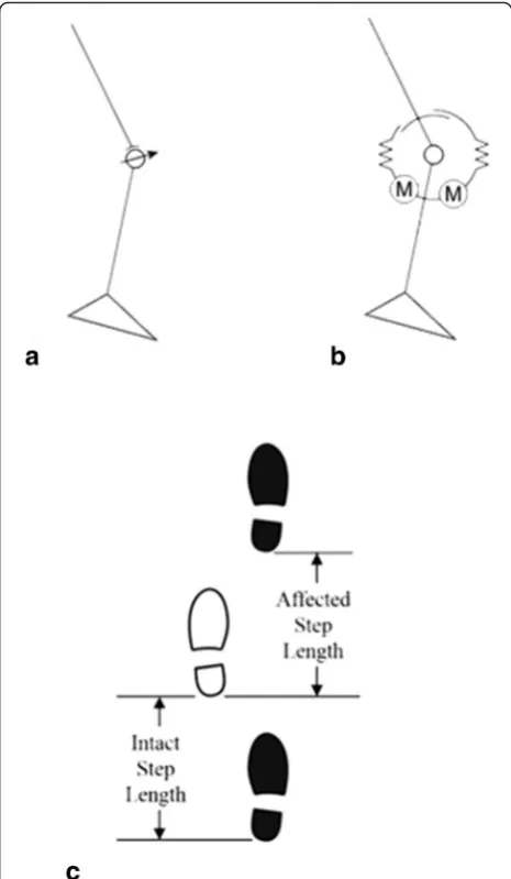
along with clutches to control the activation of these ac-tuators, have demonstrated that a large portion of the leg’s motion can be achieved using passive elastic ele-ments [24]. During level ground walking the bulk of the muscle activity at the knee is related to energy absorp-tion and storage than actively generating power [25]. Current prosthetic knees (such as the OttoBock C-Leg studied in this work) dissipate mechanical energy, only providing braking torque on the knee during swing phase (Fig. 1a) [26]. Different from this passive ap-proach, series elastic actuators can be used in prostheses to provide net positive energy with low electrical power by taking advantage of energy storage in the elastic
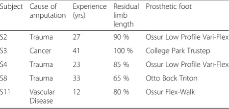
elements during gait, as well as accurately replicate the general joint kinematics and kinetics of the knee. In addition to their capability for energy storage, this type of actuator allows for more biomimetic motion and has inherent impact tolerance due to their elastic components (critical for a walking device) and have been used in a number of walking robots and robotic prostheses [27, 28].
The work of Martinez-Villalpando and Herr has devel-oped a variable impedance knee prosthesis (VI Knee) that uses an agonist-antagonist arrangement of two series elastic actuators to control knee position and im-pedance in addition to providing propulsive power in gait (Fig. 1b) [22]. This opposed actuation method en-ables each actuator to operate often unloaded, chiefly to control joint stiffness. This results in a significant reduc-tion in the electrical demands of the device. By using series elastic actuators, the device can not only store and release mechanical energy (via the elastic elements of the actuators) during walking, but can also help to lift the center of mass by producing net positive power in late stance. Variable impedance control adjusts the knee output torque (either braking or producing positive torque) as a function of its rotational velocity, thereby producing a more biomimetic gait. In early studies, indi-viduals with above knee amputations walking on level ground at their normal, customary walking speed were observed to have a 6.8 % reduction in their metabolic ef-fort due to using this device compared to their conven-tional prosthesis [29].
This study investigated the biomechanical differences between using the VI Knee compared to a C-Leg (Otto Bock, Duderstadt, Germany), the current standard of care device for active walkers. It was hypothesized that by using a knee with active flexion and extension, the user’s gait would be improved at speeds both above and below their customary walking speed due to increased joint symmetry as well as reduced involvement of the af-fected side hip in controlling the motion of the lower leg.
Methods
The device tested in this study was the VI Knee [29], a powered prosthetic knee developed at The Massachusetts Institute of Technology (MIT). Eleven unilateral, PWAs above the knee were recruited through connections with local prosthetists based on the following inclusion criteria: Height over 1.5 m, weight between 83 and 113 kg, age be-tween 20 and 70 years, and rated as a level K3 or above ambulator (defined as being able to voluntarily vary walk-ing speed and walk without hand-held assistive devices), and be experienced (greater than 1 year) C-Leg users. Weight and height criteria were set by the weight and length of the VI Knee so that it approximately matched the weight and length of the missing lower leg. Recruited
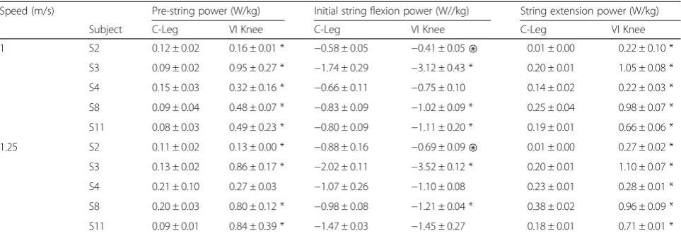
subjects covered a breadth of years since amputation and represented a span of residual limb lengths (defined as the proportion (listed as a percentage in this work) of intact femur length remaining after amputation). Of those re-cruited, five subjects completed the experiments with the other six dropping out shortly after screening due to in-creased time demands at work (n= 3), injuries at home not associated with the study (n= 2) and one exceptionally tall and heavy participant who despite meeting the inclu-sion criteria was unable to properly walk with the VI Knee due to over-torqueing the device when turning corners. Subject demographics of the participants who completed the study can be seen in Table 1. All subjects gave in-formed consent to participate and experimental protocols were approved by the Providence VA Medical Center (PVAMC) IRB.
Subjects participated in three sessions: An initial fit-ting of the VI Knee, a practice walking session and the biomechanics testing session. Each session was per-formed on different days to avoid fatigue. All sessions were performed at the PVAMC Gait Lab.
In the initial fitting session, the VI Knee was attached to subjects’ existing socket and foot. All subjects used similar low-profile, carbon fiber energy storage and re-turn feet (Table 1), which is not considered to cause sig-nificant differences in gait between subjects [30]. The prosthesis was attached and aligned by a certified pros-thetist. The knee was then adjusted to the subject’s gait by tuning the initial swing acceleration, maximum swing velocity and terminal swing deceleration to produce an optimal gait that was comfortable for the subject and natural in appearance as determined by the prosthetist. Behavior of the VI Knee in stance was fixed across sub-jects and allowed for stance flexion of the device. These tuning values were recorded and used for the later prac-tice and experimental sessions. Tuning was performed at each subject’s customary walking speed (1.13 ± 0.15 m/s across subjects). The fitting session lasted about an hour, at the end of which, subjects were able to assuredly walk with the VI Knee at variable speeds over clear, level ground. At this session, each subject’s height and weight was measured. The length and weight of their current
Table 1Subject Demographics
Subject Cause of amputation
Experience (yrs)
Residual limb length
Prosthetic foot
S2 Trauma 27 90 % Ossur Low Profile Vari-Flex
S3 Cancer 41 100 % College Park Trustep
S4 Trauma 23 85 % Ossur Low Profile Vari-Flex
S8 Trauma 33 65 % Otto Bock Triton
S11 Vascular Disease
prosthesis was also measured and its center of mass de-termined using the knife-edge balance technique by finding the balance point of the prosthesis with shank and the prosthetic foot [31]. This information was ne-cessary for later use in the biomechanical model of each subject.
The practice sessions involved continuous walking at customary walking speed as well as the speeds to be tested - 1 and 1.25 m/s. The speeds tested were selected as they were fixed speeds slightly outside the subjects’ customary walking speed but were not overly laborious due to being too slow or too fast. Subjects started with a 30 min “free walk” where they were asked to walk at their customary walking speed in a large, cleared hallway around the building (approximately 120 m/lap, 1.25 m corner radius) to become further acquainted with the device. Rest periods were available as needed during the “free walk”portion of the experiment. After a 5 min rest, they then walked with the new device for 7 min at 1 and 1.25 m/s (randomly determined) with a 5 min rest be-tween practice trials. This was repeated twice for a total experiment duration of about 2 h. The fixed walking speeds were controlled by having subjects keep pace with a member of the research team who used a metro-nome (via earphones so only they could hear it) cali-brated to their gait such that they maintained a fixed speed with respect to their paced cadence. Subjects were instructed to keep pace with the pace-setter and to maintain a forward gaze so that they did not visually en-train to the pace-setter’s cadence.
In the testing session, 64 IR reflective markers were placed on the limb segments and joint centers of the subjects’ limbs and torso. The testing involved subjects walking along a 10 m walkway while their gait was re-corded at 100 Hz using an infrared (IR) video motion capture system (Qualisys, Gotenberg, Sweden, Oqus 500 cameras). Force plates (AMTI, Watertown, MA) in the middle of the walkway recorded ground reaction forces and moments. Motion capture data was processed using Visual3D (C-Motion, Germantown, MD). Data process-ing of marker signals consisted of 10 sample (0.1 s) 3rd order polynomial spline-fit gap filling, applying a 6 Hz low-pass filter, and using automatic gait event detection [32]. Force plate data was used to compute gait kinetics by using the standard human body model provided by Visual3D, modified for each subject to reflect the differ-ences in weight and location of center of mass of the prostheses investigated. Five of these walking trials with each prosthesis at both speeds tested were used to calcu-late mean values of parameters characterizing each sub-ject’s performance.
Comparisons across subjects between the two knees tested were tested using a mixed model design ANOVA with the knees investigated as the fixed effects and
subjects the random effects. Each subject’s individual performance difference with each knee was tested using Welch’s t-tests (to account for differences in variance be-tween the conditions tested), with two tails andα= 0.05 were used to test significance between conditions (pros-thesis). In all tests, p-values less than 0.05 were consid-ered significant. Correlation comparisons were tested for significance as per [33]. Normality of data was checked using normal probability plots and was found to be nor-mally distributed.
Results
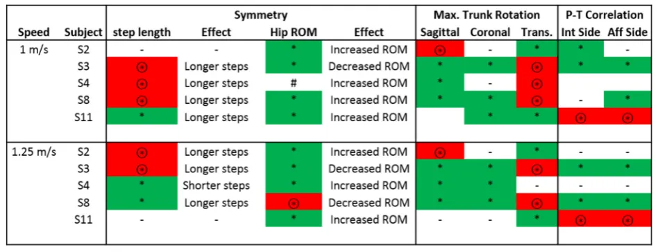
Significant differences between prosthetic conditions were observed across all subjects (p values ranging from <0.0001 to 0.04) in the following: Step length symmetry between intact and affected sides (Fig. 2), hip range of mo-tion symmetry between intact and affected sides (Fig. 3), affected leg hip extension moment (Fig. 4), prosthesis knee power (Table 2), upper body rotation (Fig. 5) and pelvis-torso twist correlation (Fig. 6). The significant observa-tions of step length and hip ROM symmetry, trunk rotation and pelvis-torso twist correlations for each sub-ject are summarized in Table 3. Each subsub-ject responded
differently to the VI Knee, resulting in a large variability across subjects. However, statistically significant differences between the knees tested were observed. Throughout the text, values which represent improved performance while using the VI Knee compared to the C-Leg are denoted by a star (*). Similarly, those values that indicate worse per-formance while using the VI Knee compared to the C-Leg are indicated by a circled start (⊛).
Step length is defined as the forward distance of the heel at footfall from the preceding contra-lateral heel at its footfall (Fig. 1c). Step length symmetry is defined as the ratio of the affected side step length to that of the in-tact side. To better illustrate the differences, the devi-ation in symmetry (difference from 100 %) is presented. More positive values indicate a longer affected side step length, while more negative deviations from symmetry indicate a shorter affected side step. Ideally, the magni-tude of the deviation from 100 % is as small as possible. At 1 m/s, one subject (S11) showed improvement in symmetry by taking longer steps with the VI Knee than with the C-Leg (Fig. 2a, starred). Three subjects showed decreased step length symmetry (S3, 4 and 8) while using the VI Knee at this speed, the result of taking lon-ger steps with the affected leg (Fig. 2a, circled star). One subject (S2), showed no overall change in symmetry
though they did exhibit a shift from shorter affected side steps with the C-Leg to longer steps with the VI Knee. Four of five subjects showed significant differences in step length symmetry at 1.25 m/s. Two subjects exhib-ited improved symmetry (Fig. 2b, starred), one by taking shorter steps with the VI Knee (S4) and one by taking longer ones (S8). Two subjects showed decreased step length symmetry (Fig. 2b, circled star) by increasing their step length with the VI Knee (S2 and 3) and one subject (S11) showed no significant difference between devices at the faster speed.
Hip range of motion (ROM) symmetry is defined as the ratio of the differences (maximum flexion–maximum ex-tension) between affected and intact sides. Similar to step
Fig. 3Deviation from 100 % hip range of motion (max flexion– max extension) symmetry between affected and intact sides for walking at 1 m/s (a) and 1.25 m/s (b). Significant differences (p≤0.05) in symmetry magnitude are denoted by a star (improved symmetry due to using the VI Knee) or a circled star (reduced symmetry). Positive values indicate greater hip range of motion on the affected side, negative values, a smaller range of motion
length, more positive values indicate a larger affected side hip range of motion, more negative values, a smaller range of motion. Again, closer to zero deviation from 1 is ideal. At 1 m/s, four of five subjects (S2, 3, 8 and 11) showed an improvement in hip ROM symmetry (Fig. 3a, starred). The subject who showed no statistical difference in sym-metry (S4) did show a significant increase in ROM (but not symmetry) while using the VI Knee. At 1.25 m/s, four of five subjects (S2, 3, 4 and 11) exhibited improved gait symmetry (Fig. 3b, starred), while one had reduced sym-metry (S8) while using the VI Knee at this speed (Fig. 3b, circled star).
Hip flexion moment during terminal stance is associ-ated with sagittal plane (forward) torso lean, with less lean producing a larger flexion moment [34]. While greater moment is required by the hip, maintaining a more vertical posture is more beneficial to overall com-fort. At both speeds tested, subjects exhibited increased affected side hip flexion moment in late stance while using the VI Knee (Fig. 4). Subjects typically exhibited both decreased hip extension shortly after heel strike and greater maximum flexion moment in terminal stance (Fig. 4). At 1 m/s, all five subjects showed signifi-cant increases in maximum hip flexion moment (Fig. 4b, starred). At the faster 1.25 m/s, four of five subjects (S2, 3, 4 and 8) showed significantly increased maximum hip flexion moment (Fig. 4c, starred). Subject S11 showed no significant difference in hip flexion moment between conditions at this speed.
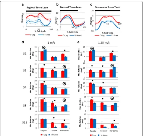
The robotic VI Knee showed increased knee power generation across most subjects and both speeds during pre-swing, and swing extension and absorbed more energy during initial flexion compared to the passive C-Leg (Table 2). At 1 m/s, the VI Knee generated sig-nificantly greater power during pre-swing and swing extension (starred values). Maximum power absorbed during initial swing at this speed was significantly
greater for three subjects (S3, 8 and 11, starred values) and less for one subject (S2, denoted on Table 2 with a ‘⊛’). Walking at 1.25 m/s, the knee generated signifi-cantly more power during pre-swing for four of five sub-jects (S2, 3, 8 and 11) and all subsub-jects during swing extension. During initial swing flexion, two subjects had significantly greater power absorption (S3, and 8) at this speed and one subject had less (S2).
Across speeds, subjects showed differences in the max-imum torso lean angle in both the sagittal (forward) and coronal (lean toward the affected side) planes as well as torso twist relative to the pelvis while using the VI Knee. Typical torso lean and twist angles as a function of the gait cycle for one subject is shown in Fig. 5a–c. Across all subjects, at both 1 and 1.25 m/s, three of five subject exhibited reduced forward lean while using the VI Knee (S3, 4 and 8, Fig. 5d & d, starred). One subject signifi-cantly increased their forward lean (S2, Fig. 5d and e, circled star) and one subject (S11) showed no difference.
At 1 m/s, three subjects had a reduced lean to the af-fected side while using the VI Knee (S3. 8, and 11, Fig. 5d, starred). Two subjects showed no significant dif-ference due to using the powered device at this speed (S2 and 4). At the faster speed, again three subjects showed significantly reduced affected side lean (S3, 4 and 8, Fig. 5e, starred) and two subjects exhibited no sig-nificant difference (S2 and 11). No subjects exhibited in-creased coronal plane lean while using the VI Knee at either speed tested.
At both speeds, two subjects exhibited a reduction in maximum torso twist (transverse plane rotation) while walking with the VI Knee (S2 and 11, Fig. 5d and e, starred). At the slower walking speed, three subjects generated a larger maximum torso twist when using the robotic device (S3, 4 and 8, Fig. 5d, right column, circled star). Similarly, walking with the VI Knee caused an in-crease in maximum torso twist in two subjects at the Table 2Maximum knee power during three sub-phases of gait normalized to body weight by prosthesis
Speed (m/s) Pre-string power (W/kg) Initial string flexion power (W//kg) String extension power (W/kg)
Subject C-Leg VI Knee C-Leg VI Knee C-Leg VI Knee
1 S2 0.12 ± 0.02 0.16 ± 0.01 * −0.58 ± 0.05 −0.41 ± 0.05⊛ 0.01 ± 0.00 0.22 ± 0.10 *
S3 0.09 ± 0.02 0.95 ± 0.27 * −1.74 ± 0.29 −3.12 ± 0.43 * 0.20 ± 0.01 1.05 ± 0.08 *
S4 0.15 ± 0.03 0.32 ± 0.16 * −0.66 ± 0.11 −0.75 ± 0.10 0.14 ± 0.02 0.22 ± 0.03 *
S8 0.09 ± 0.04 0.48 ± 0.07 * −0.83 ± 0.09 −1.02 ± 0.09 * 0.25 ± 0.04 0.98 ± 0.07 *
S11 0.08 ± 0.03 0.49 ± 0.23 * −0.80 ± 0.09 −1.11 ± 0.20 * 0.19 ± 0.01 0.66 ± 0.06 *
1.25 S2 0.11 ± 0.02 0.13 ± 0.00 * −0.88 ± 0.16 −0.69 ± 0.09⊛ 0.01 ± 0.00 0.27 ± 0.02 *
S3 0.13 ± 0.02 0.86 ± 0.17 * −2.02 ± 0.11 −3.52 ± 0.12 * 0.20 ± 0.01 1.10 ± 0.07 *
S4 0.21 ± 0.10 0.27 ± 0.03 −1.07 ± 0.26 −1.10 ± 0.08 0.23 ± 0.01 0.28 ± 0.01 *
S8 0.20 ± 0.03 0.80 ± 0.12 * −0.98 ± 0.08 −1.21 ± 0.04 * 0.38 ± 0.02 0.96 ± 0.09 *
S11 0.09 ± 0.01 0.84 ± 0.39 * −1.47 ± 0.03 −1.45 ± 0.27 0.18 ± 0.01 0.71 ± 0.01 *
faster walking speed (S3 and 8, Fig. 5e, circled star) and no significant difference in one subject (S4).
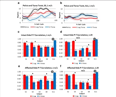
Figure 6a and b shows the transverse rotation angles (twist) between the pelvis and torso for single subjects over the gait cycle while using each knee prosthesis. In Fig. 6a, the subject demonstrates decreased correlation between the pelvis and torso twist. The subject in Fig. 6b illustrates increased correlation in the direction of twist of the pelvis and torso while using the VI Knee. The
Pearson’s correlation coefficient between the torso and pelvis twist for all subjects over each step (heel strike to heel strike) is shown in Fig. 6c–f. In these graphs, values closer to 1 represent tighter coupling between pelvis and torso twist and that they are rotating together (in phase), whereas values closer to zero represent looser coupling,, and negative values approaching−1 (anti-correlated) indi-cate the pelvis and torso are twisting in opposite direc-tions (out of phase). For reference, able-bodied individuals
tend to have a pelvis-torso correlation of about−0.7 and are thus anti-correlated during normal gait with the pelvis and torso generally rotating out of phase in opposite di-rections [7]. At 1 m/s, for steps taken with the intact leg, two subjects show a significant reduction in pelvis-torso correlation (S2 and 3, Fig. 6c, starred) while one subject exhibited a significant increase in pelvis-torso correlation (S11, Fig. 6c, circled star). Two subjects (S4 and 8) showed no significant difference in intact side pelvis-torso correl-ation at this speed. Similarly, at 1.25 m/s two subjects (S3 and 8) had reduced correlation and one (S11) increased (Fig. 6d) and two subjects (S2 and 4) showing no signifi-cant differences between devices. For steps taken with the
affected side, at both 1 and 1.25 m/s, two subjects (S3 and 8) showed significantly reduced pelvis-torso correlation (Fig. 6e and f, starred) and one subject (S11) significantly increased their pelvis-torso coupling (Fig. 6e and f, circled star). Two subjects (S2 and 4) displayed no significant dif-ference in affected side pelvis-torso correlation across both speeds.
Discussion
This study investigated the biomechanical differences between using the variable impedance VI Knee com-pared to the current standard of care for active walkers (OttoBock C-Leg). Significant differences were observed
between conditions, with most of the five subjects tested showing improvements due to using the VI Knee in in-creased hip range of motion symmetry, inin-creased pros-thesis knee power and reduced sagittal and coronal plane lean as well as increased terminal stance hip flexion moment (an indicator of more upright gait pos-ture). Mixed results were observed regarding the impact of using the VI Knee on pelvis-torso twist correlation, with two of five subjects showing improvement and one a worsening of performance. Significant disadvantages to using the VI Knee, where most subjects exhibited worse performance were observed in decreased step length symmetry and increased torso twist at the slower speed.
Individuals with an above knee amputation tend to ex-hibit a greater amount of torso rotation (in all three planes) and decreased step-length symmetry (by taking shorter steps on the affected side) compared to able-bodied subjects [7, 35] in part to compensate for weak hip abductors on their affected side due to disuse atro-phy. By leaning (primarily toward the affected side), they are able to use the upper body to help lift the intact side hip to assist in clearing the toe. This population also shows a significant increase in in-phase coupling of the pelvis and torso compared to able-bodied individuals, due to an overall stiffening of the entire upper body dur-ing gait [7, 35]. Reasons for this stiffendur-ing range from a psychological/guarding behavior to the use of the upper body to control the motion of the pelvis and leg due to hip weakness [7]. This coupling of body segment rotation can have negative impacts on gait by increasing mechan-ical energy required for walking [36] and potentially
reduces customary walking speed [7]. While using the VI Knee, the majority of subjects exhibited improved hip range of motion symmetry between intact and affected sides, (Fig. 3), reduced forward and affected side torso lean (Fig. 5). Mixed results were seen in the amount of coup-ling between the torso and pelvis twist with two of five subjects showing improvement and one worse perform-ance (Fig. 6). The ability of the VI Knee to produce greater power compared to passive knees (Table 2) is potentially one difference between it and passive knees like the C-Leg that enables these improvements and allows for a more natural gait, unburdens the upper body from the gait task, and helps to lift the lower leg to clear the toe. As the bio-mechanical changes often associated with prosthetic use can lead to chronic hip and back pain and osteoarthritis of the hip and intact-side knee [8, 9], it is possible that im-provements in hip ROM symmetry, with long term use of the VI Knee compared to a standard of care, passive knee prosthesis, may lead to reductions in the likelihood of these conditions arising. While some subjects exhibited increased torso twist, which has been linked to back pain in individuals with an above knee amputation [35], none of the subjects increased their torso twist to a magnitude or by an amount shown to cause back pain (15.4° ± 4.2° vs. maximum 10.85° ±0.68° observed in this study).
Across both speeds, most subjects increased their af-fected side step length when using the VI Knee (Fig. 2, Table 3), however in some cases this resulted in a de-crease in symmetry. At the slower speed, most of those who took longer steps with the affected side leg were also the ones who showed an increase in maximum Table 3Summary of the significant effects on step length and hip range of motion (ROM) symmetry, maximum trunk rotation and pelvis-torso (P-T) rotation correlation due to using the VI Knee instead of the C-Leg
torso twist (Table 3). At the faster speed, this is also shown by two subjects (though one showed an improve-ment in symmetry by taking shorter affected side steps). In these subjects, increased rotation of the pelvis (rela-tive to the torso) serves to increase step length. At both speeds, of the five instances of reduced step-length sym-metry, four also occurred with increased torso twist (the fifth occurring with a reduction in maximum twist, but an increase in hip range of motion, Table 3). Further training may allow them to reduce their torso twist and thus improve step length symmetry.
Across speeds, most to all subjects increased their af-fected side maximum stance phase hip flexion moment (Fig. 4). This is consistent with the reduced forward torso lean observed in most subjects (Fig. 5). While not a direct assessor of overall gait performance itself, it is a result of walking with a more upright posture [34]. In-creased terminal stance hip flexion moment is a result of needing to balance the ground reaction force which shifts posteriorly with less forward torso lean such that the line of action is more behind the hip. This allows the walker to maintain the center of pressure under the fore-foot prior to weight transfer to the other fore-foot. Comparing Figs. 4 and 6, those subjects who showed more reduced forward torso lean while using the VI Knee also exhibited more increased terminal stance hip flexion moment.
Based on the data collected, the VI Knee demonstrates potential advantages in knee power over a passive device (Table 2) including the generation of net positive power during pre-swing to help raise the center of mass. The de-vice also absorbed more power than a passive dede-vice during initial swing flexion and generated positive power during swing, such that while being heavier than the conventional device, subjects tended to feel that the VI Knee was more responsive and had“less weight”than their C-Leg.
It should be noted that while significant differences were observed in the biomechanics of gait while using the two devices compared in this study, limitations do exist. Given the relatively small number of subjects stud-ied and the initial findings from this work, a future study using the described methods with a larger population of PWAs above the knee, as well as age-height-weight matched able-bodied controls would be beneficial to more fully explore the biomechanical effects of using the VI Knee. While a variety of subjects with different re-sidual limb lengths participated in this study, additional subjects would also allow for better observation of how limb length impacts VI Knee walking performance. An-other particular issue with the current work is the amount of practice subjects received with the VI Knee. While all subjects walked with the VI Knee for several hours to become acquainted with the device, a longer practice time lasting days to weeks, including at home use, with testing before, during and at the end of this
period would be a good next step for future work. It’s possible, that with a longer habituation time, users could become more comfortable with the VI Knee and better take advantage of its capabilities with the end result of reducing or eliminating the detriments to performance observed and further improving walking performance over the C-Leg. Another application to investigate with the VI Knee would be its combined use with a powered foot-ankle prosthesis. Such a combined system, with syn-chronized operation between the two devices could poten-tially allow for more complete replication of natural gait further enhancing the benefits observed in this work.
Conclusion
PWAs above the knee tend to walk with marked gait asymmetry and with increased upper body involvement compared to able-bodied individuals. Over time, these gait changes can lead to chronic hip and lower back pain and osteoarthritis. While current prostheses allow users to walk with improved gait over older devices and re-cently developed powered devices have shown increased knee power, they have not been able to fully restore typ-ical gait to PWAs above the knee. Towards this end, the preliminary data on five subjects from this study demon-strate that the VI Knee may have the potential to improve gait symmetry and reduce upper body involvement in PWAs above the knee compared to current standard of care devices such as the C-Leg. While using the VI Knee, most subjects demonstrated significant improvement in reducing upper body involvement in the walking task by decreasing forward and affected side lean, and by increas-ing hip range of motion symmetry between affected and intact sides. Subjects also exhibited detriments to correct gait including decreased step length symmetry. These ef-fects may be mitigated through longer term training and experience with the device. Based on the observed benefits shown in this preliminary work and the potential to re-duce or eliminate detrimental effects through training, the data suggest that using a powered device like the VI Knee, particularly over an extended period of time, may help to improve walking comfort and reduce the likelihood of chronic hip and back pain and osteoarthritis due to using a knee prosthesis.
Abbreviations
IR, infrared; PWA, person with amputation; ROM, range of motion; VI Knee, variable impedance knee
Acknowledgements
Authors’contributions
MRW, SD and HMH conceptualized and designed the study. MRW and SD conducted the experiments. MRW and SD, performed the primary data analysis with review by HMH. MRW and SD prepared the manuscript with final review and editing by HMH. All authors read and approved the final manuscript.
Competing interests
One of the authors, Hugh Herr, holds a position, stock and stock options with a company he founded (iWalk) to manufacture and market a powered ankle-foot prosthesis. At current, iWalk does not market a powered prosthetic knee and the design of the knee tested and described in the manuscript is in no way being considered for commercial development.
Author details
1
Louis Stokes Cleveland VA Medical Center, Cleveland, OH, USA.2Department of Biomedical Engineering, Case Western Reserve University, Cleveland, OH, USA.3Providence VA Medical Center, Providence, RI, USA.4Department of Orthopedics, Brown University, Providence, RI, USA.5Media Lab, Massachusetts Institute of Technology, Cambridge, MA, USA.
Received: 10 July 2015 Accepted: 1 June 2016
References
1. Dillingham TR, Pezzin LE, MacKenzie EJ. Limb amputation and limb deficiency: epidemiology and recent trends in the United States. South Med J. 2002;95:875–83.
2. Mayfield JA, Reiber GE, Maynard C, Czerniecki JM, Caps MT, Sangeorzan BJ. Trends in lower limb amputation in the Veterans Health Administration, 1989–1998. J Rehabil Res Dev. 2000;37:23–30.
3. Krueger CA, Wenke JC, Ficke JR. Ten years at war: comprehensive analysis of amputation trends. J Trauma Acute Care Surg. 2012;73:S438–44.
4. Waters RL, Perry J, Antonelli D, Hislop H. Energy cost of walking of amputees: the influence of level of amputation. J Bone Joint Surg Am. 1976;58:42–6. 5. Jaegers SM, Arendzen JH, de Jongh HJ. Prosthetic gait of unilateral
transfemoral amputees: a kinematic study. Arch Phys Med Rehabil. 1995;76:736–43.
6. Waters RL, Mulroy S. The energy expenditure of normal and pathologic gait. Gait Posture. 1999;9:207–31.
7. Goujon-Pillet H, Sapin E, Fodé P, Lavaste F. Three-dimensional motions of trunk and pelvis during transfemoral amputee gait. Arch Phys Med Rehabil. 2008;89:87–94.
8. Ehde DM, Smith DG, Czerniecki JM, Campbell KM, Malchow DM, Robinson LR. Back pain as a secondary disability in persons with lower limb amputations. Arch Phys Med Rehabil. 2001;82:731–4.
9. Gailey R, Allen K, Castles J, Kucharik J, Roeder M. Review of secondary physical conditions associated with lower-limb amputation and long-term prosthesis use. J Rehabil Res Dev. 2008;45:15–29.
10. Taylor MB, Clark E, Offord EA, Baxter C. A comparison of energy expenditure by a high level trans-femoral amputee using the Intelligent Prosthesis and conventionally damped prosthetic limbs. Prosthet Orthot Int. 1996;20:116–21. 11. Johansson JL, Sherrill DM, Riley PO, Bonato P, Herr H. A clinical comparison
of variable-damping and mechanically passive prosthetic knee devices. Am J Phys Med Rehabil. 2005;84:563–75.
12. Segal AD, Orendurff MS, Klute GK, McDowell ML, Pecoraro JA, Shofer J, et al. Kinematic and kinetic comparisons of transfemoral amputee gait using C-Leg and Mauch SNS prosthetic knees. J Rehabil Res Dev. 2006;43:857–70. 13. Flowers WC, Mann RW. An electrohydraulic knee-torque controller for a
prosthesis simulator. J Biomech Eng. 1977;99:3–8.
14. Grimes DL, Flowers WC, Donath M. Feasibility of an active control scheme for above knee prostheses. J Biomech Eng. 1977;99:215–21.
15. Stein JL, Flowers WC. Stance phase control of above-knee prostheses: Knee control versus SACH foot design. J Biomech. 1987;20:19–28.
16. Popovic D, Tomovic R, Tepavac D, Schwirtlich L. Control aspects of active above-knee prosthesis. Int J Man Mach Stud. 1991;35:751–67.
17. PopovićD, Oǧuztöreli MN, Stein RB. Optimal control for an above-knee prosthesis with two degrees of freedom. J Biomech. 1995;28:89–98. 18. Sup F, Bohara A, Goldfarb M. Design and control of a powered transfemoral
prosthesis. Int J Robot Res. 2008;27:263–73.
19. Sup F, Varol H, Goldfarb M. Upslope walking with a powered knee and ankle prosthesis: initial results with an amputee subject. IEEE Trans Neural Syst Rehabil Eng. 2011;19:71–8.
20. Wolf EJ, Everding VQ, Linberg AL, Schnall BL, Czerniecki JM, Gambel JM. Assessment of transfemoral amputees using C-Leg and Power Knee for ascending and descending inclines and steps. J Rehabil Res Dev. 2012;49:831–42. 21. Wolf EJ, Everding VQ, Linberg AA, Czerniecki JM, Gambel COL. Comparison
of the power knee and C-Leg during step-up and sit-to-stand tasks. Gait Posture. 2013;38:397–402.
22. Martinez-Villalpando EC, Herr H. Agonist-antagonist active knee prosthesis: a preliminary study in level-ground walking. J Rehabil Res Dev. 2009;46:361–73. 23. Winter DA, Robertson DG. Joint torque and energy patterns in normal gait.
Biol Cybern. 1978;29:137–42.
24. Endo K, Paluska D, Herr H. A quasi-passive model of human leg function in level-ground walking. In: 2006 IEEERSJ Int. Conf. Intell. Robots Syst. 2006. p. 4935–9.
25. DeVita P, Helseth J, Hortobagyi T. Muscles do more positive than negative work in human locomotion. J Exp Biol. 2007;210:3361–73.
26. Buckley JG, Spence WD, Solomonidis SE. Energy cost of walking: Comparison of“intelligent prosthesis”with conventional mechanism. Arch Phys Med Rehabil. 1997;78:330–3.
27. Pratt G, Williamson MM. Series elastic actuators. In: 1995 IEEERSJ Int. Conf. Intell. Robots Syst. 95 Hum. Robot Interact. Coop. Robots Proc, vol. 1. 1995. p. 399–406.
28. Robinson DW, Pratt JE, Paluska DJ, Pratt G. Series elastic actuator development for a biomimetic walking robot. In: 1999 IEEEASME Int. Conf. Adv. Intell. Mechatron. 1999 Proc. 1999. p. 561–8.
29. Martinez-Villalpando EC, Mooney L, Elliott G, Herr H. Antagonistic active knee prosthesis. A metabolic cost of walking comparison with a variable-damping prosthetic knee. Conf Proc Annu Int Conf IEEE Eng Med Biol Soc IEEE Eng Med Biol Soc Conf. 2011;2011:8519–22.
30. Schmalz T, Blumentritt S, Jarasch R. Energy expenditure and biomechanical characteristics of lower limb amputee gait: the influence of prosthetic alignment and different prosthetic components. Gait Posture. 2002;16:255–63. 31. Smith JD. Effects of prosthesis inertia on the mechanics and energetics of
amputee locomotion. Ann Arbor: ProQuest; 2008.
32. Stanhope SJ, Kepple TM, McGuire DA, Roman NL. Kinematic-based technique for event time determination during gait. Med Biol Eng Comput. 1990;28:355–60.
33. Cohen J, Cohen P, West SG, Aiken LS. Applied multiple regression/ correlation analysis for the behavioral sciences. Abingdon: Routledge; 2013. 34. Simonsen EB, Cappelen KL, Skorini Rί, Larsen PK, Alkjær T, Dyhre-Poulsen P. Explanations pertaining to the hip joint flexor moment during the stance phase of human walking. J Appl Biomech. 2012;28:542–50.
35. Morgenroth DC, Orendurff MS, Shakir A, Segal A, Shofer J, Czerniecki JM. The relationship between lumbar spine kinematics during gait and low-back pain in transfemoral amputees. Am J Phys Med Rehabil Assoc Acad Physiatr. 2010;89:635–43.
36. van Emmerik RE, Wagenaar RC. Effects of walking velocity on relative phase dynamics in the trunk in human walking. J Biomech. 1996;29:1175–84.
• We accept pre-submission inquiries
• Our selector tool helps you to find the most relevant journal • We provide round the clock customer support
• Convenient online submission • Thorough peer review
• Inclusion in PubMed and all major indexing services • Maximum visibility for your research
Submit your manuscript at www.biomedcentral.com/submit