of complement predispose to HELLP syndrome
Arthur J. Vaught, … , C. John Sperati, Robert A. Brodsky
JCI Insight. 2018;
3(6)
:e99128.
https://doi.org/10.1172/jci.insight.99128
.
BACKGROUND.
HELLP (hemolysis, elevated liver enzymes, and low platelets) syndrome
is a severe variant of hypertensive disorders of pregnancy affecting approximately 1% of all
pregnancies, and has significant maternal and fetal morbidity. Previously, we showed that
upregulation of the alternative pathway of complement (APC) plays a role in HELLP
syndrome. We hypothesize that HELLP syndrome follows a 2-hit disease model similar to
atypical hemolytic uremic syndrome (aHUS), requiring both genetic susceptibility and an
environmental risk factor. Our objective was to perform a comparative analysis of the
frequency of APC activation and germline mutations in affected women and to create a
predictive model for identifying HELLP syndrome.
METHODS.
Pregnant women with HELLP syndrome, and healthy controls after 23 weeks of
gestation were recruited, along with aHUS and thrombotic thrombocytopenic purpura
participants. We performed a functional assay, the mHam, and targeted genetic sequencing
in all groups.
RESULTS.
Significantly more participants with rare germline mutations in APC genes were
present in the HELLP cohort compared with controls (46% versus 8%, P = 0.01). In addition,
significantly more HELLP participants were positive for the mHam when compared with
controls (62% versus 16%, P = […]
Clinical Medicine
Genetics
Hematology
Find the latest version:
Authorship note: AJV and EMB contributed equally to this work.
Conflict of interest: RAB is currently serving as an Alexion Pharmaceuticals Scientific Advisory Board member, an Achillion Pharmaceuticals Scientific Advisor (or consultant), and an Apellis Pharmaceuticals Scientific Advisor (or consultant).
Submitted: December 8, 2017
Accepted: February 7, 2018
Published: March 22, 2018
Reference information:
JCI Insight. 2018;3(6):e99128. https:// doi.org/10.1172/jci.insight.99128.
Germline mutations in the alternative
pathway of complement predispose to
HELLP syndrome
Arthur J. Vaught,1 Evan M. Braunstein,2 Jagar Jasem,2 Xuan Yuan,2 Igor Makhlin,2
Solange Eloundou,1 Andrea C. Baines,2 Samuel A. Merrill,2 Shruti Chaturvedi,2 Karin Blakemore,1 C. John Sperati,3 and Robert A. Brodsky2
1Division of Maternal Fetal Medicine, Department of Gynecology and Obstetrics, 2Division of Hematology, Department of Medicine, and 3Division of Nephrology, Department of Medicine, Johns Hopkins University School of Medicine, Baltimore, Maryland, USA.
Introduction
Preeclampsia is a devastating multisystem disorder of pregnancy that occurs in 3%–5% of all pregnan-cies, manifesting as hypertension with or without proteinuria and/or end-organ damage. Notably, not only does preeclampsia account for maternal morbidity, but it also accounts for 30% of all preterm deliveries, which results in neonatal intensive care unit admissions, increased health care costs, severe neonatal morbidity, and neonatal mortality (1–3). HELLP (hemolysis, elevated liver enzymes, and low platelets) syndrome is the most severe variant of this disorder, and affects approximately 1% of all preg-nancies (4). While thought to be due to endothelial cell dysfunction, the precise etiology of both pre-eclampsia and HELLP syndrome remains unclear, resulting in treatment with supportive regimens such as fetal monitoring, steroids for fetal lung maturity, magnesium for seizure prophylaxis, management of hypertension and ultimately delivery that results in iatrogenic preterm birth (1, 5–7). Furthermore, the diagnostic criteria for HELLP syndrome are ambiguous, with 2 distinct sets of criteria (Mississippi and Tennessee) based on clinical testing that can be confused by differences in laboratory assays (8).
BACKGROUND. HELLP (hemolysis, elevated liver enzymes, and low platelets) syndrome is a severe variant of hypertensive disorders of pregnancy affecting approximately 1% of all pregnancies, and has significant maternal and fetal morbidity. Previously, we showed that upregulation of the alternative pathway of complement (APC) plays a role in HELLP syndrome. We hypothesize that HELLP syndrome follows a 2-hit disease model similar to atypical hemolytic uremic syndrome (aHUS), requiring both genetic susceptibility and an environmental risk factor. Our objective was to perform a comparative analysis of the frequency of APC activation and germline mutations in affected women and to create a predictive model for identifying HELLP syndrome.
METHODS. Pregnant women with HELLP syndrome, and healthy controls after 23 weeks of gestation were recruited, along with aHUS and thrombotic thrombocytopenic purpura participants. We performed a functional assay, the mHam, and targeted genetic sequencing in all groups.
RESULTS. Significantly more participants with rare germline mutations in APC genes were present in the HELLP cohort compared with controls (46% versus 8%, P = 0.01). In addition, significantly more HELLP participants were positive for the mHam when compared with controls (62% versus 16%, P = 0.009). Testing positive for both a germline mutation and the mHam was highly predictive for the diagnosis of HELLP syndrome.
CONCLUSION. HELLP syndrome is characterized by both activation of the APC and frequent germline mutations in APC genes. Similar to aHUS, treatment via complement inhibition to mitigate maternal and fetal morbidity and mortality may be possible.
Complement plays a crucial role in host immunity secondary to the opsonization of pathogens, the recruitment and activation of inflammatory cells, and the initiation of membrane attack complexes (MACs) (9, 10). It consists of an enzymatic cascade of over 30 proteins that are activated by the classi-cal pathway, the lectin pathway, and the alternative pathway (9). While the classiclassi-cal pathway depends on antigen-antibody complexes (e.g., lupus) for activation, the alternative pathway of complement (APC) is antibody independent and has various triggers including infection, trauma, and pregnancy (11). In disease states, dysregulation of the APC is frequently secondary to inactivation of regulatory proteins, resulting in amplification loops that increase proinflammatory cytokines and activation of C3 and C5, leading to an increase in formation of MACs and subsequent endothelial damage.
Although integral in host survival, the overactivation of complement can cause highly morbid dis-eases, exemplified by atypical hemolytic uremic syndrome (aHUS). aHUS is caused by APC dysregula-tion and presents with signs of thrombocytopenia, hemolytic anemia, acute kidney injury, hypertension, and neurologic abnormalities, a clinical presentation phenotypically similar to HELLP syndrome (12, 13). Heterozygous germline mutations in genes that function in the APC are found in approximately 50% of aHUS patients, leading to loss of function of complement inhibitors or occasionally gain of function in complement activators. Mutations in APC genes have also been identified in small cohorts of patients with preeclampsia and HELLP syndrome (14–21). While genetic alterations confer susceptibility to disease in aHUS, an additional risk factor, or trigger, is required for disease onset (12). Importantly, treatment with terminal complement inhibitors such as eculizumab are effective in treating aHUS (22).
Previously, we assessed activation of the APC in patients with HELLP syndrome using the modi-fied Ham (mHam) test (13). This assay measures the ability of a patient’s serum to induce death of cells sensitive to complement activity due to a lack of glycosylphosphatidylinositol-modified (GPI-modified) proteins. Without GPI, the cells cannot anchor crucial complement regulator proteins of the APC to their membrane, such as CD55 and CD59, and are unable to regulate complement activation (20). Thus, the mHam is a functional assay that can be used to delineate diseases of APC dysregulation, and has been vali-dated in its ability to distinguish aHUS from thrombotic microangiopathies (TMAs) that have similar clini-cal presentations but differ in pathophysiology (13, 20). We previously reported that like aHUS, a majority of HELLP syndrome patients display dysregulation of the APC in their serum, evidenced by increased cell killing in the mHam assay (13).
Based on our data, we hypothesized that HELLP syndrome follows a 2-hit disease model similar to aHUS, requiring both a germline susceptibility mutation as well as an environmental risk factor (pregnan-cy). Here, we show that the APC is more likely to be active in the serum of patients with HELLP syndrome compared with healthy pregnant females. In addition, we find that patients with HELLP syndrome harbor germline mutations in APC genes at a similar prevalence to that seen in aHUS. We envision this informa-tion to be useful in identifying women that have APC dysregulainforma-tion that may respond to complement inhibition, much like other diseases of the APC.
Results
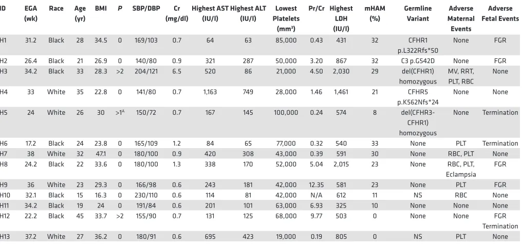
Participant demographics. To test our hypothesis, women with HELLP syndrome, partial HELLP syndrome, and healthy pregnant controls were recruited from 2015 to 2016. Our cohort consisted of 13 participants with HELLP syndrome (H1–H13, Table 1), 14 participants with partial HELLP syndrome (PH1–PH14, Table 2), and 19 healthy pregnant controls (HPC1–HPC19, Table 3). In order to compare these cohorts to patients with TMA, we also recruited 18 participants diagnosed with aHUS (aHUS1–aHUS18, Table 4), and 8 participants diagnosed with thrombotic thrombocytopenic purpura (TTP1–TTP8, Table 4). The par-ticipant demographics among HELLP, partial HELLP, and HPC cohorts did not differ in body mass index (BMI), race, or parity. However, the HELLP group had a statistically significant younger age when com-pared with the HPC group (27 versus 32 years, P < 0.030), but not when comcom-pared with partial HELLP syndrome. We also found that the mean estimated gestational age (EGA) of HELLP participants was significantly younger compared with the HPC group (30 versus 37 weeks, P < 0.001).
syndrome group. Three participants terminated their pregnancies secondary to the diagnosis of HELLP syndrome in the setting of a nonviable fetus (less than 23 weeks and/or estimated fetal weight less than 500 grams). The other 5 fetuses survived but were diagnosed with fetal growth restriction (FGR), which manifested in the third trimester.
There was one adverse maternal outcome in the partial HELLP group, which was an eclamptic sei-zure. Further, 4 of 14 participants (29%) experienced the adverse fetal event of FGR. There were no fetal terminations in the partial HELLP group. In our HPC cohort, there was one participant who was later diagnosed with preeclampsia (not HELLP or partial HELLP syndrome). At the time of recruitment, the participant was normotensive without signs or symptoms of preeclampsia and had an EGA of 38 weeks. She was diagnosed with preeclampsia at 39 weeks and 5 days and this necessitated induction of labor. There was one adverse fetal outcome, which was FGR in the HPC group. All HELLP or partial HELLP syndrome resolved postpartum or after termination in all cases, and no cases of pregnancy-associated aHUS or TTP were observed.
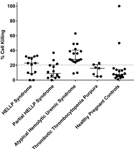
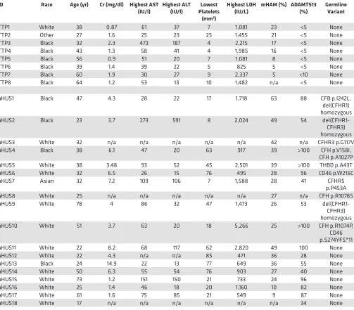
Complement activation in HELLP and aHUS. The mHam assay was performed for all participants with available serum samples in order to assess activation of the APC. A positive test is defined as greater than 20.5% cell killing, based on our previous study suggesting that HELLP patients are positive in the mHam assay (13). In this analysis, 8 of 13 HELLP participants (62%) were positive, compared with only 2 of 18 participants (11%) in the HPC group (Figure 1). In addition, only 3 of 14 (21%) partial HELLP participants were positive in the mHam assay. For participants with aHUS, 15 of 17 (88%) were positive, while only 1 of 7 participants (14%) with TTP were positive, confirming as expected that the mHam is a useful discrimina-tor between these 2 phenotypes (20).
While the mHam assesses complement activation as a measure of relative cell death, the magnitude of a result above the 20.5% threshold has unclear significance and has not been found to be associated with clinical outcomes. Thus, statistical analysis was performed using the mHam as a binary measure (positive or negative). When compared with the HPC group, HELLP syndrome participants were signifi-cantly more likely to test positive for the mHam assay (62% versus 11%, P = 0.006) (Table 5). We also
Table 1. HELLP syndrome participants
ID EGA (wk)
Race Age (yr)
BMI P SBP/DBP Cr (mg/dl) Highest AST (IU/l) Highest ALT (IU/l) Lowest Platelets
(mm3)
Pr/Cr Highest LDH (IU/l) mHAM (%) Germline Variant Adverse Maternal Events Adverse Fetal Events
H1 31.2 Black 28 34.5 0 169/103 0.7 64 63 85,000 0.43 431 32 CFHR1
p.L322Rfs*50
None FGR
H2 26.4 Black 21 26.9 0 140/80 0.9 321 287 50,000 3.20 867 32 C3 p.G542D None FGR
H3 34.2 Black 33 28.3 >2 204/121 6.5 520 86 21,000 4.50 2,030 29 del(CFHR1)
homozygous
MV, RRT, PLT, RBC
None
H4 33 White 35 22.8 0 141/80 0.7 1,163 749 28,000 1.46 1,461 21 CFHR5
p.K562Nfs*24
None None
H5 24 White 26 30 >1A 150/72 0.7 167 145 100,000 0.24 574 8
del(CFHR3-CFHR1) homozygous
None Termination
H6 17.2 Black 24 23.8 0 165/109 1.2 84 65 77,000 0.32 540 33 None PLT Termination
H7 38 White 32 47.1 0 180/100 0.9 420 308 43,000 0.39 591 30 None RBC, PLT None
H8 24.2 Black 22 33.6 0 180/100 1.3 338 170 52,000 5.04 2,015 23 None RBC, PLT,
Eclampsia FGR
H9 36 White 23 29.3 0 166/98 0.6 243 181 42,000 12.35 581 23 None PLT FGR
H10 32.1 Black 15 16.3 0 230/110 0.6 114 81 42,000 N/A 612 11 NS RBC None
H11 34.2 Black 19 24 0 191/84 0.6 201 101 63,000 6.93 325 10 None None None
H12 22.2 Black 45 33.7 >2 155/90 0.7 131 125 68,000 9.77 503 0 None None FGR
Termination
H13 37.2 White 27 36.2 0 180/91 0.6 695 423 19,000 0.19 805 0 NS PLT None
generated a control cohort for a TMA during pregnancy by combining the HPC and TTP groups, as both are expected to have normal complement regulation, and the TTP participants are a confirmed negative control in our previous study (20). HELLP participants were significantly more likely to test positive in the mHam assay (62% versus 16%, P = 0.009) compared with the composite control cohort. This was also true for aHUS patients compared with this cohort (88% versus 16%, P < 0.001). Conversely, mHam positivity in the HELLP syndrome and aHUS groups was not significantly different. These data indicate that HELLP syndrome, similar to aHUS, is marked by activation of complement.
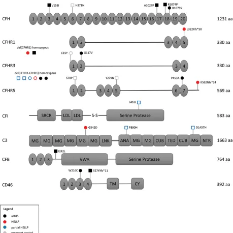
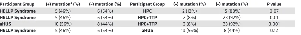
Identification of rare germline variants in HELLP syndrome. Approximately 50% of aHUS patients harbor germline mutations in genes that regulate the APC; thus, we performed targeted sequencing of participant DNA to identify germline variants in 9 genes known to have a functional role in the APC. To minimize the contribution of likely benign germline polymorphisms, only rare heterozygous germline variants with a minor allele frequency less than 0.01 (1%) were included in our analysis (detailed in methods). In addition, homozy-gous deletion of CFHR1 and CFHR3, reported to occur in approximately 2% of the population, was included in our analysis owing to its association with aHUS and likely functional significance (21). In our pregnant cohorts, 2 participants in the HELLP syndrome group and 2 participants in the HPC group did not give con-sent for gene mutation testing. We identified a rare germline variant or homozygous deletion in 5 of 11 HELLP participants (46%), compared with only 3 of 14 partial HELLP participants (21%), and 2 of 17 participants (12%) in the HPC group (Tables 1–3 and Figure 2). Further, in our TMA cohorts, germline variants were found in 10 of 18 participants with aHUS (56%), while no variants were identified any of the 8 TTP participants (Table 4). When compared with the HPC group alone, there was a nonsignificant trend towards the presence of a genetic variant in the HELLP syndrome cohort (46% versus 12%, P = 0.07). When compared with the composite controls (combined HPC and TTP groups), HELLP patients were significantly more likely to have a rare germline variant in an APC gene (46% versus 8%, P = 0.01). In addition, there was no difference between the HELLP syndrome and aHUS cohorts for the presence of a genetic variant (Table 6).
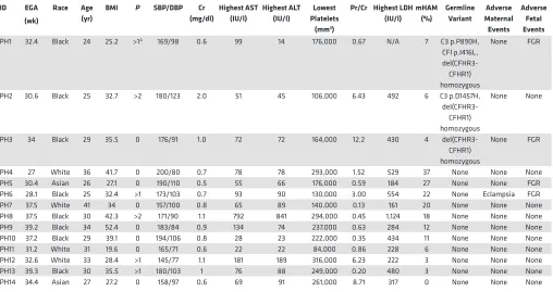
Table 2. Partial HELLP syndrome participants
ID EGA (wk)
Race Age (yr)
BMI P SBP/DBP Cr (mg/dl) Highest AST (IU/l) Highest ALT (IU/l) Lowest Platelets
(mm3)
Pr/Cr Highest LDH (IU/l) mHAM (%) Germline Variant Adverse Maternal Events Adverse Fetal Events
PH1 32.4 Black 24 25.2 >1A 169/98 0.6 99 14 176,000 0.67 N/A 7 C3 p.P890H,
CFI p.I416L,
del(CFHR3-CFHR1) homozygous
None FGR
PH2 30.6 Black 25 32.7 >2 180/123 2.0 51 45 106,000 6.43 492 6 C3 p.D1457H,
del(CFHR3-CFHR1) homozygous
None None
PH3 34 Black 29 35.5 0 176/91 1.0 72 72 164,000 12.2 430 4
del(CFHR3-CFHR1) homozygous
None FGR
PH4 27 White 36 41.7 0 200/80 0.7 78 78 293,000 1.52 529 37 None None None
PH5 30.4 Asian 26 27.1 0 190/110 0.5 55 66 176,000 0.59 184 27 None None FGR
PH6 28.1 Black 25 32.4 >1 173/103 0.7 93 90 130,000 3.00 554 22 None Eclampsia FGR
PH7 37.5 White 41 34 0 157/100 0.8 65 89 140,000 0.13 161 20 None None None
PH8 37.5 Black 30 42.3 >2 171/90 1.1 792 841 294,000 0.45 1,124 18 None None None
PH9 39.2 Black 34 52.4 0 183/84 0.9 134 74 237,000 0.63 284 12 None None None
PH10 37.2 Black 29 39.1 0 194/106 0.8 28 23 222,000 0.35 434 11 None None None
PH11 31.2 White 31 19.6 0 165/71 0.6 22 22 84,000 0.86 228 6 None None None
PH12 32.6 White 33 28.4 >1 145/77 1.1 181 189 316,000 6.23 222 3 None None None
PH13 39.3 Black 30 35.5 >1 180/103 1 76 88 249,000 0.20 480 3 None None None
PH14 34.4 Asian 27 27.2 0 158/97 0.6 69 91 261,000 8.71 317 0 None None None
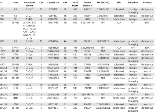
Detailed characteristics of all identified genetic variants, along with pathogenicity prediction outcomes for single-nucleotide variants (SNVs) via 3 different algorithms, are provided in Table 7. There were 2 HELLP participants with frameshift mutations, assumed to be loss of function, and 1 participant with a missense mutation predicted to be pathogenic in 2 out of 3 algorithms. Each of these participants also had a positive mHam test. Another HELLP participant with a positive mHam test harbored a homozygous dele-tion of the CFHR1 gene. There was one HELLP participant with a negative mHam assay and a germline variant. This individual (H5) was found to have homozygous deletion of both CFHR1 and CFHR3 and car-ried a history of HELLP syndrome in a prior pregnancy. Interestingly, homozygous deletion of CFHR1 and CFHR3 was found in 3 participants with partial HELLP, all with negative mHam tests. One of these partici-pants (PH1) also had a history of HELLP syndrome in a prior pregnancy. Of note, PH1 was found to have 2 additional missense variants in other APC genes as well. There were 2 HPC participants identified with rare genetic variants, both negative in the mHam assay. One individual (HPC1) carried a diagnosis of sickle cell trait and α-thalassemia and harbored multiple missense mutations, with one predicted to be pathogenic by all 3 algorithms. By comparison, all aHUS participants with identified germline variants were also found to be positive in the mHam assay. Two participants were found to have deletions of both CFHR1 and CFHR3, while 1 participant harbored a deletion of CFHR1 only. Deletion of CFHR genes were not identified in any HPC or TTP participants, supporting the functional significance of this variant. However, this variant by itself does not appear to sufficiently activate the APC in order to produce a positive mHam test.
Discussion
In this study, we demonstrate that HELLP syndrome is part of the phenome that results from germline muta-tions in genes that regulate the APC. Using phenotypic, functional (mHam assay demonstrating impaired complement regulation), and genetic (mutations in genes that regulate the APC) data we found striking similarities to aHUS. Our genetic analysis was limited to rare germline variants in order to increase the likeli-hood of identifying pathogenic mutations that alter the function of APC genes. Using this strict criterion, significantly more APC germline mutations were identified in the HELLP syndrome cohort compared with
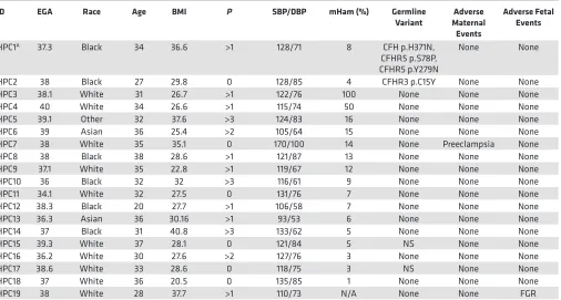
Table 3. Healthy pregnant control (HPC) participants
ID EGA Race Age BMI P SBP/DBP mHam (%) Germline
Variant Maternal Adverse Events
Adverse Fetal Events
HPC1A 37.3 Black 34 36.6 >1 128/71 8 CFH p.H371N,
CFHR5 p.S78P, CFHR5 p.Y279N
None None
HPC2 38 Black 27 29.8 0 128/85 4 CFHR3 p.C15Y None None
HPC3 38.1 White 31 26.7 >1 122/76 100 None None None
HPC4 40 White 34 26.6 >1 115/74 50 None None None
HPC5 39.1 Other 32 37.6 >3 124/83 16 None None None
HPC6 39 Asian 36 25.4 >2 105/64 15 None None None
HPC7 38 White 35 35.1 0 170/100 14 None Preeclampsia None
HPC8 38 Black 38 28.6 >1 121/87 13 None None None
HPC9 37.1 White 35 22.8 >1 119/67 12 None None None
HPC10 36 Black 32 32 >3 116/61 9 None None None
HPC11 34.1 White 32 27.5 0 131/76 7 None None None
HPC12 38.3 Black 20 27.7 >1 106/58 7 None None None
HPC13 36.3 Asian 36 30.16 >1 93/53 6 None None None
HPC14 37 Black 31 40.8 >3 133/62 5 None None None
HPC15 39.3 White 37 28.1 0 121/84 5 NS None None
HPC16 36.2 White 30 27.6 >2 127/76 3 None None None
HPC17 38.6 White 33 28.6 0 118/75 3 NS None None
HPC18 37 White 36 20.5 0 135/85 1 None None None
HPC19 38 White 28 37.7 >1 110/73 N/A None None FGR
controls (46% versus 8%, P = 0.01), while no difference was found when compared with aHUS. We also confirmed our previous data demonstrating that significantly more HELLP participants test positive in the mHam assay compared with controls (62% versus 16%, P = 0.009). Further, we did not identify any individ-uals in our control groups that tested positive for both the mHam assay and a germline variant (0 of 23 HPC and TTP participants, compared with 4 of 11 HELLP participants). Conversely, both tests were negative in 17 of 23 control participants and only 2 of 11 HELLP participants. Thus, this combination of testing may identify individuals with HELLP syndrome who could potentially benefit from therapeutic intervention.
Our findings agree with previous studies suggesting that complement plays an important role in the pathophysiology of HELLP. The APC activation fragment Bb was found to be a predictor of acquiring pre-eclampsia (23). In addition, a prospective study of 40 pregnant women with lupus and/or antiphospholipid antibodies who developed preeclampsia identified 7 (17.5%) that had germline heterozygous mutations likely to be deleterious in MCP, CFI, or CFH (14). Other studies have performed targeted sequencing of various APC genes in patients with HELLP syndrome and identified heterozygous mutations in 9% to 36% of cases (18, 19).
The major limitation of this study is the small sample sizes of the participant groups. Further, mul-tiple HELLP and partial HELLP participants tested negative for APC activation in the mHam assay. The most likely explanation for this finding is that the pathophysiology of HELLP syndrome (and par-tial HELLP even more so) is due to multiple factors. However, this study builds on our previous data and that of others demonstrating that a significant subset of HELLP syndrome is due to a reduced ability to regulate the APC, analogous to aHUS (13, 18, 20). Indeed, the clinical manifestations of hypertension, renal insufficiency, thrombocytopenia, elevated lactate dehydrogenase, elevated aspar-tate aminotransferase, and even the presence of schistocytes are common to both disorders. Roughly 50% of aHUS patients harbor germline mutations, usually heterozygous, that increase susceptibility of endothelial cells to injury from activated complement (12, 15, 16). These patients are often asymp-tomatic for decades until they encounter a strong complement trigger such as major surgery, infection, malignancy, autoimmunity, or pregnancy. In HELLP syndrome, pregnancy is the likely trigger (11). Previous studies have shown that complement proteins such as CFH and C5b-9, as well as associated contributors of increased complement (e.g., C-reactive protein) are significantly increased during preg-nancy, possibly explaining why phenotypic manifestations of HELLP syndrome almost exclusively occur in the late second or third trimester (9, 24–27). This may also explain why 2 cases in the HPC cohort tested positive in the mHam assay.
In our cohort, HELLP syndrome participants were associated with more frequent adverse maternal and fetal outcomes compared with the partial HELLP syndrome or HPC participants. Women with HELLP syn-drome are more likely to have increased morbidity and mortality secondary to disseminated intravascular coagulation and hepatic rupture compared with other hypertensive disorders of pregnancy (28, 29). However, 6 of the 7 women with HELLP syndrome who had maternal adverse events were either positive by the mHam assay or had a rare variant in an APC gene. Our data suggest that the increased morbidity or mortality in HELLP syndrome is associated with dysregulation of the APC. In the partial HELLP group, there was only one participant (PH6) with an adverse maternal outcome (an eclamptic seizure); she was also positive in the
mHam, indicating increased APC activation. There was one participant in the HPC group with a negative mHam and genetic analysis who was later diagnosed with preeclampsia.
In the HELLP group there were 7 participants who had adverse fetal outcomes, 3 of which were terminations of pregnancy secondary to early gestational age and fetal weight less than 500 grams at time of diagnosis. Two of these had either a positive mHam or a rare germline variant. In the partial HELLP group there were 4 participants with adverse fetal outcomes, all of whom had either a positive mHam test or a rare germline variant, further indicating that abnormal complement regulation is asso-ciated with both poor maternal and fetal outcomes. There were 2 HPC participants with positive mHam assays, potentially secondary to the normal upregulation of the complement system during pregnancy, particularly in the third trimester (9). Further, there were 2 HPC participants with rare variants of the APC. Similar to multiple other genetic diseases (hemochromatosis, for example), rare variants of the APC are associated with incomplete penetrance and variable expressivity (30–35). Therefore, genetic
Table 4. aHUS and TTP participants
ID Race Age (yr) Cr (mg/dl) Highest AST
(IU/l) Highest ALT (IU/l) Platelets Lowest (mm3)
Highest LDH
(IU/L) mHAM (%) ADAMTS13 (%) Germline Variant
TTP1 White 38 0.87 61 37 7 1,081 23 <5 None
TTP2 Other 27 1.6 25 23 25 1,455 21 <5 None
TTP3 Black 32 2.3 473 187 4 2,215 17 <5 None
TTP4 Black 43 1.3 58 41 4 1,985 16 <5 None
TTP5 Black 56 0.9 51 20 7 1,081 8 <5 None
TTP6 Black 39 1.4 39 22 5 825 5 <5 None
TTP7 Black 60 1.9 30 27 9 2,337 5 <10 None
TTP8 Black 64 1.2 53 13 10 1,482 n/a <5 None
aHUS1 Black 47 4.3 28 22 17 1,718 63 88 CFB p.I242L,
del(CFHR1) homozygous
aHUS2 Black 23 3.7 273 591 8 2,024 49 54
del(CFHR1-CFHR3) homozygous
aHUS3 White 32 n/a n/a n/a n/a n/a 42 n/a CFHR3 p.G117V
aHUS4 Black 38 6.1 47 20 63 917 39 >100 CFH p.V158I,
CFH p.A1027P
aHUS5 White 38 3.48 93 52 45 2,501 39 >100 THBD p.A43T
aHUS6 White 32 6.5 26 15 76 495 28 96 CD46 p.W216C
aHUS7 Asian 32 7.2 109 106 7 1,588 28 41 CFHR5
p.P453A
aHUS8 White 25 n/a n/a n/a n/a n/a 27 n/a CFH p.R1078S
aHUS9 White 78 4 86 32 47 1,473 26 53
del(CFHR1-CFHR3) homozygous
aHUS10 White 51 3.7 63 20 18 5,266 25 >100 CFH p.R1074P,
CD46 p.S274YFS*11
aHUS11 White 22 8.2 68 117 62 2,820 49 100 None
aHUS12 White 22 4.3 n/a n/a 85 471 36 28 None
aHUS13 Black 24 14.9 22 13 77 649 36 55 None
aHUS14 White 50 6.3 55 54 76 903 27 40 None
aHUS15 White 73 1.2 151 150 21 733 24 96 None
aHUS16 White 25 1.4 46 18 20 1,160 10 82 None
aHUS17 White 61 1.6 75 85 21 549 9 87 None
aHUS18 White 17 n/a n/a n/a n/a n/a n/a 34 None
variants are best characterized as risk factors, with disease onset occurring in the setting of additional environmental stress causing complement activation.
Our findings have important clinical implications for the diagnosis and treatment of HELLP syn-drome. Reliable biomarkers and genetic mutations that predispose to disease states are critical for planning future clinical trials and for targeted approaches to therapy. Paroxysmal nocturnal hemo-globinuria (PNH) and aHUS are diseases caused by mutations that predispose cells to complement-mediated destruction (16, 36–38). Both of these conditions are dramatically and rapidly improved by blocking terminal complement with the humanized monoclonal antibody, eculizumab (22, 39–41). Prior to FDA approval of eculizumab, pregnancy was relatively contraindicated in PNH due to exces-sive complement activation that increased fetal and maternal mortality. Eculizumab is now routinely recommended for PNH patients who become pregnant since the drug does not effectively cross the placenta or enter the breast milk (42). Complement inhibition has also greatly improved the outcome of patients with aHUS. Recent studies in aHUS show that this treatment can be safely discontinued in most patients once a complete remission is achieved and the complement trigger has resolved (43). Our study confirms that aHUS and HELLP share similar pathophysiology. Indeed, there is a case report of eculizumab treatment leading to clinical improvement and prolongation of pregnancy in a patient with HELLP syndrome (44). Furthermore, we previously demonstrated that C5 inhibition can effectively block complement-mediated killing in HELLP serum in vitro (13). Currently, HELLP syndrome is most effectively managed by delivering the fetus (removal of the complement trigger); however, in cases of early gestational age (before 28 weeks) there is a high risk of fetal mortality and maternal morbidity (1). Based on our data and those of others, we suggest that future trials of comple-ment inhibition in HELLP syndrome include genetic testing and the mHam assay to firmly establish the diagnosis. Patients with HELLP syndrome diagnosed prior to 28 weeks gestational age may benefit from this therapeutic intervention to allow the fetus to reach a safer gestational age and achieve less neonatal morbidity.
In summary, most HELLP syndrome and likely some preeclampsia, is driven by failure to regulate the APC. Serologic assays demonstrating a reduced ability to regulate complement (mHam), germline mutations in genes that regulate the APC, or both, are found in a majority of patients with HELLP syndrome. While we do not advocate for off-label use of complement inhibitors in HELLP syndrome, our findings form the basis for the design of clinical trials to test the efficacy of complement inhibition to reduce perinatal morbidity from early prematurity.
Table 5. Comparison of mHam for all groups
Participant Group (+) mHam (%) (–) mHam (%) Participant Group (+) mHam (%) (–) mHam (%) P value
HELLP Syndrome 8 (62%) 5 (38%) HPC 2 (11%) 16 (89%) 0.006
HELLP Syndrome 8 (62%) 5 (38%) HPC+TTP 4 (16%) 21 (84%) 0.009
aHUS 15 (88%) 2 (12%) HPC+TTP 4 (16%) 21 (84%) <0.001
HELLP Syndrome 8 (62%) 5 (38%) aHUS 15 (88%) 2 (12%) 0.19
HELLP, hemolysis, elevated liver enzymes, and low platelets; mHam, modified Ham assay; aHUS, atypical hemolytic uremic syndrome; HPC, healthy pregnant control; TTP, thrombotic thrombocytopenic purpura.
Table 6. Comparison of germline mutation status for all groups
Participant Group (+) mutationA (%) (–) mutation (%) Participant Group (+) mutation (%) (–) mutation (%) P value
HELLP Syndrome 5 (46%) 6 (54%) HPC 2 (12%) 15 (88%) 0.07
HELLP Syndrome 5 (46%) 6 (54%) HPC+TTP 2 (8%) 23 (92%) 0.01
aHUS 10 (56%) 8 (44%) HPC+TTP 2 (8%) 23 (92%) 0.001
HELLP Syndrome 5 (46%) 6 (54%) aHUS 10 (56%) 8 (44%) 0.12
Methods
Study population and design. We performed a prospective case-controlled study to identify whether HELLP syndrome is associated with both activation of the APC as well as germline variants in genes that regulate the APC. We hypothesized that HELLP syndrome patients harboring genetic variants of APC genes would also display activation of the APC via a functional assay (the mHam). All partici-pants were recruited from the Johns Hopkins Medical Institute, and none had been studied previously. The Tennessee criteria were used for the diagnosis of HELLP syndrome, and partial HELLP syndrome was defined by having 1 or 2 but not all lab abnormalities of HELLP syndrome (Supplemental Table 1; supplemental material available online with this article; https://doi.org/10.1172/jci.insight.99128DS1) (1, 45). We recruited all participants from November 1, 2015 to December 31, 2016. Healthy pregnant controls were recruited after 23 weeks of gestation, and were excluded if they had a history of hyper-tensive disease, complement or microangiopathic disorder, or autoimmune disease. Blood was collected from all participants at the time of recruitment by venipuncture of the antecubital vein. Serum was cold centrifuged within 2 hours of collection at 4°C and stored at –80°C. Whole blood was used to generate genomic DNA for targeted gene sequencing using a Qiagen DNeasy Blood & Tissue Kit. All samples were deidentified and coded prior to laboratory testing. Patients with aHUS were recruited using stan-dard diagnostic criteria defined by Legendre et al. (39). Patients with TTP were recruited following confirmation of the diagnosis via an ADAMTS13 activity less than 10%.
Table 7. Germline variant characteristics
ID Gene Nucleotide
Variant Chr Coordinate VAF DepthRead PositionProtein MAF (ExAC) Sift PolyPhen Provean
H3 C3 C>C/T 19 6710711 56 208 G542D 0.00001682 tolerated possibly
damaging deleterious PH1 C3 G>G/T 19 6697482 48 297 P890H 0.00002471 tolerated benign deleterious PH1 CFI T>T/G 4 110667561 44 424 I416L 0.001113 deleterious benign neutral H4 CFHR5 GGAAATTTG
AATATCCTA TATGT>GGA AATTTGAAT ATCCTATAT
GT/G
1 196977785 40 609 K562Nfs*24 N/A N/A N/A N/A
PH3 C3 C>C/G 19 6680256 44 393 D1457H 0.0003626 deleterious probably
damaging deleterious H2 CFHR1 CT>CT/C 1 196801100 59 711 L322Rfs*50 N/A N/A N/A N/A HPC2 CFHR3 G>G/A 1 196744060 50 277 C15Y N/A deleterious benign deleterious HPC1 CFH C>C/A 1 196658696 49 440 H371N 0.00007426 tolerated benign deleterious HPC1 CFHR5 T>T/C 1 196952188 46 725 S78P 0.0001401 deleterious probably
damaging deleterious HPC1 CFHR5 T>T/A 1 196965196 47 554 Y279N 0.0001484 tolerated benign deleterious aHUS4 CFH G>G/A 1 196646650 59 191 V158I 0.000132 tolerated benign neutral aHUS4 CFH G>G/C 1 196711127 44 369 A1027P 0.00004119 tolerated benign neutral aHUS1 CFB A>A/C 6 31915584 50 567 I242L 0.00002559 tolerated benign neutral aHUS3 CFHR3 G>G/T 1 196749023 55 190 G117V N/A deleterious probably
damaging deleterious aHUS5 THBD C>C/T 20 23030015 59 87 A43T 0.00343 tolerated benign neutral aHUS10 CFH G>G/C 1 196712669 49 508 R1074P 0.00001647 deleterious probably
damaging deleterious aHUS10 CD46 CAG>C 1 207940331 25A 8A S274YFS*11 N/A N/A N/A N/A
aHUS6 CD46 G>G/C 1 207934766 50 701 W216C N/A deleterious probably
damaging deleterious aHUS8 CFH G>G/T 1 196712682 51 332 R1078S 0.00005765 tolerated benign neutral aHUS7 CFHR5 C>C/G 1 196973817 47 220 P453A 0.000008238 deleterious probably
damaging deleterious
mHam assay. The mHam assay was performed as previously described (20). Briefly, PNH-like (PIGA–null) TF-1 cells were used to assess complement-mediated killing via the APC. Cells were plat-ed in U-shapplat-ed wells at a density at 4,000 cells/well and incubatplat-ed in 20% serum with Gelatin Veronal Buffer (GVB, Complement Technology, Inc). Each sample was tested in triplicate for 30 minutes at 37°C. After incubation, the supernatant was removed and cell proliferation agent WST-1 (Roche) was added and incubated for 2 hours at 37°C. Absorbance was measured in an iMark Microplate Absor-bance Reader (Bio-Rad) at 490 nm with a reference wave length at 595 nm. Heat-inactivated serum was used as a negative control for each sample. Heat inactivation was performed by incubating the serum at 56°C for 30 minutes. Normal human AB serum (H4522, Sigma-Aldrich) was used as an internal control in the assay. The percentage of live cells was calculated as the ratio of the absorbance of each sample divided by the absorbance of the same sample’s heat-inactivated control. The percentage of nonviable cells (cell killing) was calculated as 1 minus this ratio.
Targeted sequencing. A custom panel of 9 genes known to function in the APC was selected for sequencing using the Illumina TruSeq Custom Amplicon (v1.5). Illumina DesignStudio software was utilized to generate custom oligonucleotides designed to amplify exonic regions of the following selected genes: CFH, CFB, CFI, CFHR1, CFHR3, CFHR5, C3, CD46 (MCP), and THBD. The panel consisted of 402 amplicons covering a total of 58.3 kb with an overall coverage of 98%. An amplicon library of each patient sample was generated according to the manufacturer’s protocol using 250 ng genomic DNA as input. Successful library amplification was verified for a subset of samples by both gel electrophoresis and Bioanalyzer (Agilent) analysis. This was followed by library normalization and pooling per manu-facturer protocol, and paired-end sequencing on the Illumina MiSeq platform via the Genetic Resources Core Facility (GRCF) at Johns Hopkins School of Medicine (https://grcf.jhmi.edu). Analysis of raw sequencing data (FASTQ) was performed using the TruSeq Amplicon application (v2.0.0) via the Illu-mina BaseSpace platform, aligning to (GRCh37/hg19) human genome reference. Average base coverage was 377× per sample. SNVs and small indels were identified using Isaac variant caller and variant call files were filtered using VariantStudio software (v3.0). Variants not passing Illumina’s variant quality filters were excluded. The following criteria were used to identify rare germline SNVs and indels: non-synonymous coding region variants with a depth greater than 20×, variant allele frequency between 40% and 60%, Exome Aggregation Consortium (ExAC v0.3.1) minor allele frequency less than 0.01 in any ethnic population (total of 60,706 samples), and minor allele frequency less than 0.01 in the Exome Variant Server (46). Large deletions were determined by complete loss of signal for multiple consecutive amplicons (Supplemental Figure 1). All genomic coordinates refer to hg19.
Adverse events. The investigators sought to observe any adverse maternal or fetal events in the HELLP group, partial HELLP group, and HPC group. Maternal adverse events were maternal mortality, inten-sive care unit admission, renal failure, blood product transfusion, and eclampsia in HELLP or partial HELLP groups, or new diagnosis of preeclampsia in a control group. Fetal adverse events were fetal ter-mination or abortion secondary to diagnosis of HELLP or partial HELLP syndrome, neonatal mortality, and fetal growth restriction. Other than worsening of HELLP syndrome in the setting of a nonviable fetus, no other factors were relevant for fetal terminations. Because neonatal intensive care unit admis-sion in preeclampsia and HELLP syndrome is usually secondary to iatrogenic preterm birth, we did not include this as an adverse event in our group.
Statistics. A 1-way ANOVA test with Bonferroni’s correction was used to calculate the P values for the difference among the means for age, BMI, estimated gestational age in HELLP, partial HELLP, and the HPC groups. Either χ2 test or Fisher’s exact test was used to calculate the rest of the P values
provided. A Fisher’s exact was used if more than 20% of the expected values in the input cells were less than 5 and/or any individual values had expected counts of less than 1. Statistical significance for the mHam assay was calculated as a binary value (positive or negative), as the mHam assay is thought to be clinically significant as a qualitative test. All analyses were performed by using SPSS 18 for Windows/MAC (PASW Statistics GradPack 18; SPSS). All tests were 2-sided with P less than 0.05 indicating significance.
Author contributions
AJV and RAB were responsible for the study conception and design. AJV, EMB, RAB, SAM, SC, SE, KB, and CJS collected and categorized study participants. AJV, EMB, XY, IM, and ACB performed experi-ments and acquired data. AJV, EMB, and RAB analyzed and interpreted the data. JJ performed statistical data analysis. AJV, EMB, and RAB drafted the manuscript and performed critical revisions. All authors approved the final version of the manuscript.
Acknowledgments
AJV is awarded the Johns Hopkins Meyerhoff Professorship Award and T32HL007525 NIH/NHLBI Training Program in Hematology. RAB is awarded the R01HL133113 NIH/NHLBI Complementopa-thies: Genotype and Phenotype. ACB, who is currently employed at the FDA, was involved with this study during a previous affiliation with Johns Hopkins School of Medicine. The views expressed in this paper do not represent the views of the FDA.
Address correspondence to: Robert A. Brodsky, Ross Research Building, Room 1025, Johns Hopkins Uni-versity School of Medicine, 720 Rutland Avenue, Baltimore, Maryland 21205, USA. Phone: 410.502.2546; Email: [email protected].
ACB’s present address is: FDA, Silver Spring, Maryland, USA.
1. Report of the American College of Obstetricians Gynecologists’ Task Force on Hypertension in Pregnancy. Hypertension in pregnancy: Executive Summary. Obstet Gynecol. 2013;122(5):1122–1131.
2. Chen XK, Wen SW, Smith GN, Yang Q, Walker MC. New-onset hypertension in late pregnancy and fetal growth: different associations between singletons and twins. Hypertens Pregnancy. 2007;26(3):259–272.
3. Iacobelli S, Bonsante F, Robillard PY. Pre-eclampsia and preterm birth in Reunion Island: a 13 years cohort-based study. Com-parison with international data. J Matern Fetal Neonatal Med. 2016;29(18):3035–3040.
4. Weinstein L. Syndrome of hemolysis, elevated liver enzymes, and low platelet count: a severe consequence of hypertension in pregnancy. 1982. Am J Obstet Gynecol. 2005;193(3 Pt 1):859.
5. Fitzpatrick KE, Hinshaw K, Kurinczuk JJ, Knight M. Risk factors, management, and outcomes of hemolysis, elevated liver enzymes, and low platelets syndrome and elevated liver enzymes, low platelets syndrome. Obstet Gynecol. 2014;123(3):618–627. 6. Uzan J, Carbonnel M, Piconne O, Asmar R, Ayoubi JM. Pre-eclampsia: pathophysiology, diagnosis, and management. Vasc
Health Risk Manag. 2011;7:467–474.
7. Martin JN, Rose CH, Briery CM. Understanding and managing HELLP syndrome: the integral role of aggressive glucocorti-coids for mother and child. Am J Obstet Gynecol. 2006;195(4):914–934.
8. Martin JN, Rinehart BK, May WL, Magann EF, Terrone DA, Blake PG. The spectrum of severe preeclampsia: comparative analysis by HELLP (hemolysis, elevated liver enzyme levels, and low platelet count) syndrome classification. Am J Obstet
Gyne-col. 1999;180(6 Pt 1):1373–1384.
9. Richani K, et al. Normal pregnancy is characterized by systemic activation of the complement system. J Matern Fetal Neonatal
Med. 2005;17(4):239–245.
10. Haeger M, Unander M, Bengtsson A. Enhanced anaphylatoxin and terminal C5b-9 complement complex formation in patients with the syndrome of hemolysis, elevated liver enzymes, and low platelet count. Obstet Gynecol. 1990;76(4):698–702.
11. Fakhouri F, et al. Pregnancy-associated hemolytic uremic syndrome revisited in the era of complement gene mutations. J Am
Soc Nephrol. 2010;21(5):859–867.
12. Loirat C, Frémeaux-Bacchi V. Atypical hemolytic uremic syndrome. Orphanet J Rare Dis. 2011;6:60.
13. Vaught AJ, et al. Direct evidence of complement activation in HELLP syndrome: A link to atypical hemolytic uremic syn-drome. Exp Hematol. 2016;44(5):390–398.
14. Salmon JE, et al. Mutations in complement regulatory proteins predispose to preeclampsia: a genetic analysis of the PROM-ISSE cohort. PLoS Med. 2011;8(3):e1001013.
15. Noris M, et al. Relative role of genetic complement abnormalities in sporadic and familial aHUS and their impact on clinical phenotype. Clin J Am Soc Nephrol. 2010;5(10):1844–1859.
16. Noris M, Bresin E, Mele C, Remuzzi G. Genetic atypical hemolytic-uremic syndrome. In: Pagon RA, et al., eds.
GeneReviews(R). Seattle, WA: University of Washington, Seattle; 1993. URL: https://www.ncbi.nlm.nih.gov/books/NBK1367.
Accessed February 27, 2018.
17. Maga TK, Nishimura CJ, Weaver AE, Frees KL, Smith RJ. Mutations in alternative pathway complement proteins in American patients with atypical hemolytic uremic syndrome. Hum Mutat. 2010;31(6):E1445–E1460.
18. Crovetto F, et al. The genetics of the alternative pathway of complement in the pathogenesis of HELLP syndrome. J Matern
Fetal Neonatal Med. 2012;25(11):2322–2325.
19. Fakhouri F, et al. Factor H, membrane cofactor protein, and factor I mutations in patients with hemolysis, elevated liver enzymes, and low platelet count syndrome. Blood. 2008;112(12):4542–4545.
20. Gavriilaki E, et al. Modified Ham test for atypical hemolytic uremic syndrome. Blood. 2015;125(23):3637–3646.
syndrome. PLoS Genet. 2007;3(3):e41.
22. Legendre CM, Licht C, Loirat C. Eculizumab in atypical hemolytic-uremic syndrome. N Engl J Med. 2013;369(14):1379–1380. 23. Lynch AM, Wagner BD, Giclas PC, West NA, Gibbs RS, Holers VM. The relationship of longitudinal levels of complement Bb
during pregnancy with preeclampsia. Am J Reprod Immunol. 2016;75(2):104–111.
24. Romem Y, Artal R. C-reactive protein in pregnancy and in the postpartum period. Am J Obstet Gynecol. 1985;151(3):380–383. 25. Ernst GD, et al. C-reactive protein levels in early pregnancy, fetal growth patterns, and the risk for neonatal complications: the
Generation R Study. Am J Obstet Gynecol. 2011;205(2):132.e1–132.12.
26. Watts DH, Krohn MA, Wener MH, Eschenbach DA. C-reactive protein in normal pregnancy. Obstet Gynecol. 1991;77(2):176–180. 27. Derzsy Z, Prohászka Z, Rigó J, Füst G, Molvarec A. Activation of the complement system in normal pregnancy and
pre-eclampsia. Mol Immunol. 2010;47(7–8):1500–1506.
28. Sibai BM. Diagnosis, controversies, and management of the syndrome of hemolysis, elevated liver enzymes, and low platelet count. Obstet Gynecol. 2004;103(5 Pt 1):981–991.
29. Sibai BM, Ramadan MK, Usta I, Salama M, Mercer BM, Friedman SA. Maternal morbidity and mortality in 442 pregnancies with hemolysis, elevated liver enzymes, and low platelets (HELLP syndrome). Am J Obstet Gynecol. 1993;169(4):1000–1006. 30. Richards A, et al. Implications of the initial mutations in membrane cofactor protein (MCP; CD46) leading to atypical
hemo-lytic uremic syndrome. Mol Immunol. 2007;44(1–3):111–122.
31. Fremeaux-Bacchi V, et al. Genetic and functional analyses of membrane cofactor protein (CD46) mutations in atypical hemo-lytic uremic syndrome. J Am Soc Nephrol. 2006;17(7):2017–2025.
32. Esparza-Gordillo J, et al. Insights into hemolytic uremic syndrome: segregation of three independent predisposition factors in a large, multiple affected pedigree. Mol Immunol. 2006;43(11):1769–1775.
33. Esparza-Gordillo J, et al. Predisposition to atypical hemolytic uremic syndrome involves the concurrence of different suscepti-bility alleles in the regulators of complement activation gene cluster in 1q32. Hum Mol Genet. 2005;14(5):703–712.
34. Papanikolaou G, et al. Genetic heterogeneity underlies juvenile hemochromatosis phenotype: analysis of three families of northern Greek origin. Blood Cells Mol Dis. 2002;29(2):168–173.
35. Piperno A, et al. Heterogeneity of hemochromatosis in Italy. Gastroenterology. 1998;114(5):996–1002.
36. Hill A, DeZern AE, Kinoshita T, Brodsky RA. Paroxysmal nocturnal haemoglobinuria. Nat Rev Dis Primers. 2017;3:17028. 37. Brodsky RA. Complement in hemolytic anemia. Blood. 2015;126(22):2459–2465.
38. Brodsky RA. Paroxysmal nocturnal hemoglobinuria. Blood. 2014;124(18):2804–2811.
39. Legendre CM, et al. Terminal complement inhibitor eculizumab in atypical hemolytic-uremic syndrome. N Engl J Med. 2013;368(23):2169–2181.
40. Brodsky RA, et al. Multicenter phase 3 study of the complement inhibitor eculizumab for the treatment of patients with parox-ysmal nocturnal hemoglobinuria. Blood. 2008;111(4):1840–1847.
41. Hillmen P, et al. The complement inhibitor eculizumab in paroxysmal nocturnal hemoglobinuria. N Engl J Med. 2006;355(12):1233–1243.
42. Kelly RJ, et al. Eculizumab in Pregnant Patients with Paroxysmal Nocturnal Hemoglobinuria. N Engl J Med. 2015;373(11):1032–1039.
43. Merrill SA, Brittingham ZD, Yuan X, Moliterno AR, Sperati CJ, Brodsky RA. Eculizumab cessation in atypical hemolytic ure-mic syndrome. Blood. 2017;130(3):368–372.
44. Burwick RM, Feinberg BB. Eculizumab for the treatment of preeclampsia/HELLP syndrome. Placenta. 2013;34(2):201–203. 45. Aydin S, Ersan F, Ark C, Arıoğlu Aydın C. Partial HELLP syndrome: maternal, perinatal, subsequent pregnancy and long-term
maternal outcomes. J Obstet Gynaecol Res. 2014;40(4):932–940.