R E S E A R C H
Open Access
Full-length amelogenin influences the
differentiation of human dental pulp stem
cells
Iris Frasheri
1*, Christina Ern
1, Christian Diegritz
1, Reinhard Hickel
1, Michael Hristov
2and Matthias Folwaczny
1Abstract
Background:Amelogenin is an extracellular matrix protein well known for its role in the organization and mineralization of enamel. Clinically, it is used for periodontal regeneration and, due to its finding also in predentin and intercellular spaces of dental pulp cells, it has recently been suggested for pulp capping procedures. The aim of this study was to analyse in vitro the effect of the recombinant human full-length amelogenin on the growth and differentiation of human dental pulp stem cells (hDPSCs).
Methods:Human DPSCs were treated with a supplement of amelogenin at a concentration of 10 ng/ml, 100 ng/ ml and 1000 ng/ml. The groups were compared to the unstimulated control in terms of cell morphology and proliferation, mineralization and gene expression forALP(alkaline phosphatase),DMP1(dentin matrix protein-1) andDSPP(dentin sialophosphoprotein).
Results:Amelogenin affects hDPSCs differently than PDL (periodontal ligament) cells and other cell lines. The proliferation rate at two weeks is significantly reduced in presence of the highest concentration of amelogenin as compared to the unstimulated control. hDPSCs treated with low concentrations present a downregulation of DMP1andDSPP,which is significant for DSPP (p = 0.011), but not for DMP1 (p = 0.395).
Conclusions:These finding suggest that the role of full-length amelogenin is not restricted to participation in tooth structure. It influences the differentiation of hDPSC according to various concentrations and this might impair the clinical results of pulp capping.
Keywords:Amelogenin, Cell signaling, Pulp biology, Gene expression, Stem cells, Extracellular matrix
Background
In 1963 Eastoe identified a protein in the tooth struc-ture, which presented a unique amino-acid compos-ition, different from the collagenous enamel proteins known to this date [1]. He coined the name Amelo-genin for it, and since then it has been considered a tissue-specific protein. In the next four decades, since it was identified, it was investigated in its role as a protein involved in the nucleation of crystallites. Now this structural role of amelogenin in tooth development has been well-established. Self-assembling into nanosphere aggregates and with high affinity for hydroxyapatite and
collagen, amelogenin controls the structural organization and mineralization of enamel, being eventually replaced by apatite mineral [2, 3]. Until recently it was thought to be produced only from ameloblasts, being thus specifically present only in enamel, but in 1997 Hammerstrom showed its presence also in the apical area of developing roots. A few years later, several studies reported amelo-genin mRNA and protein in mineralized tissues, in cells such as osteoblasts, osteoclasts, chondrocytes and cemen-toblasts [4]. In 2006 Deutsch revealed the expression of amelogenin not just in other mineralized tissues, but also in soft tissues (brain and eye) [5]. Since then, its involve-ment in signaling pathways has come into the limelight.
Focusing on the pulp-dentin microenvironment, Inai et al. localized amelogenin in the predentin and in the intercellular spaces of odontoblasts and dental pulp cells, * Correspondence:irisfrasheri@yahoo.it
1Department of Conservative Dentistry and Periodontology,
Ludwig-Maximilian-University, Goethestraße, 70, D-80336 Munich, Germany Full list of author information is available at the end of the article
using the technique of immunolabeling [6]. Thereafter it was suggested that amelogenin, penetrating toward the pulp, might play a role in the interaction between amelo-blasts and odontoamelo-blasts. Although the pathway and the downstream action of these events remain unknown, its clinical application has advanced rapidly. Amelogenin has been effectively used in wound healing (Xelma®, Molnlycke Health Care, Gothenburg, Sweden) [7], and in regenerative periodontal treatment. It has also been propagated for off label use in pulp capping procedures. Moreover, in vivo studies in humans and animals have focused on enamel matrix derivatives (EMD) as pulp capping material [8, 9]. It is derived from the porcine extracellular enamel matrix and is constituted for the most part by the full-length protein, though other compo-nents such as low-density isoforms, enamelin and trans-forming growth factor beta (TGF-beta) are also present in the gel of propylene glycol alginate. Histological analyses in animals following pulp capping procedures evidence the formation of an irregular dentinal bridge, which ap-pears to be more an osteodentin-like structure with cells present in the heterogeneous tissue [10].
The ability of human teeth to produce partial reparative dentin in response to different stimuli was noted before the conception of tissue engineering. In particular, calcium hydroxide has been successfully exploited in pulp capping procedures since the 1920s. This suggests that progenitor cells, present in fully developed tooth pulp, retain the abil-ity to form functional odontoblasts. The presence of stem cells in dental pulp was first proposed by Fitzgerald [11] and demonstrated a decade later, in a pioneering study by Gronthos et al. These are mesenchymal cells which have shown the potential to differentiate toward mesenchymal lineages (osteoblasts, smooth muscle cells, adipocytes, chondrocytes).
Upon these findings, our main goal was to determine if the recombinant human full-length protein, acting as a signaling protein, has an effect on the pulp at a genetic level, in the view of possible implications in regenerative treatments. This study aims to analyze the potential role of different concentrations of the full-length amelogenin on the stem cells present in different niches within the dental pulp and its purported influence on their growth and differentiation.
Materials and methods
Cell culture
The human dental pulp stem cells hDPSCs were obtained from AllCells (Alameda, CA, USA). They had been char-acterized by the company by means of flow cytometry, revealing a positivity for surface markers CD73, CD90, CD105 and CD166, and negativity for CD34, CD45 and CD133. These cells were provided at the second pas-sage and cultivated upon arrival until the fifth paspas-sage
in minimal essential medium, α-modification (α-MEM) with GlutaMAX™(Life Technologies, Carlsbad, CA, USA), supplemented with 10 % Fetal Bovine Serum Gold (PAA, Pasching, Austria), 100 U/mL Penicillin and 100 μg/mL Streptomicin (Sigma Aldrich, St. Louis, MO, USA). In order to eliminate the variability of hDPSCs culture, they were used in the same passage for all experiments. Cells in the fifth passage were chosen, as studies report that, at around the fifth passage, the dentinogenic markers appear to present peak levels of expression, suggesting a greater differentiation potential [12].
The hDPSCs were assigned to four different groups: a control, and three groups with a serial dilution of ame-logenin. All the experiments were carried out in bio-logical triplicates, while PCR analyses were carried out in technical triplicates. The control group was cultivated in α-MEM supplemented as mentioned above, without other additions. The same supplemented medium was used in the amelogenin groups, adding the amelogenin protein at a concentration of 10 ng/mL, 100 ng/mL and 1000 ng/mL. Three samples were included in each group.
The amelogenin was provided by Abnova Biotec GmbH (Taipei, Taiwan): it is a human recombinant pro-tein, full-length product of the gene Amelx (1-191a.a.), weighting approximately 48 kDa, which primary se-quence [NX_Q99217-1] is MGTWILFACLLGAAFAMP LPPHPGHPGYINFSYEVLTPLKWYQSIRPPYPSYGYEP MGGWLHHQIIPVLSQQHPPTHTLQPHHHIPVVPAQ QPVIPQQPMMPVPGQHSMTPIQHHQPNLPPPAQQP YQPQPVQPQPHQPMQPQPPVHPMQPLPPQPPLPPM FPMQPLPPMLPDLTLEAWPSTDKTKREEVD.
The cells were seeded in T25 flasks (BD Falcon, San Jose, CA, USA), at a density of 2*104cells/cm2and cul-tured in a humidified atmosphere containing 5 % CO2 at 37 °C, with the medium changed twice a week.
Cell morphology, proliferation and viability
The specimens were examined daily under inverted light microscopy (AXIO, Zeiss, Jena, Germany). The population doubling (PD) time and viability were evaluated passaging the cells weekly, re-plating them in T25 flasks at the start-ing concentration of 2*104 cells/cm2 and counting them with an automated cell analyzer (Cedex XS, Innovatis, Ba-sel, Switzerland), using Trypan Blue staining (Gibco, Thermo Fisher Scientific, Karlsruhe, Germany) in a 1:2 di-lution, according to the manufacturer’s instructions. The PD and cumulative PD were calculated at days 7, 14 and 21 using the following formula:
PD ¼ log2ðP=P0Þ
DT¼CT=PD
with CT being the time in culture.
Flow cytometry analysis of surface antigens
Cells were detached with Accutase (Sigma-Aldrich; 5 min at 37 °C) and then incubated with the following mouse anti-human mAbs for 30 min on ice: CD73-Pacific Blue™, CD90-FITC, CD105-FITC, CD166-PE, CD34-APC, CD45-PerCP, CD117-PE (all from BioLe-gend) and CD133-APC (Miltenyi Biotec). After two washing steps (PBS with 2 % FBS), the sample tubes were acquired on a BD FACSAria III (BD Biosciences) and at least 10.000 events were recorded. Data were an-alyzed with BD FACSDiva and FlowJo V10 software.
Immunofluorescence analysis
hDPSC were cultured on glass slides and fixed with methanol (Carl Roth, Karlsruhe, Germany) at–20 °C for 10 min. For DMP-1 and DSPP labelling, cells were washed with phosphate-buffered saline (PBS) with Tween-20 (Carl Roth, Karlsruhe, Germany), and blocked with 10 % horse serum for 1 h at room temperature. The cells were incubated overnight at 4 °C with the pri-mary antibodies diluted 1:200 and 1:50 to detect respect-ively DMP-1 and DSPP (Santa Cruz Biotechnology, Santa Cruz, CA, USA). Secondary antibodies were AlexaFluor 488 anti-rabbit and anti-goat IgG (Invitro-gen, Eugene, OR, USA) used in a dilution of 1:500. For alkaline phosphatase, cells were treated with Hyaluroni-dase (Sigma Aldrich, Steinheim, Germany) for 30 min. After washing with PBS with Tween the same protocol was used as for DMP-1 and DSPP. The primary antibody was diluted 1:20. The nuclear counterstaining was per-formed with 4’,6-diamidino-2-phenylindole (DAPI) (Invi-trogen, Eugene, OR, USA). Negative controls for all antibodies were carried out on the same slide omitting the primary antibody. The staining was evaluated under fluorescence microscope LSM 510 using a digital cam-era, AxioCam MRc (both Carl Zeiss, Jena, Germany).
Metabolic activity
WST-1 assays were used for determining the metabolic activity. hDPSC were seeded in 24-well plates with 3*103cells/well and cultured for 21 days with the above mentioned concentrations of amelogenin. Every 7 days WST-1 (Roche Diagnostics GmbH, Mannheim, Germany) was added according to manufacturer’s protocols and the medium of each well was transferred in a 96-well plate. The cells in the 24-well plates were washed three times with PBS and further cultured in supplemented medium. Varioskan 3.00.7 was used for spectrophoto-metric analyses.
Polymerase chain reaction
hDPSCs in the fourth passage were harvested at conflu-ence, and RNA was extracted. This was considered day 0. The cells were then seeded in T75 flasks (BD Falcon, San Jose, CA, USA) at a density of 2*104cells/cm2and further cultured without passaging. After 21 days cell samples underwent a process of lysation with Trizol® Reagent (Invitrogen, Carlsbad, CA, USA). The total RNA was extracted using the RNeasy Mini Kit (Qiagen, Hilden, Germany) following the manufacturer’s protocol. To exclude contamination, RNA samples with an optical density A260/A280 ratio between 1.8 and 2.1 were used.
Hexamer primer-based reverse transcription was per-formed for all the samples simultaneously using the First-Strand cDNA Synthesis Kit (Invitrogen, Carlsbad, CA, USA). For each sample, cDNA was synthetized using 500 ng RNA /mL.
Gene expression of alkaline phosphatase (ALP), dentin matrix protein-1 (DMP1) and dentin sialopho-sphoprotein (DSPP) was detected using a semiquanti-tative PCR method. Normalisation was performed using glyceraldehyde-6-phosphate dehydrogenase (GAPDH) as housekeeping gene. The master mix was prepared using FastStart DNA Master PLUS SYBR Green I dye.
The primers GAPDH and ALP were provided by TIB-MOLBIOL (Berlin, Germany). The primers DMP1 and DSPP for the amplification of cDNA were designed ac-cording to the published sequences, and provided by Biomol GmbH (Hamburg, Germany). The primer se-quences and the thermocycling conditions are listed in Table 1.
The samples of the obtained cDNA for the two latter primers were sequenced after the gel electrophoresis run, confirming the sequence of the genes. In order to avoid variability, the samples from all the groups in the study were processed concurrently for each analysed gene. To separate the cDNA fragments obtained from the PCR, they were loaded into agarose gel with eth-idium bromide staining. The primers used were of max-imum 238 bp, so a 1.5 % agarose gel in Tris-Borate EDTA (TBE) buffer and GeneRuler Low Range DNA Ladder 25 to 700 bp (Thermo Scientific) were used. The PCR products were then separated by electrophoresis at 70 mV and visualized under ultraviolet light. InfinityCapt software (Vilber Lourmat, Marne-la-Valée, France) was used as a gel documentation and quantification system. This represents a limitation of our study. We are aware that our data are semiquantitative and treat them as such.
Alizarin red staining
Red Staining (Sigma-Aldrich-A5533). In order to detect the alleged role as an inducer, the hDPSCs were plated in 12-well plates at a concentration of 104cells/well and cul-tured for 21 days with the same media as described above. The respective media were replaced every 3–4 days. Stain-ing protocol as described by Prockop et al. was used. Briefly, the wells were washed three times with PBS, fixed in 70 % Ethanol at–20 °C for 1 h. They were subsequently washed with H2O, stained for 10 min with Alizarin Red, washed again with PBS and kept at a temperature of 4 °C.
Statistical analyses
Statistical analyses of quantitative data were performed using SPSS Software Program (version 22, SPSS Inc, Chicago, IL, USA). All datasets have been tested for normal distribution using the Kolmogoroff-Smirnov test or Shapiro-Wilk test and for homogeneity of vari-ances using Levene test. Comparisons between different stimulation conditions have been performed using Wilcoxon test or Games-Howell test. Where appropriate (comparisons between two groups) all test procedures were two-tailed. P values <0.05 have been considered significant.
Results
Phenotypic expression
Cell morphology and proliferation
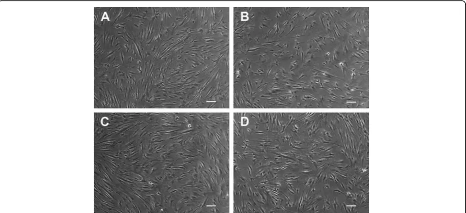
The monitoring of morphological changes in response to different amelogenin concentrations revealed no sub-stantial differences between the control and the stimu-lated groups. The cells presented a spindle shape and conserved a high nucleus:cytoplasm ratio 1:2 and prom-inent nucleoli. Attention was also given to the pattern formation as a differentiation index of the cells, as lately published [13]. In all the flasks the plated cells were cap-able of forming a herringbone pattern at ×5 view, with characteristic parallel arrays observed under a magnifica-tion of ×10 and ×20 (Fig. 1). These characteristics were constant in all the groups at all time points.
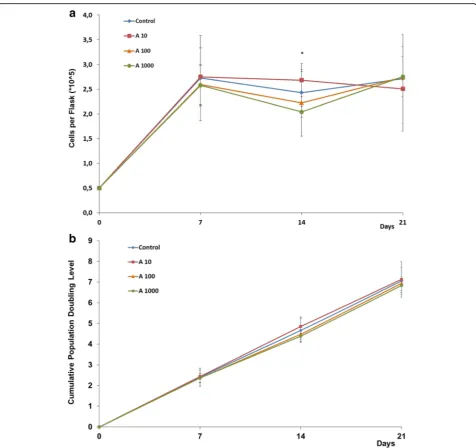
From the results we obtained regarding proliferation, the full-length amelogenin does not seem to significantly
affect the proliferation rate of this dental pulp cell line (p<0.05) (Fig. 2).
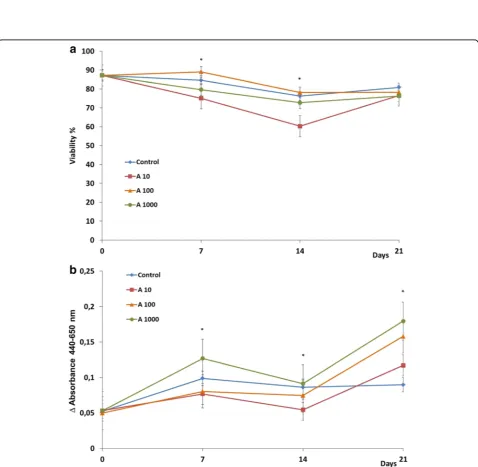
The exposure of cells to 10 ng/mL human full-length amelogenin resulted in a slight increase of the growth rate (10 % compared to the control, p= 0.814) during the second week of cultivation. The exposure of cells to 100 and 1000 ng/mL human full-length amelogenin re-sulted, on the contrary, in a slight decrease of the growth rate (8 %;p= 0.195 and 16 %; p= 0.014 respect-ively, compared to the control) during the same week. Presence of higher concentrations of amelogenin has sig-nificantly reduced the proliferation rate. Also the differ-ence in proliferation as observed for stimulation with 10 ng/mL vs. 1000 ng/mL was shown to be significantly different. There was no statistically significant difference in the cumulative PD (Fig. 2b), which indicated a con-stant trend during the three weeks. Analysis on cell via-bility showed a significant decrease after a stimulation with 10 ng/mL and 100 ng/mL compared to the control and to 1000 ng/mL, particularly at day 7 (p= 0.028) and 14 (p= 0.046).
Characterization of hDPSCs by flow cytometry
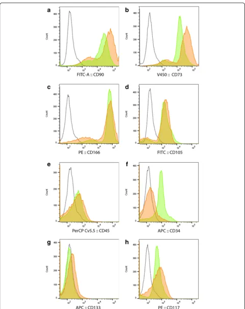
Freshly cultured (day 0) hDPSCs showed high expression of CD90, CD73, CD166 and CD105 (Fig. 3a, b, c, d). These cells also express CD34, some CD117 and CD45 but are negative for CD133. After 21 days in culture the hDPSCs still maintained high expression of CD90, CD73, CD166 and CD105 but were no longer positive for CD34 and had lower expression of CD45, while the expression levels of CD133, CD117 remained similar to day 0 (Fig. 3). No variability was evidenced between the study groups.
Immunofluorescence analysis
Immunofluorescence staining showed a relatively homo-geneous pattern of protein labeling in different cells of the same hDPSC population. Labeling for DMP1 and ALP revealed a fibrillary intracellular pattern relatively homogeneous throughout the whole cytoplasm (Fig. 4a and c), while assuming a more granular appearance for
Table 1Primer sequences and thermocycling conditions used for target cDNA
Primer Length (bp) Sequence 5′-3′ Annealing conditions for PCR
GAPDH 238 Forward: GAG TCA ACG GAT TTG GTC GT 60 °C 30 sec; × 45cy
Reverse: TTG ATT TTG GAG GGA TCT CG
ALP 196 Forward: CCA CGT CTT CAC ATT TGG TG 65 °C 30 sec; × 40cy
Reverse: AGA CTG CGC CTG GTA GTT GT
DMP1 235 Forward: ACA GCA GCT CAG CAG AGA GT 60 °C 30 sec; × 40cy
Reverse: TAA TAG CCG TCT TGG CAG TC
DSPP 237 Forward: GTC GCT GTT GTC CAA GAA GA 60 °C 30 sec; × 40cy
DSPP (Fig. 4b). Positive reactions to all antibodies tested were observed irrespective of the group analyzed.
Metabolic activity
The WST-1 assays (Fig. 5b) showed the conversion of tetrazolium salts in formazan products to be lower in the presence of lower quantities of amelogenin 10 and 100 ng/mL, compared to the control. This constant trend during the first 14 days swaps at day 21. The low viability of the cells stimulated with 10 ng/mL amelogenin is asso-ciated with a lower metabolism of tetrazolium.
Alizarin red staining
During 21 days, the cells formed a tightly packed mono-layer culture but without deposition of mineral aggre-gates, as seen in the control and in the amelogenin groups, regardless of the concentration of supplement used. There was no addition of any osteogenic media, and amelogenin alone does not seem to be sufficient to elicit mineral nodule formation (Fig. 6).
Effect of rhAmelogenin on odontogenic gene expression
At PCR analyses, hDPSCs in the control group show, after 21 day in culture, a significant upregulation of all tested differentiation marker genes, i.e.,ALP(mature osteoblast/ odontoblast marker), DMP-1 and DSPP (odontoblast markers) (Fig. 7).
In the 3-week-prolonged culture conditions there was a tendency for confluent growth of the hDPSCs, with a presumable increase of the cell-cell adhesion forces, thus
explaining the observed response in DNA expression (Figs. 7 and 8, day 0 versus control day 21).
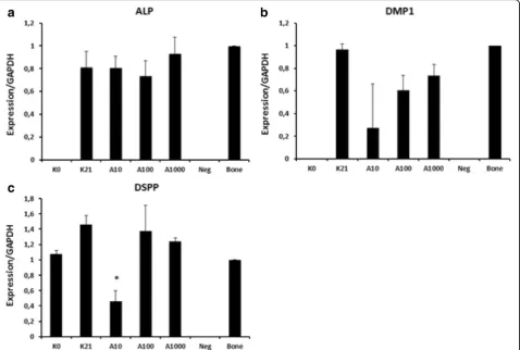
The expression ofALPat day 21 was relatively similar for all four groups. The full-length amelogenin had little influence on the gene expression, even at the highest concentration tested (1000 ng/mL), (p= 0.938) (Fig. 7, lanes 2–4).
Both, the expressions of DMP1 and DSPP were re-duced in the presence of the lowest concentration of amelogenin (10 ng/mL) compared to the unstimulated control group. However, this difference was significant for DSPP(p= 0.011) but not forDMP1 (p= 0.395). The quantification of the results shows a downregulation that is inversely related to the concentration of amelogenin (Fig. 8).
Discussion
different functions [20–22], so we should not refer to the role of amelogenin in general, but to the role of each spe-cific isoform. Moreover, different concentrations are re-ported to be used ranging from 10 ng/mL to 100μg/mL. EMD components have been isolated from the tooth germs in their secretory phase, and used at a concentra-tion of 1–100μg/mL [23]. In any case, being composed of a mixture of several enamel proteins and amelogenin iso-types, the composition is likely to vary qualitatively as well as quantitatively. In vitro studies like those of Huang [24], Izumikawa [25], and Ye [21], which have focused on single isotypes, have actually seen a greater effect on MSC and
DPC at an optimal concentration of 100 ng/mL. This is also why we chose the tested concentrations, and this also permits comparability of the results with these studies. Our results on the influence of full-length amelogenin on cell morphology and proliferation of hDPSCs (Figs. 1 and 2) are in line with the findings of Izumikawa [25] who tested the full-length amelogenin on rat mesenchymal stem cells (MSC), showing similitude of this isotype activity in both species.
In our study, human amelogenin did not induce the for-mation of mineralized nodules in hDPSCs (Fig. 6). Our results support the findings of Ye [21] and Tanimoto [26] Fig. 2Growth curve (a) and cumulative population doubling levels (b) of human dental pulp stem cells supplemented with different
Fig. 4Immunofluorescence assay for dentin matrix protein-1 (a), dentin sialophosphoprotein (b) and alkaline phosphatase (c). Representative fluorescence microscopy photographs of human dental pulp stem cells stained with respective primary antibodies after 21 days in culture. Nuclei were stained with 4’,6-diamidino-2-phenylindole (blue).Scale bars20μm
who reported no mineralizing effect of the full-length amelogenin on human dental papilla, dental pulp cells or PDL cells. It should be mentioned that in both stud-ies, it was not used an osteogenic supplement. This in-dicates that amelogenin cannot act as an inducer of mineralization. On the other hand, studies performed using amelogenin as a supplement of osteogenic medium, show its capacity to enhance the osteogenic differentiation of BM-MSC [27], PDL cells or cementoblasts [26].
Even though amelogenin does not seem to be an in-ducer, the investigation of its role as an enhancer of the osteogenic differentiation and mineralization process might be of interest. Moreover, the role of low weight amelogenins, which are other important components of EMD, in mineralization has widely been confirmed [28], and they might be responsible for the clinical ef-fect of EMD.
Regarding the ‘self-differentiation’of this stem cell line (Figs. 7 and 8, day 0 vs. day 21), it might be due to the pro-moting role of fetal bovine serum supplement as sug-gested by Lindroos in 2009 [29]. In fact, STRO-1+ hDPSCs have been shown to consist of several interrelated subpopulations which can spontaneously differentiate into
odontoblasts, osteoblasts, and chondrocytes [30]. From our FACS results, this corresponds to a selection of a sub-population of hDPSC CD34- or to a loss of expression of CD34. As reviewed by Sidney et al. [31], there is increas-ing evidence that this is a marker not specifically linked to hematopoietic origin; it is also present on progenitor cells of mesenchymal cell lines and its loss suggests lineage commitment. The expression of different CD marker phe-notypes correspond to a different protein setting. In con-cordance with our results, Yu et al. observed increased gene expression ofALP, DSPPand other osteoblast differ-entiation markers, concluding that we do not have yet an optimal medium to avoid spontaneous cell differentiation, while permitting their amplification [32]. Another possible theory, which does not exclude the first, involves mechan-ical reasons: the cytoskeletal tension related to cell adhe-sion and extracellular forces have been shown to influence stem cell (SC) commitment [33, 34]. A remarkable finding is the expression of odontoblast-related genes,DMP1and DSPPin bone, which we used afterwards as a positive con-trol. Until nowDMP1andDSPPhave been considered as odontoblasts markers.DMP1has already been detected in osteocytes [35]. We confirm these findings forDMP1, and Fig. 6Alizarin Red staining. Human dental pulp stem cells seeded in 12-well plates at a concentration of 104cells/well and cultured for 21 days
without amelogenin supplement (a, control) and with 10 (b) or 100 ng/mL (c) full-length amelogenin
Fig. 7Expressions of alkaline phosphatase (ALP), dentin matrix protein-1 (DMP1), dentin sialophosphoprotein (DSPP) in human dental pulp stem cells (hDPSCs). Total RNAs were extracted and reversely transcribed. The cDNAs were amplified by PCR using the primers shown in Table 1. The PCR products were run on 1.5 % agarose gel and stained with ethidium bromide.Lane 1hDPSC expression at the initial state;lane 2after 21-day cultivation in medium without amelogenin supplements;lane 3after 21 days with 10 ng/mL amelogenin supplement in the medium;lane 4after 21 days with 100 ng/mL amelogenin supplement in the medium;lane 5after 21 days with 1000 ng/mL amelogenin supplement in the medium;
for the first time proved that DSPP is also expressed in bone. This means that we in fact, do not have a specific marker for odontoblasts. There is a need for further inves-tigations into the diversification between osteoblasts and odontoblasts and how this impacts pulp regeneration therapies.
From our results, low concentrations of amelogenin determine a lower expression of odontoblast differenti-ation genes (Figs. 7 and 8). This is suggestive of main-tenance of an undifferentiated status of the cells, similar to the starting cells at day 0, even preventing spontan-eous differentiation visible in the control cultures. The higher effectiveness of lower concentrations of the pro-tein can be explained by its three-dimensional structure and its capacity to assembly into nanospheres as de-scribed by Fang in 2011 [36]. In particular, the full-length protein has an internal structure constituted of dodecamers, not present in other isotypes. Furthermore, it is widely known that self-assembly is a common prop-erty of many extracellular organic matrix macromole-cules [37], facilitated by intermolecular interactions and
specifically assembly of amelogenins into nanosphere in-creases at increasing concentrations.
We explained the results with the presence of the pro-tein in a monomeric state at lower concentrations, having thus a higher impact on cell signaling. On the contrary an increasing assembly into nanospheres at higher concentra-tions leads to its structural role. Our results support the previously reported role of amelogenin as a signaling molecule, adding the hDPSCs as another target of this protein.
and splicing products of amelogenin, with the use of different animal or human sources of this protein and dif-ferent cell types and also source of stem cells (embryonic [28], apical papilla, periodontal ligament). A comprehen-sive review focusing on this topic, specifically limited to in vitro studies, has been published by Grandin in 2012 and Bosshardt in 2008 [14, 40]. Recently a systematic review has summarized and compared the results of EMD cap-ping in animal and in human studies [41], evidencing con-troversial results.
Conclusions
While the effects of amelogenin have been largely studied with bone mesenchymal stem cells (BMSCs) and PDL cells [42], to our knowledge, this is the first in vitro study that analyzes the effect of full-length amelogenin on hDPSCs. The results obtained suggest that amelogenin af-fects hDPSCs differently than it does in PDL cells and other cell lines. We show that the full-length amelogenin seems to have a negative mitogenic impact on hDPSCs at higher concentrations but shows a tendency for positive mitogenic effects at lower concentrations.
This study permits a further understanding of the sig-naling role of the full-length amelogenin. Considering the present result on the inhibition ofDMP1and DSPP, it seems plausible that amelogenin impairs the clinical results of pulp capping; hence previous positive clinical observations might have been random effects.
Further studies should focus on the molecular mecha-nisms behind the effects of amelogenin, taking into con-sideration that different isoforms have different roles, and considering also the cell type used as a target of the protein. For a possible application of amelogenin in pulp regeneration, odontoblasts and other cell types present in the pulp should also be analyzed.
Abbreviations
ALP:alkaline phosphatase; BM-MSC: bone marrow-mesenchymal stem cells; bp: base pairs; DAPI: 4’,6-diamidino-2-phenylindole;DMP1: dentin matrix protein-1;DSPP: dentin sialophosphoprotein; DT: doubling time; EGF: epidermal growth factor; EMD: enamel matrix derivatives; FACS: fluorescence-activated cell sorting;GAPDH: glyceraldehyde-6-phosphate dehydrogenase; hDPSCs: human dental pulp stem cells; kDa: kiloDalton; PBS: phosphate-buffered saline; PD: population doubling; PDL: periodontal ligament; TGF: transforming growth factor; WST-1: water-soluble tetrazolium salt 1;α-MEM: minimal essential medium,α-modification.
Competing interests
The authors declare that they have no competing interests.
Authors’contributions
IF contributed to conception and design, to acquisition, analysis and interpretation of the data and drafted the manuscript. CE contributed to the study design, acquisition and interpretation of the data. CD contributed to the conception of the study, analysis and interpretation of the data, RH contributed to conception of the study and interpretation of the data. MH contributed to acquisition and interpretation of the data. MF conceived and designed the study, performed the statistical analyses and drafted the manuscript. All authors critically revised the manuscript and
approved its final version and agree to be accountable for all aspects of the work.
Acknowledgements
We are grateful to B. Hackl for her continuous technical support. We gratefully acknowledge the following funding sources: Forschungsgemeinschaft Dental e.V grant: 08/2012; Friedrich-Baur Foundation: 76/13. The funders had no role in study design, data collection and analysis, decision to publish, or preparation of the manuscript.
Author details
1
Department of Conservative Dentistry and Periodontology,
Ludwig-Maximilian-University, Goethestraße, 70, D-80336 Munich, Germany. 2
Institute for Cardiovascular Prevention, Ludwig-Maximilian-University, Munich, Germany.
Received: 26 May 2015 Revised: 30 November 2015 Accepted: 21 December 2015
References
1. Eastoe JE. Enamel protein chemistry–past, present and future. J Dent Res. 1979;58(Spec Issue B):753–64.
2. Fincham AG, Moradian-Oldak J. Recent advances in amelogenin biochemistry. Connect Tissue Res. 1995;32(1-4):119–24.
3. Tanimoto K, Huang YC, Tanne K. Proliferation of Bone Marow-Derived Human Mesenchymal Stem Cells: Role of Enamel Matrix Proteins. In: Stem Cells and Cancer Stem Cells, Volume 4: Therapeutic Applications in Disease and Injury. Netherlands: Springer; 2012.
4. Hatakeyama J, Sreenath T, Hatakeyama Y, Thyagarajan T, Shum L, Gibson CW, et al. The receptor activator of nuclear factor-kappa B ligand-mediated osteoclastogenic pathway is elevated in amelogenin-null mice. J Biol Chem. 2003;278(37):35743–8. doi:10.1074/jbc.M306284200.
5. Deutsch D, Haze-Filderman A, Blumenfeld A, Dafni L, Leiser Y, Shay B, et al. Amelogenin, a major structural protein in mineralizing enamel, is also expressed in soft tissues: brain and cells of the hematopoietic system. Eur J Oral Sci. 2006;114 Suppl 1:183–9. doi:10.1111/j.1600-0722.2006.00301.x. discussion 201-2, 381.
6. Inai T, Kukita T, Ohsaki Y, Nagata K, Kukita A, Kurisu K. Immunohistochemical demonstration of amelogenin penetration toward the dental pulp in the early stages of ameloblast development in rat molar tooth germs. Anat Rec. 1991;229(2):259–70. doi:10.1002/ar.1092290213.
7. Romanelli M, Dini V, Vowden P, Agren MS. Amelogenin, an extracellular matrix protein, in the treatment of venous leg ulcers and other hard-to-heal wounds: experimental and clinical evidence. Clin Interv Aging. 2008;3(2):263–72.
8. Garrocho-Rangel A, Flores H, Silva-Herzog D, Hernandez-Sierra F, Mandeville P, Pozos-Guillen AJ. Efficacy of EMD versus calcium hydroxide in direct pulp capping of primary molars: a randomized controlled clinical trial. Oral Surg Oral Med Oral Pathol Oral Radiol Endod. 2009;107(5):733–8. doi:10.1016/j. tripleo.2008.12.017.
9. Kiatwateeratana T, Kintarak S, Piwat S, Chankanka O, Kamaolmatyakul S, Thearmontree A. Partial pulpotomy on caries-free teeth using enamel matrix derivative or calcium hydroxide: a randomized controlled trial. Int Endod J. 2009;42(7):584–92. doi:10.1111/j.1365-2591.2009.01552.x.
10. Goldberg M, Six N, Decup F, Buch D, Soheili Majd E, Lasfargues JJ, et al. Application of bioactive molecules in pulp-capping situations. Adv Dent Res. 2001;15:91–5.
11. Fitzgerald M, Chiego Jr DJ, Heys DR. Autoradiographic analysis of odontoblast replacement following pulp exposure in primate teeth. Arch Oral Biol. 1990; 35(9):707–15.
12. Min JH, Ko SY, Cho YB, Ryu CJ, Jang YJ. Dentinogenic potential of human adult dental pulp cells during the extended primary culture. Hum Cell. 2011; 24(1):43–50. doi:10.1007/s13577-011-0010-7.
13. Warmflash A, Sorre B, Etoc F, Siggia ED, Brivanlou AH. A method to recapitulate early embryonic spatial patterning in human embryonic stem cells. Nat Methods. 2014;11(8):847–54. doi:10.1038/nmeth.3016. 14. Grandin HM, Gemperli AC, Dard M. Enamel matrix derivative: a review
15. Lyngstadaas SP, Risnes S, Nordbo H, Flones AG. Amelogenin gene similarity in vertebrates: DNA sequences encoding amelogenin seem to be conserved during evolution. J Comp Physiol B Biochem Syst Environ Physiol. 1990;160(5):469–72.
16. Sasaki S, Shimokawa H. The amelogenin gene. Int J Dev Biol. 1995;39(1):127–33. 17. Toyosawa S, O’HUigin C, Figueroa F, Tichy H, Klein J. Identification and
characterization of amelogenin genes in monotremes, reptiles, and amphibians. Proc Natl Acad Sci U S A. 1998;95(22):13056–61. 18. Assaraf-Weill N, Gasse B, Al-Hashimi N, Delgado S, Sire JY, Davit-Beal T.
Conservation of amelogenin gene expression during tetrapod evolution. J Exp Zool B Mol Dev Evol. 2013;320(4):200–9. doi:10.1002/jez.b.22494. 19. Fincham AG, Belcourt AB, Termine JD, Butler WT, Cothran WC. Amelogenins.
Sequence homologies in enamel-matrix proteins from three mammalian species. Biochem J. 1983;211(1):149–54.
20. Tompkins K, Alvares K, George A, Veis A. Two related low molecular mass polypeptide isoforms of amelogenin have distinct activities in mouse tooth germ differentiation in vitro. J Bone Miner Res. 2005;20(2):341–9. doi:10.1359/JBMR.041107.
21. Ye L, Le TQ, Zhu L, Butcher K, Schneider RA, Li W, et al. Amelogenins in human developing and mature dental pulp. J Dent Res. 2006;85(9):814–8. 22. Lacerda-Pinheiro S, Septier D, Tompkins K, Veis A, Goldberg M, Chardin H. Amelogenin gene splice products A + 4 and A-4 implanted in soft tissue determine the reorientation of CD45-positive cells to an osteo-chondrogenic lineage. J Biomed Mater Res A. 2006;79(4):1015–22. doi:10.1002/jbm.a.30912. 23. Amin HD, Olsen I, Knowles J, Dard M, Donos N. A tyrosine-rich amelogenin
peptide promotes neovasculogenesis in vitro and ex vivo. Acta Biomater. 2014;10(5):1930–9. doi:10.1016/j.actbio.2013.11.027.
24. Huang YC, Tanimoto K, Tanne Y, Kamiya T, Kunimatsu R, Michida M, et al. Effects of human full-length amelogenin on the proliferation of human mesenchymal stem cells derived from bone marrow. Cell Tissue Res. 2010; 342(2):205–12. doi:10.1007/s00441-010-1064-7.
25. Izumikawa M, Hayashi K, Polan MA, Tang J, Saito T. Effects of amelogenin on proliferation, differentiation, and mineralization of rat bone marrow mesenchymal stem cells in vitro. ScientificWorldJournal. 2012;2012:879731. doi:10.1100/2012/879731.
26. Tanimoto K, Kunimatsu R, Tanne Y, Huang YC, Michida M, Yoshimi Y, et al. Differential effects of amelogenin on mineralization of cementoblasts and periodontal ligament cells. J Periodontol. 2012;83(5):672–9. doi:10.1902/jop. 2011.110408.
27. Tanimoto K, Huang YC, Tanne Y, Kunimatsu R, Michida M, Yoshioka M, et al. Amelogenin enhances the osteogenic differentiation of mesenchymal stem cells derived from bone marrow. Cells Tissues Organs. 2012;196(5):411–9. doi:10.1159/000335912.
28. Warotayanont R, Frenkel B, Snead ML, Zhou Y. Leucine-rich amelogenin peptide induces osteogenesis by activation of the Wnt pathway. Biochem Biophys Res Commun. 2009;387(3):558–63. doi:10.1016/j.bbrc.2009.07.058. 29. Lindroos B, Boucher S, Chase L, Kuokkanen H, Huhtala H, Haataja R, et al.
Serum-free, xeno-free culture media maintain the proliferation rate and multipotentiality of adipose stem cells in vitro. Cytotherapy.
2009;11(7):958–72. doi:10.3109/14653240903233081.
30. Yu J, He H, Tang C, Zhang G, Li Y, Wang R, et al. Differentiation potential of STRO-1+ dental pulp stem cells changes during cell passaging. BMC Cell Biol. 2010;11:32. doi:10.1186/1471-2121-11-32.
31. Sidney LE, Branch MJ, Dunphy SE, Dua HS, Hopkinson A. Concise review: evidence for CD34 as a common marker for diverse progenitors. Stem Cells. 2014;32(6):1380–9. doi:10.1002/stem.1661.
32. d’Aquino R, Graziano A, Sampaolesi M, Laino G, Pirozzi G, De Rosa A, et al. Human postnatal dental pulp cells co-differentiate into osteoblasts and endotheliocytes: a pivotal synergy leading to adult bone tissue formation. Cell Death Differ. 2007;14(6):1162–71. doi:10.1038/sj.cdd.4402121. 33. Nava MM, Raimondi MT, Pietrabissa R. Controlling self-renewal and
differentiation of stem cells via mechanical cues. J Biomed Biotechnol. 2012; 2012:797410. doi:10.1155/2012/797410.
34. Miyashita S, Ahmed NE, Murakami M, Iohara K, Yamamoto T, Horibe H, et al. Mechanical forces induce odontoblastic differentiation of mesenchymal stem cells on three-dimensional biomimetic scaffolds. J Tissue Eng Regen Med. 2014. doi:10.1002/term.1928.
35. Rios HF, Ye L, Dusevich V, Eick D, Bonewald LF, Feng JQ. DMP1 is essential for osteocyte formation and function. J Musculoskelet Neuronal Interact. 2005;5(4):325–7.
36. Fang PA, Conway JF, Margolis HC, Simmer JP, Beniash E. Hierarchical self-assembly of amelogenin and the regulation of biomineralization at the nanoscale. Proc Natl Acad Sci U S A. 2011;108(34):14097–102. doi:10.1073/pnas.1106228108.
37. Paine ML, Krebsbach PH, Chen LS, Paine CT, Yamada Y, Deutsch D, et al. Protein-to-protein interactions: criteria defining the assembly of the enamel organic matrix. J Dent Res. 1998;77(3):496–502.
38. Matsuda N, Horikawa M, Watanabe M, Kitagawa S, Kudo Y, Takata T. Possible involvement of extracellular signal-regulated kinases 1/2 in mitogenic response of periodontal ligament cells to enamel matrix derivative. Eur J Oral Sci. 2002;110(6):439–44.
39. Cattaneo V, Rota C, Silvestri M, Piacentini C, Forlino A, Gallanti A, et al. Effect of enamel matrix derivative on human periodontal fibroblasts: proliferation, morphology and root surface colonization. An in vitro study. J Periodontal Res. 2003;38(6):568–74.
40. Bosshardt DD. Biological mediators and periodontal regeneration: a review of enamel matrix proteins at the cellular and molecular levels. J Clin Periodontol. 2008;35(8 Suppl):87–105. doi:10.1111/j.1600-051X.2008.01264.x. 41. Al-Hezaimi K, Javed F, Al-Fouzan K, Tay F. Efficacy of the enamel matrix
derivative in direct pulp capping procedures: a systematic review. Aust Endod J. 2013;39(3):171–5. doi:10.1111/j.1747-4477.2012.00357.x. 42. Matsuzawa M, Sheu TJ, Lee YJ, Chen M, Li TF, Huang CT, et al. Putative
signaling action of amelogenin utilizes the Wnt/beta-catenin pathway. J Periodontal Res. 2009;44(3):289–96.
• We accept pre-submission inquiries
• Our selector tool helps you to find the most relevant journal • We provide round the clock customer support
• Convenient online submission • Thorough peer review
• Inclusion in PubMed and all major indexing services • Maximum visibility for your research
Submit your manuscript at www.biomedcentral.com/submit

