Diabetic Nephropathy
BARRY B. KIRSCHBAUM, M.D.
Associate Professor of Medicine, Division of N ephrology, Medical College of Virginia, Health Sciences Division of Virginia Commonwealth University, Richmond, Virginia
Any discussion of morbid events in diabetes is going to emphasize cardiovascular and peripheral vascular problems. Renal disease accounts for a rela-tively small percentage of the mortality in diabetes; yet the overall incidence of diabetes mellitus in the population so greatly exceeds that of the various types of glomerulonephritis that it has become one of the most common causes of end-stage renal failure in this country. It is difficult to assign exact numbers because the figures in the medical literature vary con-siderably; however, it is estimated that in the United States today there are some 10 million people who are known to be diabetic, who would be found to be diabetic if tested, or who during the course of their lives will develop overt diabetes. In autopsy series of diabetics the prevalence of glomerulosclerosis has varied from 15% to 82%, with a mean of approxi-mately 30%. This figure will vary depending on how carefully the kidneys are studied, particularly with respect to the use of special stains for identifying glomerulosclerosis; the incidence will also increase with the proportion ofjuvenile to adult onset diabet-ics included in the sample. Renal failure is listed as the cause of death of 6% to 12% of diabetics, and its incidence is increased seventeenfold in the diabetic population as compared to the nondiabetic. These figures are probably applicable to those diabetics who have many complications and who require referral to major medical centers for treatment. In terms of the general diabetic population renal failure is probably in the range of 1 % to 2%. This means that the preva-lence of glomerulosclerosis is severalfold higher than
Correspondence and reprint requests to Dr. Barry B. Kirsch-baum, Division of Nephrology, Box 197, Medical College of Vir-ginia, Richmond, VA 23298.
148
the prevalence of renal failure, and that simply find-ing the lesion on biopsy does not indicate that kidney function itself has been impaired or that there will be any abnormalities present on urinalysis. As a cause of end-stage renal failure, diabetes now accounts for some 15% to 25% of all new cases.
Five years' survival after onset of proteinuria in diabetics compared to age-matched controls is 65% opposed to 73% for diabetics who do not develop proteinuria and 83% for non-diabetics. This does not mean that these people are dying of renal disease but that proteinuria heralds increased mortality from all causes. For juvenile diabetics it is extremely unusual for proteinuria to develop before 10 years' duration of the disease or to appear after a protein-free inter-val greater than 30 years. This means that there are essentially two populations of juvenile diabetics with regard to the kidney; those who will have their dis-ease for an unlimited amount of time and never de-velop renal complications, and those who during the period between IO and 30 years will note the onset of renal problems. Finally, in juvenile diabetes mellitus, azotemia defined as BUN in excess of 30 mg percent implies end-stage renal failure within three years. An important point to note is the rarity of a decline in the glomerular filtration rate before the onset of protein-uria when the cause is diabetic nephropathy. Once renal failure does ensue it has been calculated that the glomerular filtration rate falls at an average rate of 1 ml/min each month. With other forms of renal dis-ease, hypertension greatly accelerates the rate of de-cline in the glomerular filtration rate, an important point in treatment.
Diabetic nephropathy and microangiopathy are the main topics under discussion here, but there are
. several other factors which contribute in varying
KIRSCHBAUM: DIABETIC NEPHROPATHY
gree to the progression of renal failure in diabetics. As there is no therapy for diabetic nephropathy itself, these other factors assume an importance in line with their response to treatment. Infection, in particular pyelonephritis and papillary necrosis, becomes an ex-tremely important factor in the management of the diabetic. This is true because the autonomic neuropa-thy that may complicate diabetes favors development of a neurogenic bladder with high residual urine which may lead to reflux up the ureter, if the urine is infected, this reflux will result in secondary pyelone-phritis and subsequent papillary necrosis with rapid loss of renal function. In patients who already have some type of renal failure, infection, even without involvement of the renal parenchyma, can result in a further decline in renal function. One of the theories that has been advocated to explain this is that certain types of bacteria can attach themselves to the cells of the ureter and thereby compromise the peristaltic action of the ureter which is important in propelling urine from the renal pelvis into the bladder. This results in a type of functional obstruction which acts in the same way as a true obstruction. Treatment of these patients with antibiotics to eradicate the bacte-riuria improves ureteral peristalsis and renal function as well.
Diabetes is also frequently complicated by hy-perlipidemia and hyperuricemia. Uric acid crystals are a cause of interstitial nephritis, and high uric acid levels which lead to increased uric acid excretion can set the stage for uric acid calculi, obstruction, and subsequent infection. In the evaluation of a diabetic patient, the potential adverse effect of uric acid should be considered as this cause of renal failure is subject to therapy either by alkalinization of the urine or the use of allopurinol.
A number of cardiovascular problems will also contribute to the declining glomerular filtration rate. Hypertension has already been mentioned; this can accelerate the vascular degenerative changes in the kidney, leading to nephrosclerosis which in turn can accelerate the hypertension, creating a cycle which may be interrupted if the hypertension is successfully controlled through the careful use of antihypertensive medication. Another complication of hypertension is its deleterious effect on the myocardium, leading to hypertensive cardiomyopathy and congestive heart failure. This will have an adverse effect on renal func-tion, for as the kidney is damaged secondary to dia-betes or other processes and as the vascular disease in the kidney progresses, autoregulation of blood flow
149
in the face of a reduced cardiac output is com-promised; thw; low-output congestive heart failure will result in prerenal azotemia superimposed on the preexisting ren.il failure. In general, this situation is handled by the combination of digitalization and di-uretics; however, it is worth pointing out the prob-lems associated with excessive use of diuretics in these patients. They may get too dehydrated, a factor that will superimpose a prerenal type of azotemia upon their already preexisting level of renal failure. A com-promise has to be worked out in terms of controlling the symptoms of congestive heart failure without, at the same time, contributing further to the impairment of renal function. Diabetics are prone to develop atherosclerosis, and particularly those patients who have extremely severe atheromatous disease of the aorta are subject to embolization to the renal vessels. This can present as episodic elevations of blood pres-sure associated with fluctuations in the level of renal function reflected by BUN and creatinine and accom-panied by microscopic or even gross hematuria, de-pending on how much of the kidney is damaged or destroyed by infarction.
TABLE
Percent Aggregation Four Minutes After the Addition of Each of the Aggregating Agents
AGGREGATION(%)
AGGREGATING DIABETIC
AGENT CONTROLS SUBJECTS P VALUE*
ADP:
lµM 22 ± 7t 80 ± 4 (9):j: <0.001
2µM 69 ± 4 88 ± 4(9) <0.005
5µM 76 ± 4
Epinephrine:
86 ± 3 (9) NS§
lµM 39 ± 8 82 ± 5 (IO) <0.001 2µM 60 ± 6 85 ± 3(9) <0.01 5µM 66 ± 6 84 ± 2(10) <0.005 Collagen:
I µg/ml 53 ± 10 80 ± 3 (8) <0.05
2µg/ml 69 ± 8 84 ± 4 (8) NS
IOµg/ml 77 ± 8 86±3(8) <0.05
* Difference between % aggregation seen in the platelet-rich
plasma obtained from control and that in diabetic subjects. t Mean± SEM.
:j: Figures in parentheses denote no. of subjects § Not significant.
resulting in glomerular ischemia and glomeruloscle-rosis. There is some further evidence that involves platelets: thromboglobulin, a protein which is derived from the platelet, has been found to be increased in
the circulation of those diabetics who have
micro-angiopathy. There are also tests for the presence of
circulating platelet aggregates, and those diabetics
who have the most severe small vessel disease are also the ones who show the highest levels of these circulat-ing platelet aggregates. A recent study2 reported on
the efficacy of sulfinpyrazone in the prevention of morbid events in patients who have severe coronary artery disease. There are active studies in progress to evaluate antiplatelet drugs in a variety of diseases that affect blood vessels, particularly coronary artery disease, atherosclerosis, and different types of renal disease.
Amyloidosis is always listed as associated with
diabetes mellitus. Certainly the onset of nephrotic
syndrome or heavy proteinuria in a patient with long-standing diabetes does raise the possibility that
sec-ondary amyloidosis may be present. With respect to immunologic factors which crop up in consideration of almost every type of renal disease,
immuno-globulins and complements are found within the
glomerulus of the diabetic kidney. Their presence in a
rather nonspecific pattern together with several other
serum proteins has been interpreted not to represent
a specific immunologic event such as immune com-plex disease or anti-glomerular basement membrane
(anti-GBM) disease. Still, there are some lines of evidence which do not exclude completely the idea
that immunologic problems are present in the
dia-betic and may be contributing to the renal disease. It was recently reported that juvenile _diabetes mellitus can be viewed as a genetic disease transmitted as an
autosomal recessive gene.3 More interesting, perhaps,
is the close association of the diabetic gene with the HL-A locus. This area of the chromosome is
con-cerned with a variety of immunologic responses, and
many other diseases which have a close association
with the HL-A locus have been more firmly
estab-lished as being the result of an immunologic
abnor-mality. Based on this there is still speculation that the diabetic may have altered immunologic responses
and that this in turn may lead to primary or
second-ary renal damage.
Drug-induced disease of the kidney, while not con-fined to the diabetic, may assume increased signficance
for these patients. There have been several reports of
acute renal failure following the use of iodine
con-trast agents; for the most part, this can be prevented
in the nondiabetic population by avoiding
dehy-40
..,,
I-w..J
w
I-<
30
..J
~
0 0 0
0
0 in
'
~
20
a:: ~
e
'
C)• Diabetic c
w
l?
10
• Control
~
0.5 1
.
0
2.0
3.0
4.0
MINUTES AFTER ADDITION OF ARACHIDONIC ACID
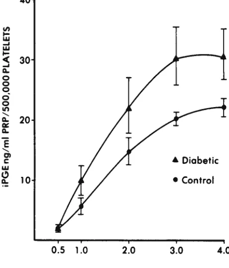
Figure-Time course for the production of iPG E in platelet-rich plasma (PRP) from exogenous arachidonic acid (0.5mM). The
KIRSCHBAUM: DIABETIC NEPHROPATHY
dration which is part of the preparation of patients for radiologic examinations. Diabetics, however, tend to develop acute renal failure with a greater frequency than nondiabetics; moreover, avoiding de-hydration does not seem to be protective. This sug-gests that something about the diabetic kidney pre-disposes it to damage in the face of a quantity of hyperosomotic material. The question arises whether the diabetic kidney is more susceptible to other neph-rotoxic agents such as the aminoglycoside antibiotics and a host of other medications which have potential nephrotoxicity. The lack of a definite answer suggests that in the management of diabetics both diagnostic and therapeutic agents should be selected for their reduced incidence of nephrotoxicity.
Several studies have attempted to identify fac-tors of importance in determining the development and progression of diabetic nephropathy. The data to be presented were derived from a small number of patients with biopsy-proven glomerulosclerosis who were followed for several years at which time a repeat biopsy was performed.' As far as the type of diabetes is concerned, of the 11 adult onset diabetics, 9 showed no progression of their glomerulosclerosis,
and 2 became worse. In contrast, of the 6 juvenile diabetics, l showed no change and 5 showed progres-sion of the disease. With regard to blood glucose control, 6 of the patients maintained good sugar con-trol for the whole length of the study and none of the 6 showed progression of their glomerulosclerosis. By contrast, 13 patients were rated to be poorly con-trolled and IO of them showed a progression of their renal lesions. In respect to the age of onset of these diabetics, patients in the group that showed no change were considerably older at the time of the onset of their diabetes than were those in the group that became worse. Obviously these three variables are not independent since, by and large, in older patients the adult onset type of diabetes can easily be managed by diet and weight reduction. But the re-sults do suggest that blood glucose control may be important in terms of slowing down or averting the development of glomerulosclerosis.
There appear then to be at least three phases in the renal disease that accompanies diabetes. The first is a pre-proteinuria phase. Phase 2 is marked by the onset of proteinuria, and phase 3 would be develop-ment of azotemia. Several groups have tried to study the question, Is there anything abnormal about the kidney in the pre-proteinuric phase?, that is, before proteinuria becomes manifest and at a time when the
151
other renal function tests are also normal.6 If
glo-merulosclerosis progresses slowly with time, there might be a gradual increase in the excretion of albu-min in the urine as the function of the duration of the diabetic process until such time as the amount of albumin in the urine definitely reaches an abnormal quantity. In a study of 97 young male diabetics who were followed from 1-19 or more years after the diagnosis of diabetes, there was no increase in albu-min excretion throughout this period of time. Those who develop proteinuria are clearly distinguished from those who do not. Because strenuous exercise increases albumin excretion, a group of 13 juvenile diabetics were exercised at a rate which had not af-fected urinary albumin levels in 9 normal controls. The diabetics showed a statistically significant in-crease in protein excretion that returned to baseline after stopping the exercise. Another element that has been examined is the excretion of low molecular weight proteins by diabetics; these represent proteins that are considerably smaller than albumin and are freely filtered at the glomerulus. Almost none, how-ever, appear in the urine because most of these fil-tered proteins are reabsorbed by the cells of the prox-imal tubule. In the pre-proteinuric phase, diabetics show an increase in excretion of low molecular weight proteins. These results, together with addi-tional information on albumin excretion, suggest that the metabolic state of diabetes can adversely affect renal cell function so that when the diabetic is poorly controlled, the cells are less able to reabsorb filtered proteins, including albumin.
improve-ment in the appearance of the diabetic kidney. This indicates that the changes seen in the glomerulus are
at least initially reversible and that metabolic control of blood glucose, or at least the presence of insulin in appropriate quantities to keep the blood glucose nor-mal, is a contributing factor to the development of diabetic changes.
The outline for therapy of diabetic kidney dis-ease calls for the best blood glucose control circum-stances will permit. Antihypertensive therapy has to be emphasized together with optimal management of congestive cardiac failure in order to maintain a high cardiac output and avoid prerenal azotemia. The pa-tient should be evaluated for obstruction and for urinary tract infection. Nephrotoxic agents should be avoided as much as possible. As for vascular compli-cations, anticoagulation offers little value as, by and large, patients who have renal disease also have reti-nal disease which is aggravated by these drugs. The
antiplatelet drugs, based on available evidence, are quite promising, although unproven at the present time. As far as the treatment of end-stage renal dis-ease in the diabetic is concerned, the same four mo-dalities are open to these patients as anyone else:
living related transplants, cadaver transplants, hemo-dialysis and peritoneal dialysis. Because of technical
advances, greater experience in dealing with diabet-ics, and earlier acceptance of diabetics into end-stage renal failure programs, the prognosis has improved considerably during the past couple of years.6 Trans-plantation from a living related donor seems to be as good in diabetics as in the general population, so that this would seem to be the treatment of choice for a diabetic with end-stage renal disease. The results with cadaver kidney transplantation tend to be poorer than those of the general population and until we have some better methods of managing cadaver kid-ney transplants in general, this is probably the least
promising mode of therapy for the diabetic. Hemo-dialysis is the treatment that will be applied to the majority of these people. The problems are in estab-lishing a suitable blood access site because of severe
vascular disease and the requirement for
anti-coagulation during dialysis which frequently leads to a deterioration of vision. Many nephrologists now feel that the treatment of choice if a living related donor is not available is peritoneal dialysis; the major problem here is peritonitis as well as the necessity for using high-glucose containing solutions in the perito-neal cavity which can lead to severe degrees of hyper-glycemia, but the complications associated with bleeding in the eye are avoided and vascular access is not necessary.
The table and the figure are reproduced with permission from
the New England Journal of Medicine (297:1306-1310, 1977).
REFERENCES
I. HALUSHKA PV, LURIE D, COLWELL JA: Platelet prostaglandin
synthesis in diabetes mellitus. N Engl J Med 297:1306-13!0,
1977.
2. Sulfinpyrazone in the prevention of cardiac death after
myo-cardial infarction. Anturane Reinfarction Trial Research
Group. N Eng J Med 298:289-294, 1978.
3. RUBINSTEIN P, SUCIU-FOCA N, NICHOLSON JF: Close genetic
linkage of HCA and juvenile diabetes mellitus. N Engl J Med
297: 1036-1040, 1977.
4. TAKAZAKURA E, NAKAMOTO Y, HAYAKAWA H, ET AL: Onset
and progression of diabetic glomerulosclerosis. Diabetes 24:
1-9, 1975.
5. MOGENSEN CE: Renal Function changes in diabetes. Diabetes
25 (suppl 2):872-879, 1976.