(arteriogenesis), the second is based on the sprout-ing of capillaries (angiogenesis) and the third is the formation of blood vessels by de novo differenti-ation and proliferdifferenti-ation of endothelial cells from stem cells (vasculogenesis) [2, 3].
Arteriogenesis is the formation of mature blood vessels from pre-existing capillaries via en-larging of the lumen and thickening of the walls. Collateral vessels, developed during arteriogenesis, are large epicardial arteries that allow for efficient blood flow. Formation of these vessels is strictly In patients with heart failure, regardless of its
cause, cardiac hypertrophy and, usually dispro-portionate, neovascularization are observed. Cor-onary microvascular dysfunction with subsequent ischemia intensify the process of myocardial dam-age, the inhibition of which potentially depends on the development of the “vascular tree” and its abil-ity to protect tissues from ischemia [1].
There are three basic processes of new blood vessel development (neovascularization). The first one involves the formation of arterial collaterals
Małgorzata Kobusiak-Prokopowicz
A–D, Beata Jołda-Mydłowska
A–C,
Tomasz Grzebieniak
A, B, Karol Początek
A, B, Andrzej Mysiak
E, FExpression of Proinflammatory Factors,
Proangiogenic Factors and Endostatin
in Patients with Heart Failure and Different Grades
of Collateral Circulation Development*
Departmentand Clinic of Cardiology, Wroclaw Medical University, Poland
A – research concept and design; B – collection and/or assembly of data; C – data analysis and interpretation;
D – writing the article; E – critical revision of the article; F – final approval of article
Abstract
Background. The process of collateral vessel maturation is stimulated by numerous factors affecting the endothe-lium and smooth muscle cells building the vessel wall. Looking for arteriogenesis stimulating factors means look-ing for a potential innovative heart failure treatment method in the patients unresponsive to traditional therapies.
Objectives. The purpose of this study was to assess the changes in serum concentrations of pro-inflammatory factor IL-6, growth factors FGF (FGFa, FGFb, FGFbH), HGF, VEGF and endostatin in heart failure patients in relation to the coronary collaterals development stage.
Material and Methods. This study included 22 patients with chronic heart failure NYHA II or III (mean age 62.5 ± 11.6 years) and 8 control patients (mean age 58.4 ± 10.7 years). Coronary angiography was performed and the presence and grade of collateral circulation was assessed by a four-level scale proposed by Rentrop and Cohen. The level of the studied factors was determined in the blood samples collected during the angiographic procedure.
Results. The concentration of IL-6 was significantly higher in the heart failure patients than in the control group (p < 0.001) and in NYHA III vs. NYHA II patients (p < 0.02). Patients with heart failure and collaterals grade 1 or 2 exhibited higher serum concentrations of FGFbH (from p < 0.03 to p < 0.01). The serum VEGF level in NYHA III patients was significantly higher than in NYHA II individuals (from p < 0.03 to p < 0.01).
Conclusions. Higher levels of IL-6 and FGFbH were observed in patients with heart failure. Collaterals formation seems to be associated with the activation of pro-inflammatory factors, growth factors and endostatin (Adv Clin Exp Med 2015, 24, 6, 987–994).
Key words: arteriogenesis, proinflammatory factors, proangiogenic factors, endostatin, innovative treatment.
ORIGINAL PAPERS
Adv Clin Exp Med 2015, 24, 6, 987–994
DOI: 10.17219/acem/33811 © Copyright by Wroclaw Medical University ISSN 1899–5276
limited to the area surrounding the narrowed or blocked epicardial artery. This is confirmed by an-giographic images of collateral bypasses in patients with advanced occlusion of the main arteries. The role of arteriogenesis in collateral circulation de-velopment is important in ischemic and hypoxic organs affected by atherosclerosis. When an occlu-sion occurs in an existing vessel, blood is direct-ed into other vessels, filling all the patent, but not fully developed vessels. The process of their matu-ration is stimulated by a number of factors affect-ing the endothelium and smooth muscle cells of the vessel wall. The best conditions for collater-als formation involve increased fluid shear stress and stimulation of bone marrow-derived cells [4]. In the course of arteriogenesis, perivascular mac-rophages produce monocyte chemotactic protein MCP-1 and basic fibroblast growth factors, bFGF and VEGF, which are important mediators of arte-riogenesis, but probably to a lesser degree than an-giogenesis [5, 6].
Angiogenesis results in the formation of small, intramyocardially spreading vessels with thin walls. Collaterals formed in the process of arterio-genesis exhibit low resistance, while angioarterio-genesis results in the formation of a high-resistance vas-cular bed. Angiogenesis is dependent on the in-teraction of endothelial cells, angiogenic media-tors, cytokines, growth factors and cell adhesion molecules [7]. Many studies have shown that in-flammation may also contribute to the formation of new blood vessels. The influx of inflammatory cells such as macrophages, monocytes and plate-lets, leads to the release of numerous cytokines that stimulate the release of proangiogenic factors [8].
Chung et al. showed that the levels of angio-genic factors and thus the development of collater-al circulation were greater in patients with chronic ischemic heart disease [9]. In patients with isch-emic heart disease who developed collateral circu-lation, the level of anti-angiogenic factors was 40% lower than in patients lacking the collaterals [10].
With this group of patients in mind, many re-search centers are striving to develop alternative methods of restoring coronary vasculature. Col-lateral circulation plays an immensely important role in limiting the ischemic zone and myocardial infarction size. Charney and Cohen observed pa-tients undergoing coronary angioplasty and found that transient myocardial ischemia was better tol-erated in patients with collateral flow than in those without. The collateral circulation also reduced the risk of perioperative infarct rate and death dur-ing coronary artery bypass graftdur-ing [11]. Look-ing for neovascularization stimulatLook-ing factors is, in fact, looking for a potential innovative treatment method, especially for those patients in whom
traditional invasive surgical and pharmacological therapy is ineffective [12].
Aim of the Study
The purpose of this study was to assess chang-es in serum concentrations of pro-inflammato-ry factor IL-6, growth factors FGF (FGFa, FGFb, FGFbH), HGF and VEGF and endostatin in pa-tients with heart failure in relation to the coronary collaterals development stage.
Material and Methods
This study included 22 patients admitted to the Department of Cardiology, Wroclaw Medical Uni-versity, with a diagnosis of chronic heart failure NYHA stage II or III (mean age 62.5 ± 11.6 years), including 8 women aged 49–81 years (mean age 69.26 ± 9.5 years) and 14 men aged 41–86 years (mean age 60.0 ± 11.3 years). Twelve patients were diagnosed with heart failure due to ischemic heart disease and 10 patients suffered from idiopathic dilated cardiomyopathy. In the group of patients with heart failure, the mean left ventricular ejec-tion fracejec-tion (LVEF) was 33.5% ± 7.4%. The con-trol group consisted of 8 age-matched participants, 2 women and 6 men (mean age: 58.4 ± 10.7 years), without heart failure, diagnosed for ischemic heart disease. The mean LVEF in this group was 57.5% ± 2.7%.
The study excluded individuals with acute re-nal failure and stages 4 and 5 of chronic kidney dis-ease, thyroid disdis-ease, neoplastic disdis-ease, acute in-flammatory disease, treatment history of steroids and immunosuppressive drugs and extremely high blood glucose levels exceeding 400 mg/dL.
The study protocol was granted the consent of the local Ethics Committee and research was conducted in accordance with the Declaration of Helsinki.
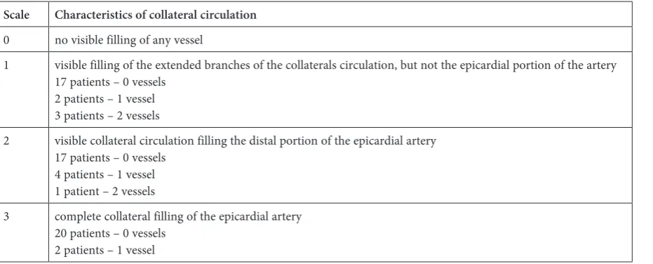
The presence and grade of collateral circula-tion were assessed by the four-level scale proposed by Rentrop and Cohen [13, 14]. Angiograms were evaluated by two doctors without access to clinical data (Table 1).
To determine the level of the studied factors, blood samples were collected from each patient during the angiographic procedure. Blood sam-ples were taken from the femoral artery (sam-ple 1), the left main coronary artery or the initial segment of the LAD (sample 2) and a peripheral vein (sample 3). After collecting blood into SST tubes, the samples were left for 30 min for clotting. The samples for IL-6, VEGF, FGFa, FGFb, FGFbH and endostatin were then centrifuged for 15 min at 1000 × g and the serum was stored at ≤ –20o C. HGF samples were centrifuged for 10 min at 1000 × g and the serum was stored at ≤ –70o C.
The concentration of IL-6 was determined using ELISA assay (R&D Systems, Inc., Cata-log Number DHG00); inter-assay precision was 1.6–4.2% and intra-assay precision was 3.3–6.4%. The concentration of VEGF was determined using ELISA assay (R&D Systems, Inc., Catalog Num-ber DVE00); inter-assay precision was 4.5–6.7% and intra-assay precision was 6.2–8.8%. The con-centration of endostatin was determined using ELISA assay (R&D Systems, Inc., Catalog Number DNST0); inter-assay precision was 3.6–6.9% and intra-assay precision was 5.7–7.9%. The concen-tration of FGF basic high sensitivity (HFGFb) was determined using ELISA assay (R&D Systems, Inc., Catalog Number HSFB00D); inter-assay pre-cision was 3.5–7.7% and intra-assay prepre-cision was 4.7–8.2%. The concentration of FGF basic (FGFb) was determined using ELISA assay (R&D Sys-tems, Inc., Catalog Number DFB50); inter-assay precision was 3.0–9.7% and intra-assay precision was 7.4–9.1%. The concentration of FGF acidic
(FGFa) was determined using ELISA assay (R&D Systems, Inc., Catalog Number DFA00B); intra-assay precision was 2.3–7.2% and inter-intra-assay pre-cision was 8.4–8.6%. The concentration of HGF was determined using ELISA assay (R&D Sys-tems, Inc., Catalog Number DHG00); inter-assay precision was 4.1–7.0% and intra-assay precision was 5.4–8.4%.
Within 48 h after admission, trans-thoracic echocardiography was performed in all of the pa-tients using a Vingmed System 5, General Elec-tric device equipped with a 2.5 MHz probe. The EF was determined using the standard formula: EF = (LVEDV - LVESV)/ LVEDV × 100%, where LVEDV is left ventricular end-diastolic volume and LVESV is left ventricular end-systolic volume. LVEDV and LVESV were calculated using the bi-plane Simpson method, in which a computer de-termined the left ventricular volume using ap-proximately perpendicular cross-section areas of the left ventricle in apical two- and four-chamber views, determined by an investigator.
Statistical Analysis
Calculations were made using the statisti-cal software, STATISTICA v. 5.0. Measurable an-alyzed parameters were characterized using the arithmetic mean and standard deviation. For pa-rameters whose normal distribution was con-firmed by the Shapiro-Wilk test, the differences between the two groups were assessed using Stu-dent’s t-test for independent variables, after testing equality of variances with the Fisher-Snedecor test. When normal distribution was not confirmed, the nonparametric Mann-Whitney U test or ANOVA Kruskal-Wallis rank test was used.
The assumed level of statistical significance was p < 0.05.
Table 1. Rentrop scale and number of vessels with collaterals Scale Characteristics of collateral circulation
0 no visible filling of any vessel
1 visible filling of the extended branches of the collaterals circulation, but not the epicardial portion of the artery 17 patients – 0 vessels
2 patients – 1 vessel 3 patients – 2 vessels
2 visible collateral circulation filling the distal portion of the epicardial artery 17 patients – 0 vessels
4 patients – 1 vessel 1 patient – 2 vessels
3 complete collateral filling of the epicardial artery 20 patients – 0 vessels
Results
The results are shown in Tables 2–4.
The concentration of IL-6 in the peripher-al blood (sample 1 and sample 3) was significant-ly higher in the heart failure patients than in the
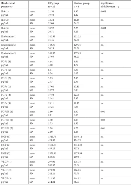
Table 2. Biochemical parameters in patients with heart failure (HF) and in the control group Biochemical
parameter HF groupn = 22 Control groupn = 8 Significance of differences – p IL6 (1)
pg/mL meanSD 11.5419.78 1.951.42 0.001
IL6 (2)
pg/mL meanSD 12.3222.06 15.1934.61 ns.
IL6 (3)
pg/mL meanSD 10.9220.71 3.255.23 0.001
Endostatin (1)
ng/mL meanSD 140.5335.46 132.3232.80 ns.
Endostatin (2)
ng/mL meanSD 143.3950.25 129.3631.30 ns.
Endostatin (3)
ng/mL meanSD 141.9537.66 137.6338.23 ns.
FGFb (1)
pg/mL meanSD 4.644.80 4.664.77 ns.
FGFb (2)
pg/mL meanSD 8.919.24 5.356.02 ns.
FGFb (3)
pg/mL meanSD 3.252.47 2.832.34 ns.
FGFa (1)
pg/mL meanSD 17.0213.72 17.856.08 ns.
FGFa (2)
pg/mL meanSD 17.7012.41 22.007.87 ns.
FGFa (3)
pg/mL meanSD 19.1115.21 19.179.04 ns.
FGFbH (1)
pg/mL meanSD 3.802.11 1.600.56 0.01
FGFbH (2)
pg/mL meanSD 3.401.73 2.081.52 0.03
FGFbH (3)
pg/mL meanSD 3.282.10 1.731.48 0.01
HGF (1)
pg/mL meanSD 1323.79429.32 1180.12474.39 ns.
HGF (2)
pg/mL meanSD 1561.83489.25 2434.39307.91 ns.
HGF (3)
pg/mL meanSD 1571.90628.89 1175.99259.81 ns.
VEGF (1)
pg/mL meanSD 297.04286.35 178.7861.06 ns.
VEGF (2)
pg/mL meanSD 270.94242.24 166.0570.70 ns.
VEGF (3)
control group (p < 0.001). In addition, IL-6 serum levels in all 3 studied blood samples were signif-icantly higher in patients with NYHA III HF as compared to the NYHA II group (p < 0.02). The serum IL-6 concentration was, however, signifi-cantly lower in patients with collaterals grade 1 or 2, as assessed by the Rentrop scale (p < 0.03 and p < 0.04).
Patients with heart failure exhibited higher se-rum concentrations of FGFbH (from p < 0.03 to p < 0.01). The serum concentration of FGFbH was also significantly higher in patients with collater-als grade 1 or 2, as assessed by the Rentrop scale (p < 0.02).
The serum levels of endostatin, FGFb, FGFa, VEGF and HGF did not differ significantly in any
of the samples for the patients with heart failure and the control group. However, the serum VEGF level in NYHA III patients was significantly high-er than in NYHA II individuals (from p < 0.03 to p < 0.01).
The ANOVA Kruskal-Wallis rank test re-vealed significant correlations between the levels of IL-6, endostatin, FGFbH, HGF and VEGF and the presence of collateral vessels.
Discussion
The main arteriogenesis-inducing factor is in-creased shear stress in vessels and the result is the formation of collateral arteries far from the isch-emic area [15].The acute phase of the systemic re-sponse is mediated by proinflammatory cytokines, particularly IL-1 and IL-6, acting in a complex manner [16]. Proinflammatory cytokines, pro-duced within inflammation areas [17, 18], are po-tent autocrine and paracrine agents and may affect vascular function [19, 20].
IL-6 was suspected of exhibiting angiogenic ac-tivity, as high levels of this interleukin were deter-mined within the areas of the endothelium where enhanced angiogenesis was observed. Such pro-cesses take place, for example, during wound heal-ing or in carcinogenesis (Kaposi’s sarcoma) [21]. Aoki et al. published the results of an experiment in which they inoculated athymic mice with cells transformed to secrete IL-6 and found that they stimulated the growth of more vascularized tu-mor-like lesions [21].
The mechanisms by which IL-6 stimulates an-giogenesis are complex, but a number of growth factors with known angiogenic properties, such as HGF, EGF and VEGF, stimulate the activation of Table 3. Biochemical parameters in patients with NYHA II and III
Biochemical
parameter HF NYHA IIn = 13 HF NYHA IIIn = 9 Significance of differences – p IL6 (1)
pg/mL meanSD 4.233.24 23.4228.56 0.02
IL6 (2)
pg/mL meanSD 7.8910.70 19.5828.98 0.02
IL6 (3)
pg/mL meanSD 7.1710.98 16.3526.20 0.02
VEGF (1)
pg/mL meanSD 192.43147.34 467.02379.01 0.02
VEGF (2)
pg/mL meanSD 171.60138.29 432.36294.56 0.01
VEGF (3)
pg/mL meanSD 218.14172.94 445.91301.80 0.03
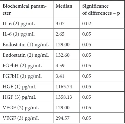
Table 4. Biochemical parameters and the presence of vas-cular collaterals in HF patients (ANOVA Kruskal-Wallis rank test)
Biochemical
param-eter Median Significance of differences – p
IL-6 (2) pg/mL 3.07 0.02
IL-6 (3) pg/mL 2.65 0.05
Endostatin (1) ng/mL 129.00 0.05 Endostatin (2) ng/mL 132.60 0.05
FGFbH (2) pg/mL 4.59 0.05
FGFbH (3) pg/mL 3.41 0.05
STAT3 factor (signal transducer and transcription 3 factor) [22, 23].
The ability of IL-6 to induce full angiogenic ac-tivity in ex vivo and in vivo models was also test-ed [17]. It was hypothesized that IL-6 may directly affect individual components of the vascular wall, thus contributing to the prevention of ischemic episodes. Our study compared the release of IL-6 and collaterals formation during the arteriogenesis process. In patients with heart failure, IL-6 serum concentrations in peripheral arteries and veins were significantly higher than in healthy subjects. Levels of IL-6 were also significantly greater in all vascular territories in patients with HF and higher NYHA classes than in those with developed collat-eral circulation. Our observations revealed that the role of IL-6 in the pathophysiology of heart fail-ure was not associated with arteriogenesis prog-ress, despite its expected effect on the vascular wall. VEGF is a potent mobilizer of stem cells and progenitor cells. In the study by El-Melegy et al., significantly higher VEGF levels were found in children with congenital heart disease and devel-oped collaterals [24]. Patel et al. reported that se-rum VEGF levels did not correlate with objective markers of left ventricular function, but were sig-nificantly lower in patients with ischemic heart disease accompanied by heart failure [25]. Furlani et al. [26] described an experimental canine mod-el of myocardial infarction in which an induction of therapeutic angiogenesis by intramuscular ad-ministration of VEGF resulted in the maintenance of left ventricular function. An increased number of capillary vessels within the myocardium was ob-served, especially in the transition region between the infarcted and normal myocardium, while the number of arterioles was significantly lower than in the control group [26].
Our study showed significantly higher levels of serum VEGF in patients with more advanced stag-es of heart failure. One of the latstag-est studistag-es eval-uated the efficacy and clinical significance of ad-ministration of the VEGF encoding gene [27]. The authors reported a significant improvement in myocardial perfusion in patients who received VEGF in combination with adenoviral vector af-ter 6 months. Enhanced VEGF concentration in patients from higher NYHA classes can be associ-ated with its repair properties, and the absence of elevated concentration in patients with collaterals seems to be associated with greater capillary circu-lation area, rather than the formation of arterioles.
One of the most important functions of FGF is the stimulation of endothelial cell proliferation and organization of the formed cells into tubu-lar structures to induce angiogenesis. FGF1 and FGF2 are more potent stimulators of angiogenesis than VEGF and PDGF. The efficacy and safety of FGF1 was evaluated in 40 patients with three-ves-sel disease undergoing CABG. Twenty patients in this group received an FGF1 injection near the left descending coronary artery anastomosis. Pecher et al. concluded that after three years, the network of blood vessels resulting from the application of FGF1 was clearly visible and the followed patients presented better exercise tolerance and increased left ventricular ejection fraction [28]. In our ob-servations, FGFbH serum levels in all three blood samples were significantly higher than in the con-trol group. Moreover, in patients with developed collaterals and heart failure, FGFbH concentration in the coronary arteries was higher, which may in-dicate not only a stimulating role of FGFbH in an-giogenesis, but also in arteriogenesis.
A meta-analysis by Meier et al. showed that the presence of collaterals in patients with isch-emic heart disease reduced mortality [29]. Accord-ing to W. Schaper, who commented on the above meta-analysis, if the prognosis in patients with col-laterals is better than with CABG, and maybe even stenting, then there is a chance for improved prog-nosis for the patients in whom heart failure pro-gresses when other treatment options have been exhausted [30].
Limitations
The main limitation is the small size of our group of patients. Additionally, we cannot exclude the effects of drug therapy on our results. Our findings help to advance knowledge on this topic and are of potential practical value.
The authors concluded that higher levels of pro-inflammatory factor IL-6 and growth factor FGFbH were observed in patients with heart fail-ure, especially in the more advanced stages of HF. The concentrations of pro-inflammatory factors, growth factors and endostatin were significantly associated with the presence of coronary collater-als; however, a direct relationship was demonstrat-ed only for higher levels of FGFbH and collaterals formation. Collaterals formation is associated with the activation of pro-inflammatory factors, growth factors and endostatin.
References
[1] Al Haj Zen A, Madeddu P: Notch signalling in ischemia-induced angiogenesis. Biochem Soc Trans 2009, 37, 1221–1229.
[2] Heilmann C, Beyersdorf F, Lutter G: Collateral growth: cells arrive at the construction site. Cardiovasc Surg 2002, 10, 570–578.
[3] Emanueli C, Madeddu P: Therapeutic angiogenesis: translating experimental concepts to medically relevant goals. Vasc Pharmacol 2006, 45, 334–339.
[4] Pipp F, Boehm S, Cai WJ, Adili F, Ziegler B, Karanovic G, Ritter R, Balzer J, Scheler C, Schaper W, Schmitz- -Rixen T: Elevated fluid shear stress enhances postocclusive collateral artery growth and gene expression in the pig hind limb. Arterioscler Thromb Vasc Biol 2004, 24, 1664–1668.
[5] Schierling W, Troidl K, Troidl C, Schmitz-Rixen T, Schaper W, Eitenmüller IK: The role of angiogenic growth factors in arteriogenesis. J Vasc Res 2009, 46, 365–374.
[6] Buschmann I, Heil M, Jost M, Schaper W: Influence of inflammatory cytokines on arteriogenesis. Microcirculation 2003, 10, 371–379.
[7] Griffioen AW, Molema G: Angiogenesis: potentials for pharmacologic intervention in the treatment of cancer, cardiovascular diseases, and chronic inflammation. Pharmacol Rev 2000, 52, 237–268.
[8] Tabibiazar R, Rockson SG: Angiogenesis and the ischaemic heart. Eur Heart J 2001, 22, 903–918.
[9] Chung NA, Lydakis C, Belgore F: Angiogenesis in myocardial infarction. An acute or chronic process? Eur Heart J 2002, 23, 1604–1608.
[10] Panchal VR, Rehman J, Nguyen AT, Brown JW, Turrentine MW, Mahomed Y, March KL: Reduced pericardial levels of endostatin correlate with collateral development in patients with ischemic heart disease. J Am Coll Cardiol 2004, 43, 1383–1387.
[11] Charney R, Cohen M: The role of the coronary collateral circulation in limiting myocardial ischemia and infarct size. Am Heart J 1993, 126, 937–945.
[12] Lahteenvuo JE, Lahteenvuo MT, Kivela A, Rosenlew C, Falkevall A, Klar J, Heikura T, Rissanen TT, Vaha-kangas E, Korpisalo P, Enholm B, Carmeliet P, Alitalo K, El-Melegy NT, Mohamed NA: Angiogenic biomarkers in children with congenital heart disease: possible implications. Ital J Pediatr 2010, 36, 32.
[13] Cohen M, Rentrop KP: Limitation of myocardial ischaemia by collateral circulation during sudden controlled coronary artery occlusion in human subjects. A prospective study. Circulation 1974, 469, 1986–1992.
[14] Rentrop KP, Feit F, Sherman W, Thornton JC: Serial angiographic assessment of coronary artery obstruction and collateral flow in acute myocardial infarction. Report from the second Mount Sinai-New York University Reperfusion Trial. Circulation 1989, 80, 1166–1175.
[15] Pipp F, Boehm S, Cai WJ, Adili F, Ziegler B, Karanovic G, Ritter R, Balzer J, Scheler C, Schaper W, Schmitz- -Rixen T: Elevated fluid shear stress enhances postocclusive collateral artery growth and gene expression in the pig hind limb. Arterioscler Thromb Vasc Biol 2004, 24, 1664–1668.
[16] Baumann H, Gauldie J: The acute phase response. Immunol Today 1994, 15, 74–80.
[17] Weyand CM, Hicok KC, Hunder GG, Goronzy JJ: Tissue cytokine patterns in patients with polymyalgia rheu-matica and giant-cell arteritis. Ann Intern Med 1994, 121, 484–491.
[18] Hernández-Rodríguez J, Segarra M, Vilardell C, Sánchez M, García-Martínez A, Esteban MJ, Queralt C, Grau JM, Urbano-Márquez A, Palacín A, Colomer D, Cid MC: Tissue production of proinflammatory cytokines (IL-1ß, TNF-{alpha}, and IL-6) correlates with the intensity of the systemic inflammatory response and with cor-ticosteroid requirements in giant-cell arteritis. Rheumatology 2004, 43, 294–301.
[19] Mantovani A, Garlanda C, Introna M, Vecchi A: Regulation of endothelial cell function by pro- and anti-inflam-matory cytokines. Transplant Proc 1998, 30, 4239–4243.
[20] Rossi V, Breviario F, Ghezzi P, Dejana E, Mantovani A: Prostacyclin synthesis induced by in vascular cells by interleukin-1. Science 1985, 225, 174–176.
[21] Aoki Y, Jaffe ES, Chang Y, Jones K, Teruya-Feldstein J, Moore PS, Tosato G: Angiogenesis and hematopoiesis induced by Kaposi-sarcoma-associated herpes-virus encoded interleukin-6. Blood 1999, 93, 4034–4043.
[22] Zhong Z, Wen Z, Darnell JE Jr: Stat3: a STAT family member activated by tyrosine phosphorylation in response to epidermal growth factor and interleukin-6. Science 1994, 264, 95–98.
[23] Valdembri D, Serini G, Vacca A, Ribatti D, Bussolino F:In vivo activation of JAK2/STAT-3 pathway during angiogenesis induced by GM-CSF. FASEB J 2002, 16, 225–227.
[24] El-Melegy NT, Mohamed NA: Angiogenic biomarkers in children with congenital heart disease: possible implica-tions. Ital J Pediatr 2010, 36, 32.
[25] Patel JV, Abraheem A, Chackathayil J, Gunning M, Creamer J, Hughes EA, Lip GY: Circulating biomarkers of angiogenesis as indicators of left ventricular systolic dysfunction amongst patients with coronary artery disease. J Intern Med 2009, 265, 562–567.
[26] Furlani AP, Kalil RA, Castro I, Canedo-Delgado A, Barra M, Prates PR, Sant`Anna RT, Nesralla IA: Effects of therapeutic angiogenesis with plasmid VEGF165 on ventricular function in a canine model of chronic myocardial infarction. Rev Bras Cir Cardiovasc 2009, 24, 143–149.
[28] Pecher P, Schumacher BA: Angiogenesis in ischemic human myocardium: clinical results after 3 years. Ann Thorac Surg 2000, 69, 1414–1419.
[29] Meier P, Hemingway H, Lansky AJ, Knapp G, Pitt B, Seiler Ch: The impact of the coronary collateral circulation on mortality: a meta-analysis. Eur Heart J 2012, 33, 614–621.
[30] Schaper W: Collateral vessels reduce mortality. Eur Heart J 2012, 33, 564–566.
Address for correspondence:
Małgorzata Kobusiak-Prokopowicz Department and Clinic of Cardiology Wroclaw Medical University Borowska 213
50-514 Wrocław Poland
Tel.: +48 71 376 42 00 E-mail: [email protected] Conflict of interest: None declared Received: 12.11.2014