O R I G I N A L R E S E A R C H A R T I C L E
Open Access
Metabolomic profile of amniotic fluid to evaluate
lung maturity: the diaphragmatic hernia lamb
model
Gloria Pelizzo
1*, Maurizio Ballico
2, Maria Chiara Mimmi
2, José Louis Peirò
3, Mario Marotta
4, Costanzo Federico
1,
Erika Andreatta
1, Ghassan Nakib
1, Maurilio Sampaolesi
5, Elisa Zambaiti
1and Valeria Calcaterra
1,6Abstract
Background:Tracheal occlusion (TO) stimulates lung growth in fetuses affected with congenital diaphragmatic hernia (CDH) although the processes involved in lung maturation still remain unknown. The objective of this study was to evaluate the metabolomic profile of amniotic fluid (AF) following TO in fetal lamb model in order to obtain an indirect view of mechanisms involved in pulmonary reversal hypoplasia and biochemical maturity in response to fetal TO.
Methods:Liquid Chromatography Mass Spectrometry was performed on lamb AF samples at: age I (70 days’ gestation); age II (102 days’gestation); age III (136 days’ gestation). CDH was induced at age I and TO at age II. Results:Betaine, choline, creatinine were found significantly increased during gestation in the control group. The CDH group showed choline (p =0.007) and creatinine (p =0.004) decreases during pregnancy. In the TO group choline and creatinine profiles were restored.
Conclusions:Alveolar tissue and fetal global growth ameliorated after TO. Metabolomics provided useful
information on biochemical details during lung maturation. Metabolomic profiling would help to identify the best time to perform TO, in order to increase survival of CDH affected patients.
Keywords:Amniotic fluid, Animal model, Congenital diaphragmatic hernia, Metabolomic, Tracheal occlusion
Background
Congenital diaphragmatic hernia (CDH) is a congenital birth defect that occurs in 1/2000 to 1/3000 newborns with a survival rate of 42-68%. Abnormal development of the pulmonary parenchyma and vasculature lead to variable degrees of respiratory insufficiency and pulmon-ary hypertension in early neonatal life [1-4].
While modern neonatal intensive care has improved the prognosis for surviving cases, comorbidities affect al-most half of live-born infants and the mortality rate re-mains high in patients with poor prenatal prognosis. Experimental studies could potentially ameliorate out-comes in pulmonary hypoplasia [5,6]. Fetal endoscopic
tracheal occlusion (FETO) may still be a possible thera-peutic procedure in case of severe CDH, even if the mortality rate is reported to be nearly 40% [7-9].
Amniotic fluid (AF) composition reflects the physio-logical status during fetal development and it may be used to detect potential pathological conditions. AF con-tains large amounts of proteins and metabolites pro-duced by the amnion epithelial cells, fetal tissues, fetal excretions and placental tissues.
Metabolomics is commonly used to measure the entire metabolomic profile in biological fluids, cells, tissues or organs and to elucidate the association between meta-bolic pathways and perturbations that arise as a result of disease or organ malformation [10-13].
The objective of this study was to determine amniotic fluid metabolic profiling changes in fetal lambs who underwent surgically induced CDH and subsequent tra-cheal occlusion (TO) which provides an excellent vision
* Correspondence:[email protected]
1Department of the Mother and Child Health, Pediatric Surgery Unit,
Fondazione IRCCS Policlinico San Matteo, Pavia and University of Pavia, Piazzale Golgi 2, 27100 Pavia, Italy
Full list of author information is available at the end of the article
© 2014 Pelizzo et al.; licensee BioMed Central Ltd. This is an Open Access article distributed under the terms of the Creative Commons Attribution License (http://creativecommons.org/licenses/by/4.0), which permits unrestricted use, distribution, and reproduction in any medium, provided the original work is properly credited. The Creative Commons Public Domain Dedication waiver (http://creativecommons.org/publicdomain/zero/1.0/) applies to the data made available in this article, unless otherwise stated.
of lung biochemical maturity and is an excellent model to determine the real benefits and best timing for TO.
Methods
The experimental protocol was approved by the National Animal Care and Ethics Committee and was conducted in accordance with Italian and European legislation (D.lgs. 116/92, European Directives 86/609/EE for the protection of animals used in scientific and experimental studies and 2010-63UE).
A preliminary study with Liquid Chromatography Mass Spectrometry (LC-MS) [14,15] was performed on 20 lamb AF samples, taken at three gestational ages: Age I (70 days’of gestation); Age II (102 days’gestation); Age III (136 days’gestation).
CDH induction was performed at Age I and TO was carried out during the 2ndtrimester at Age II.
Based on this information, AF samples were divided into:
– –CONTROL group, healthy control pregnancies: Age I, number of samples =5; Age II, number of samples =3; Age III, number of samples =3;
– –CDH group, fetuses with induced CDH: Age II, number of samples =3 and at Age III, number of samples =3;
– –CDH + TO group, fetuses with induced CDH and TO treatment: Age III, number of samples =3
Healthy control AF samples were collected to study the normal metabolomic evolution.
We report on the effect of induced CDH and TO treatment for selected metabolites at stage Age III.
Anesthesia protocol
All surgical procedures were performed under general anesthesia. After premedication with intramuscular (IM) injection of 5 mg/Kg ketamine and 0.2 mg/Kg midazo-lam (Dormicum®, Roche), intravenous access (IV) was established at the external jugular vein.
The pregnant ewes were pre-oxygenated using a face-mask and induced with 5 mg/Kg IV propophol (Propo-fol®-Lipuro 1%, B. Braun Melsungen AG). Endotracheal intubation was performed, and general anesthesia was maintained with isoflurane 2% (Isoflo, Abbott laboratories Ltd) in 100% oxygen (1.5 L/minute).
Esophageal intubation was performed to prevent ruminal bloat. A continuous infusion of Ringer lactate (B. Braun Melsungen AG) was administered at 10 ml/Kg/h during surgery. Intra-operative monitoring consisted of electrocar-diography, pulse oximetry, non invasive blood pressure and capnography. All animals received a 75μg transdermal fentanyl patch (Durogesic®, Janssen laboratories) for post-operative pain relief. For peri-operative infection
prophylaxis, the animals received a single dose of 22 mg/Kg IV cephazolin (Kurgan®, Normon Laboratories) at the time of induction and 15 mg/Kg IM amoxicillin (Duphamox L. A., Fort Dodge) every 48 h, for eight days. Fetuses were anesthetized through the placenta and additional anesthesia with fentanil (10μg/kg IM) and the muscle relaxant pan-curonium (0.3 mg/Kg IM) were administered. Meloxicam 0.5 mg/kg IM /24 h was administered as post-operative an-algesia for seven days. Maternal body temperature was monitored by using a digital probe and maintained at 36-37°C with a warming bed. Standard ECG electrodes were used for monitoring.
Surgical technique
As a first step, malformation was surgically induced (day 70 of gestation). A midline laparotomy exposed the gravid uterus. The fetus was partially extruded from the uterus with a short hysterectomy (4 cm). A left fetal thoracotomy was achieved and a diaphragmatic hernia was created. The fetal stomach was raised into the thorax, which was closed into one layer. The fetus was returned to the uterus (2 gr of amoxicillin were added to the amniotic fluid). The uterus wall and maternal lapar-otomy were closed.
The next step included the tracheal occlusion (day 102 of gestation). The uterus was externalized after a midline laparotomy. The fetal lamb’s mouth was located and also partially externalized by a short hysterectomy (2 cm). A latex detachable balloon was placed into the trachea and inflated. The fetus was returned to the uterus and 2 gr of amoxicillin were added to the amniotic fluid. Closure of the uterus and the abdomen walls was performed.
At the end of the pregnancy (day 136 of gestation), lambs were delivered via terminal caesarean section, ap-proximately 10 days before term to prevent natural par-turition. Lambs were euthanized with a bolus of Pentobarbital 200 mg/Kg IV.
Metabolomic analysis Sample preparation
Amniotic fluid samples were centrifuged after collection in order to remove cells and cellular debris. The supernatants were immediately frozen and stored at -70°C/-80°C until metabolite extraction and analysis were performed. Super-natant samples were thawed at room temperature and methanol extraction was accomplished with the protocol reported by Graca et al. [16]. An internal standard (2’ -deox-yadenosine) was added to the extracted dry aliquots which were reconstituted in ultrapure H2O (Milli-Q H2O).
HPLC-TOF-MS analysis
electrospray ionization source; acquired data were ana-lyzed using AB-SCIEX Analyst™ QS software (version 1.1). The HPLC system includes an Agilent 1100 series micro LC pump. The liquid chromatographic separation was performed on a Jupiter® 5μm 300 Å (150 × 0.5 mm) Reverse PhaseC18 column (PhenomenexInc, Torrance, CA, USA) at a flow of 10μL/min using a gradient from 0% to 90% of solvent Bover 35 min. Solvents A and Bconsisted of water and acetonitrile, respectively; both contained 0.1% (v/v) formic acid. All samples were ana-lyzed in triplicate.
Metabolites were quantified using LC-MS-TOF stand-ard runs in positive-ion mode, while LC-MS/MS runs were used for molecule identification. The assignment of the metabolites was based on analytical standards ana-lyzed under the same chromatographic conditions.
Data processing and analysis
The metabolite peaks from raw HPLC-MS chromato-grams were integrated with the software Analyst QS 1.1 (Applied Biosystems-SCIEX) by evaluating the extracted ion chromatogram (XIC) counts. All data sets were nor-malized to account for variable sample dilution and ex-perimental influences using the peak area of the internal standard (2’-deoxyadenosine).
The HPLC-MS data shown in Tables 1, 2 and 3 represent the metabolite XIC areas, which do not account for con-centrations, but can be used to evaluate the relative level of each metabolite among the three sample groups (Control Age I/II/III, CDH Age II/III and CDH + TO Age III).
A comparison of Age II groups (Control II and CDH II) and among Age III groups (Control III, CDH III and CDH + TO III) was performed and the One-Way Ana-lysis of Variance (ANOVA) was applied to determine
whether the means of groups significantly differed. We performed Welch’s ANOVA, which does not assume constant variance across all groups, with an alpha level set at 0.05. In the case of Age III, sample differences be-tween all pairs of groups were compared according to the Games-Howell method, which does not assume constant variance while controlling the multiple com-parisons error rate.
Results
The AF analysis in control samples (gestational Ages I to III) led to the identification of potentially interesting mole-cules in the amniotic fluid, including amino acids, organic acids, sugars and nucleotide metabolites. Among these, we observed that only three metabolites,betaine, choline and creatinine, continuously increased during ovine gestation, while all the other identified metabolites exhibited a vari-able trend including increasing and decreasing phases. This made the mentioned metabolites the best biomarkers of a physiological pregnancy and we therefore focused on these three compounds and analysed their variation following in-duced CDH and TO treatment.
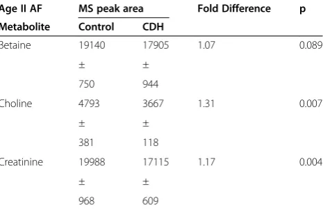
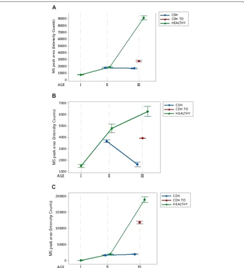
According to the ANOVA analysis of age II groups (102 days), CDH induces a significant decrease in cho-line (p 0.007) and creatinine (p 0.004), while betaine al-terations were not significant (p 0.089) (Figure 1 and Table 1).
The most significant alterations observed were in AF collected at Age III (136 days), since at this stage the ef-fect of CDH could be evaluated either with or without the TO treatment.
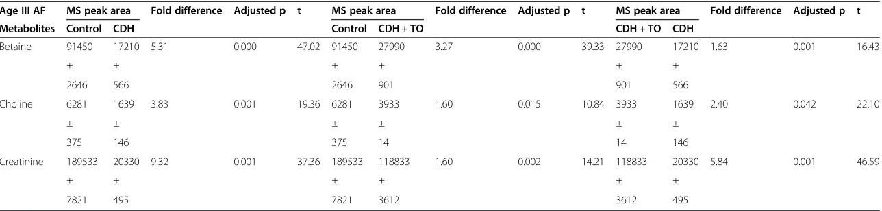
The results from non-treated CDH, in comparison with healthy controls, resulted in the following adjusted p values: choline (p 0.001), creatinine ( p 0.001) and betaine (p 0.000) (Figure 1 and Table 2).
Interestingly, the TO treatment led to a substantial re-covery in choline and creatinine levels and to a modest Table 1 Metabolite MS peak area (Intensity Counts) in
ovine amniotic fluid from a fetal lamb model of congenital diaphragmatic hernia (CDH) and tracheal occlusion (TO)
MS peak area
Age I (70 days)
Age II (102 days)
Age III (136 days)
Metabolite Control Control CDH Control CDH CDH + TO
Betaine 7931 19140 17905 91450 17210 27990
± ± ± ± ± ±
426 750 944 2646 566 901
Choline 1500 4793 3667 6281 1639 3933
± ± ± ± ± ±
179 381 118 375 146 14
Creatinine 687 19988 17115 189533 20330 118833
± ± ± ± ± ±
77 968 609 7821 495 3612
Data are expressed as group means ± standard deviation.
Table 2 Metabolite MS peak area (Intensity Counts) in Age II ovine amniotic fluid
Age II AF MS peak area Fold Difference p
Metabolite Control CDH
Betaine 19140 17905 1.07 0.089
± ±
750 944
Choline 4793 3667 1.31 0.007
± ±
381 118
Creatinine 19988 17115 1.17 0.004
± ±
968 609
Data are expressed as group means ± standard deviations. p refers to Welch’s ANOVA.
Table 3 Metabolite MS peak area (Intensity Counts) in Age III ovine amniotic fluid
Age III AF MS peak area Fold difference Adjusted p t MS peak area Fold difference Adjusted p t MS peak area Fold difference Adjusted p t
Metabolites Control CDH Control CDH + TO CDH + TO CDH
Betaine 91450 17210 5.31 0.000 47.02 91450 27990 3.27 0.000 39.33 27990 17210 1.63 0.001 16.43
± ± ± ± ± ±
2646 566 2646 901 901 566
Choline 6281 1639 3.83 0.001 19.36 6281 3933 1.60 0.015 10.84 3933 1639 2.40 0.042 22.10
± ± ± ± ± ±
375 146 375 14 14 146
Creatinine 189533 20330 9.32 0.001 37.36 189533 118833 1.60 0.002 14.21 118833 20330 5.84 0.001 46.59
± ± ± ± ± ±
7821 495 7821 3612 3612 495
Data are expressed as group means ± standard deviation. Adjusted p and t were obtained with the One-way ANOVA with Games-Howell Pairwise Comparisons.
Multidiscip
linary
Respirat
ory
Medici
ne
2014,
9
:54
Page
4
o
f
8
l.com/con
recovery in betaine (Figure 1). The ANOVA analysis of the Age III groups and the following Games Howell multiple comparison test are consistent with this trend (Figure 1 and Table 3). In particular, p obtained when comparing the control and CDH + TO groups indicate that by the end of gestation, betaine, choline and
creatinine were positively affected by the TO treatment and resulted closer to the physiological level. In fact, al-though the p indicate a significant difference between the control and CDH + TO conditions, the comparison of fold difference values between control and CDH sam-ples, with and without TO, show a significant recovery Figure 1Metabolomic data summarized in Table 1.The Interval Plots show the trend exhibited by the three metabolic markers: betaine (panelA), choline (panelB) and creatinine (panelC) during gestation in the case of“control/CDH induced/CDH induced and TO”treatedfetal lambs. Bullets represent the group mean values, while the interval bars extend 2 standard errors away from the mean.
of the biomarkers associated with the TO treatment (Table 3).
Discussion
Amniotic fluid volume and composition are the result of the dynamic interaction between the secretion of fetal lung liquid, fetal urine and the removal of fluid by fetal swallow-ing and/or resorption through the fetal membranes [17]. A great amount of AF comes from epithelial lung. Between 47-54% of the lung secreted fluids comes out of fetal tra-chea and are swallowed and the rest is mixed with the am-niotic fluid during breathing movements [17].
Metabolomics of human AF has been recently used to detect different fetal pathologies in pregnancy such as chromosomal disorders [18-20], renal dysfunction [10,21], metabolic disease [10], neural tube anomalies [10,22-24] and defects of intrauterine growth [25-28]. Recent advances in metabolomics [10-13,29] also introduce AF evaluation as a new approach to fetal pulmonary develop-ment studies.
Preliminary data of our study show an interesting in-crease in choline, betaine and creatinine levels during physiologic pregnancy. The profile of these metabolites in-creases constantly during gestational age. These molecules are detectable in AF in different concentrations, according to the gestational period [11,12]. Changes in the gestational choline profile have already been correlated with the fetal lung maturity [30]. Choline seems to be involved in the syn-thesis of phospholipid, a major surfactant component. The role of betaine is unclear; it is formed by choline oxidation catalyzed by the mitochondrial enzyme choline dehydro-genase [16,30,31]. Betaine is also engaged in pulmonary de-velopment in the same metabolic pathway as choline. Creatinine has also been used long as a test of fetal matur-ity. Bock showed that in relation to maturity of the fetus, the relative intensity of the creatinine peak correlated best with conventional tests of fetal lung maturity [12]. Choline and creatinine are involved in development of different fetal organs. The possibility that the profile of these metabolites may be influenced by other variables, such as the fetal renal system, cell membrane turnover, cannot be excluded.
Newborns with CDH have hypoplastic lung. These pa-tients have normal maturation values for alveolar epithelial type II. At birth, surfactant and phospholipid production is not delayed [31,32]; the total amount of cells is reduced, as expected in a hypoplasia condition. Prenatal TO to treat se-vere CDH seems to improve pulmonary growth [1-4,33,34]. TO prevents egress of pulmonary fluid leading to lung tissue stretching and reversal of lung hypoplasia. The lung paren-chymal structure increases in alveolar growth and inter-alveolar wall thickness. In the animal model, sustained FETO causes type II cell depletion resulting in significantly low surfactant production. Temporary FETO fails to cause lung growth in the CDH model, but it preserves type II cells,
surfactant production, and partially corrects abnormal mus-cularization of pulmonary arterioles seen in pulmonary hypertension [31-36].
Our study confirms the perturbation of lung maturity in CDH. Certain AF metabolites stop increasing from stage II to stage III in CDH fetuses. The intensity peaks of creatin-ine and betacreatin-ine at stage III, at the end of gestation, were similar to those found at stage II but reduced in average compared to controls at the same gestational age (stage II). Choline intensity peaks in CDH fetuses were even found to be significantly lower during gestation than control speci-mens; growth curve did not increase from the beginning of gestation and values found at term were lower than those found in controls at stage I.
In this study, sustained TO was performed to repro-duce the same conditions inrepro-duced by FETO in CDH hu-man fetuses. Surprisingly, fetal TO seems to improve the profile of the three metabolites: immediately at Age II after TO induction and at stage III gestation. Creatinine had the most improved metabolomic recovery profile, supporting the role of this metabolite as a marker of lung involvement in fetal development [12,30].
The mild response of betaine, with respect to creatinine and choline, could be related to the involvement of this me-tabolite in simultaneous methylation reactions which also include the development of fetal respiratory system.
Nuclear magnetic resonance (NMR) spectroscopy or mass spectrometry (MS) are employed to characterize metabolomic profiles and quantify all small molecules in biological samples. Thus a comprehensive global moni-toring of metabolites and their fluctuations in response to various stimuli can be achieved. Metabolomic profil-ing can provide information on what is actually changprofil-ing in a biological system, and serves as a crucial link be-tween phenotype and genetics [13-16]. Simultaneous analysis of different groups of metabolic markers like amino acids (ie alanine), sugars (ie glucose) and muscle catabolites (ie creatinine) in a single MR spectrum al-lows indirect assessment of function and development of fetal organs [11-13]. The observed variation could also reflect the AFS cell composition in the AF. In this view, congenital diaphragmatic hernia could trigger AFS cell maturation according to tissue regeneration needs, resulting in a AF metabolic signature in the CHD lamb model [9].
There are no previous studies on the metabolomic profile of the amniotic fluid in the CDH lamb model after TO.
Although this is a preliminary study, our findings support the hypothesis that the best biomarkers of a physiological pregnancy may be considered as suggestive indexes of lung maturity also in CDH fetuses. Increased knowledge of pul-monary maturation may be useful in defining biochemical mechanisms which are at the basis of lung hypoplasia in CDH fetuses. The effects of TO confirm their role in re-storing the processes involved in surfactant-mediated lung maturation. This study also supports novelinvestigations in potential pathways implicated in AF regulatory mecha-nisms. Intra-tracheal pulmonary absorption could be con-sidered an influent pathway in the amniotic fluid dynamics.
Conclusions
AF metabolomic profiles may be considered indirect markers of lung growth and could be useful in defining the prognosis of CDH fetuses. Metabolomic analysis in AF in a CDH animal model, provides useful information on fetal lung biochemical mechanisms involved in pul-monary development processes.
Further investigations are still needed to identify new biochemical macromolecules involved in fetal lung mat-uration to improve survival of severe CDH fetuses.
Abbreviations
AF:Amniotic fluid; AFS: Amniotic fluid-derived stem; CDH: Congenital diaphragmatic hernia; FETO: Fetal endoscopic tracheal occlusion; NMR: Nuclear magnetic resonance; MS: mass spectrometry; TO: Tracheal occlusion; VEGF: Vascular endothelial growth factor.
Competing interests
The authors declare that they have no competing interests.
Authors’contributions
GP, surgical support, writing of the final manuscript. MB, MCM,metabolomic analysis. JLP, MM, surgical support, preparation of the manuscript draft, GN, surgical support, preparation of the manuscript draft, MS, preparation of the manuscript draft. FC, EA, EZ, surgical support. VC, preparation of the manuscript draft, revision of the literature. All authors read and approved the final manuscript.
Author details 1
Department of the Mother and Child Health, Pediatric Surgery Unit, Fondazione IRCCS Policlinico San Matteo, Pavia and University of Pavia, Piazzale Golgi 2, 27100 Pavia, Italy.2Department of Medical and Biological Sciences, University of Udine, Udine, Italy.3Cincinnati Fetal Center. Pediatric
Surgery Division, CCHMC, Cincinnati, OH, USA.4Fetal Surgery Program, Congenital Malformations Research Group, Research Institute of Hospital Universitari Vall d’Hebron, Edifici Infantil, Barcelona, Spain.5Laboratory of Translational Cardiomyology, Stem Cell Interdepartmental Institute, KU Leuven and Human Anatomy, University of Pavia, Pavia, Italy.6Department of Internal Medicine, University of Pavia, Pavia, Italy.
Received: 8 August 2014 Accepted: 23 September 2014 Published: 4 November 2014
References
1. Badillo A, Gingalewski C:Congenital diaphragmatic hernia: treatment and outcomes.Semin Perinatol2014,38:92–96.
2. Haroon J, Chamberlain RS:An evidence-based review of the current treatment of congenital diaphragmatic hernia.Clin Pediatr (Phila)2013,
52:115–124.
3. Leeuwen L, Fitzgerald DA:Congenital diaphragmatic hernia.J Paediatr Child Health2014,50:667–673.
4. Deprest JAM, Veerle A, Evrard K, Verbeken EK, Perales AJ, Delaere PR, Lerut TE, Flageole H:Tracheal side effects of endoscopic balloon tracheal occlusion in the fetal lamb model.Eur J Obstet Gynecol Reprod Biol2000,92:119–126. 5. Chiu PP:New Insights into Congenital Diaphragmatic Hernia - A
Surgeon’s introduction to CDH animal models.Front Pediatr2014,2:36. 6. Gallot D, Coste K, Jani J, Roubliova X, Marceau G, Velemir L, Verheyen A, Lemery
D, Sapin V, Deprest J:Effects of maternal retinoic acid administration in a congenital diaphragmatic hernia rabbit model.Pediatr Pulmonol2008,
43:594–603.
7. Ruano R, Yoshisaki CT, da Silva MM, Ceccon ME, Grasi MS, Tannuri U, Zugaib M, Ceccon ME, Grasi MS, Tannuri U, Zugaib M:A randomized controlled trial of fetal endoscopic tracheal occlusion versus postnatal management of severe isolated congenital diaphragmatic hernia. Ultrasound Obstet Gynecol2012,39:20–27.
8. Jani JC, Nicolaides KH, Gratacós E, Valencia CM, Doné E, Martinez JM, Gucciardo L, Cruz R, Deprest JA:Severe diaphragmatic hernia treated by fetal endoscopic tracheal occlusion.Ultrasound Obstet Gynecol2009,34:304–310.
9. Deprest J, Gucciardo L, Eastwood P, Zia S, Jimenez J, Russo F, Lesage F, Lewi L, Sampaolesi M, Toelen J:Medical and regenerative solutions for congenital diaphragmatic hernia: a perinatal perspective.Eur J PediatrSurg2014,24:270–277.
10. Fanos V, Atzori L, Makarenko K, Melis GB, Ferrazzi E:Metabolomics application in maternal-fetal medicine.Biomed Res Int2013,2013:720514. 11. Cohn BR, Fukuchi EY, Joe BN, Swanson MG, Kurhanewicz J, Yu J, Caughey
AB:Calculation of gestational age in late second and third trimesters by ex vivo magnetic resonance spectroscopy of amniotic fluid.Am J Obstet Gynecol2010,203:76.e1–76.e10.
12. Cohn BR, Joe BN, Zhao S, Kornak J, Zhang VY, Iman R, Kurhanewicz J, Vahidi K, Yu J, Caughey AB, Swanson MG:Quantitative metabolic profiles of 2nd and 3rd trimester human amniotic fluid using (1) H HR-MAS spectros-copy.MAGMA2009,22:343–352.
13. Joe BN, Vahidi K, Zektzer A, Chen MH, Clifton MS, Butler T, Keshari K, Kurhanewicz J, Coakley F:Swanson MG: (1) H HR-MAS spectroscopy for quantitative measurement of choline concentration in amniotic fluid as a marker of fetal lung maturity: inter- and intraobserver reproducibility study.J Magn Reson Imaging2008,28:1540–1545.
14. Dunn WB, Broadhurst D, Begley P, Zelena E, Francis-McIntyre S, Anderson N, Brown M, Knowles JD, Halsall A, Haselden JN, Nicholls AW, Wilson ID, Kell DB, Goodacre R:Human Serum Metabolome (HUSERMET) Consortium: Procedures for large-scale metabolic profiling of serum and plasma using gas chromatography and liquid chromatography coupled to mass spectrometry.Nat Protoc2011,6:1060–1083.
15. Zhu ZJ, Schultz AW, Wang J, Johnson CH, Yannone SM, Patti GJ, Siuzdak G:Liquid chromatography quadrupole time-of-flight mass spectrometry characterization of metabolites guided by the METLIN database.Nat Protoc2013,8:451–460. 16. Graça G, Goodfellow BJ, Barros AS, Diaz S, Duarte IF, Spagou K, Veselkov K,
Want EJ, Lindon JC, Carreira IM, Galhano E, Pita C, Gil AM:UPLC-MS metabolic profiling of second trimester amniotic fluid and maternal urine and comparison with NMR spectral profiling for the identification of pregnancy disorder biomarkers.Mol Biosyst2012,8:1243–1254. 17. Brace RA, Cheung CY:Regulation of amniotic fluid volume: evolving
concepts.Adv Exp Med Biol2014,814:49–68.
18. Bahado-Singh RO, Akolekar R, Chelliah A, Mandal R, Dong E, Kruger M, Wishart DS, Nicolaides K:Metabolomic analysis for first-trimester trisomy 18 detection.Am J Obstet Gynecol2013,209:65.e1-9.
19. Bahado-Singh RO, Akolekar R, Mandal R, Dong E, Xia J, Kruger M, Wishart DS, Nicolaides K:Metabolomic analysis for first-trimester Down syndrome prediction.Am J Obstet Gynecol2013,208:371.e1-8.
20. Diaz SO, Barros AS, Goodfellow BJ, Duarte IF, Galhano E, Pita C, Almeida Mdo C, Carreira IM, Gil AM:Second trimester maternal urine for the diagnosis of trisomy 21 and prediction of poor pregnancy outcomes. J Proteome Res2013,12:2946–2957.
21. Zhang A, Sun H, Qiu S, Wang X:Metabolomics insights into pathophysiological mechanisms of nephrology.Int Urol Nephrol2014,46:1025–1030.
22. Zheng X, Su M, Pei L, Zhang T, Ma X, Qiu Y, Xia H, Wang F, Zheng X, Gu X, Song X, Li X, Qi X, Chen G, Bao Y, Chen T, Chi Y, Zhao A, Jia W:Metabolic signature of pregnant women with neural tube defects in offspring. J Proteome Res2011,10:4845–4854.
23. Coen M, Wevers RA, Lindon JC, Blom HJ:High-resolution 1H NMR spectroscopic investigation of a chick embryo model of neural tube development.MagnResonChem2009,47(Suppl 1):S62–67.
24. Hansler A, Chen Q, Gray JD, Ross ME, Finnell RH, Gross SS:Untargeted metabolite profiling of murine embryos to reveal metabolic perturbations associated with neural tube closure defects.Birth Defects Res A Clin Mol Teratol2014,100:623–632.
25. Sanz-Cortés M, Carbajo RJ, Crispi F, Figueras F, Pineda-Lucena A, Gratacós E:
Metabolomic profile of umbilical cord blood plasma from early and late intrauterine growth restricted (IUGR) neonates with and without signs of brain vasodilation.PLoS One2013,8:e80121.
26. van Vliet E, Eixarch E, Illa M, Arbat-Plana A, González-Tendero A, Hogberg HT, Zhao L, Hartung T, Gratacos E:Metabolomics reveals metabolic alterations by intrauterine growth restriction in the fetal rabbit brain. PLoS One2013,8:e64545.
27. O’Connor C, Stuart B, Fitzpatrick C, Turner MJ, Kennelly MM:A review of contemporary modalities for identifying abnormal fetal growth.J Obstet Gynaecol2013,33:239–245.
28. Dessì A, Ottonello G, Fanos V:Physiopathology of intrauterine growth retardation: from classic data to metabolomics.J Matern Fetal Neonatal Med2012,25:13–8. 23025763.
29. Mimmi MC, Ballico M, Nakib G, Calcaterra V, Peiró JL, Marotta M, Pelizzo P:
Altered metabolic profile in congenital lung lesions revealed by 1H nuclear magnetic resonance spectroscopy.ISRN Analytical Chemistry2014,
2014:ID 391836. http://dx.doi.org/10.1155/2014/391836.
30. Wu BT, Dyer RA, King DJ, Richardson KJ, Innis SM:Early second trimester maternal plasma choline and betaine are related to measures of early cognitive development in term infants.PLoS One2012,7:e43448. 31. Sullivan KM, Hawgood S, Flake AW, Harrison MR, Adzick NS:Amniotic fluid
phospholipid analysis in the fetus with congenital diaphragmatic hernia. J Pediatr Surg1994,29:1020–1023.
32. Davey M:Surfactant levels in congenital diaphragmatic hernia.PLoS Med 2007,31:4:e243.
33. Ruano R, Peiro JL, da Silva MM, Campos JA, Carreras E, Tannuri U, Zugaib M:
Early fetoscopic tracheal occlusion for extremely severe pulmonary hypoplasia in isolated congenital diaphragmatic hernia: preliminary results.Ultrasound Obstet Gynecol2013,42:70–76.
34. Sanz-López E, Maderuelo E, Peláez D, Chimenti P, Lorente R, Muñoz MA, Sánchez-Luna M:Changes in the expression of vascular endothelial growth factor after fetal tracheal occlusion in an experimental model of congenital diaphragmatic hernia.Crit Care Res Pract2013,2013:958078. 35. Cogo PE, Zimmermann LJ, Verlato G, Midrio P, Gucciardi A, Ori C, Carnielli
VP:A dual stable isotope tracer method for the measurement of surfactant disaturated-phosphatidylcholine net synthesis in infants with congenital diaphragmatic hernia.Pediatr Res2004,56:184–190. 36. Luks FI, Wild YK, Piasecki GJ, De Paepe ME:Short-term tracheal occlusion
corrects pulmonary vascular anomalies in the fetal lamb with diaphragmatic hernia.Surgery2000,128:266–272.
doi:10.1186/2049-6958-9-54
Cite this article as:Pelizzoet al.:Metabolomic profile of amniotic fluid
to evaluate lung maturity: the diaphragmatic hernia lamb model.
Multidisciplinary Respiratory Medicine20149:54.
Submit your next manuscript to BioMed Central and take full advantage of:
• Convenient online submission
• Thorough peer review
• No space constraints or color figure charges
• Immediate publication on acceptance
• Inclusion in PubMed, CAS, Scopus and Google Scholar
• Research which is freely available for redistribution