R E S E A R C H
Open Access
Haploinsufficiency of the autism-associated
Shank3
gene leads to deficits in synaptic
function, social interaction, and social
communication
Ozlem Bozdagi
1,2†, Takeshi Sakurai
1,2†, Danae Papapetrou
3, Xiaobin Wang
4, Dara L Dickstein
3, Nagahide Takahashi
2,
Yuji Kajiwara
2, Mu Yang
6, Adam M Katz
6, Maria Luisa Scattoni
6,7, Mark J Harris
6, Roheeni Saxena
6, Jill L Silverman
6,
Jacqueline N Crawley
6, Qiang Zhou
4,8, Patrick R Hof
3, Joseph D Buxbaum
1,2,3,5*Abstract
Background:SHANK3 is a protein in the core of the postsynaptic density (PSD) and has a critical role in recruiting many key functional elements to the PSD and to the synapse, including components ofa -amino-3-hydroxyl-5-methyl-4-isoxazole-propionic acid (AMPA), metabotropic glutamate (mGlu) andN-methyl-D-aspartic acid (NMDA) glutamate receptors, as well as cytoskeletal elements. Loss of a functional copy of theSHANK3gene leads to the neurobehavioral manifestations of 22q13 deletion syndrome and/or to autism spectrum disorders. The goal of this study was to examine the effects of haploinsufficiency of full-lengthShank3in mice, focusing on synaptic
development, transmission and plasticity, as well as on social behaviors, as a model for understandingSHANK3
haploinsufficiency in humans.
Methods:We used mice with a targeted disruption ofShank3in which exons coding for the ankyrin repeat domain were deleted and expression of full-length Shank3 was disrupted. We studied synaptic transmission and plasticity by multiple methods, including patch-clamp whole cell recording, two-photon time-lapse imaging and extracellular recordings of field excitatory postsynaptic potentials. We also studied the density of
GluR1-immunoreactive puncta in the CA1 stratum radiatum and carried out assessments of social behaviors.
Results:InShank3heterozygous mice, there was reduced amplitude of miniature excitatory postsynaptic currents from hippocampal CA1 pyramidal neurons and the input-output (I/O) relationship at Schaffer collateral-CA1 synapses in acute hippocampal slices was significantly depressed; both of these findings indicate a reduction in basal neurotransmission. Studies with specific inhibitors demonstrated that the decrease in basal transmission reflected reduced AMPA receptor-mediated transmission. This was further supported by the observation of reduced numbers of GluR1-immunoreactive puncta in the stratum radiatum. Long-term potentiation (LTP), induced either withθ-burst pairing (TBP) or high-frequency stimulation, was impaired inShank3heterozygous mice, with no significant change in long-term depression (LTD). In concordance with the LTP results, persistent expansion of spines was observed in control mice after TBP-induced LTP; however, only transient spine expansion was observed inShank3heterozygous mice. MaleShank3heterozygotes displayed less social sniffing and emitted fewer
ultrasonic vocalizations during interactions with estrus female mice, as compared to wild-type littermate controls. Conclusions:We documented specific deficits in synaptic function and plasticity, along with reduced reciprocal social interactions inShank3heterozygous mice. Our results are consistent with altered synaptic development and
* Correspondence: [email protected]
†Contributed equally
1
Seaver Autism Center for Research and Treatment, Mount Sinai School of Medicine, New York, NY 10029, USA
Full list of author information is available at the end of the article
function inShank3haploinsufficiency, highlighting the importance of Shank3 in synaptic function and supporting a link between deficits in synapse function and neurodevelopmental disorders. The reduced glutamatergic
transmission that we observed in theShank3 heterozygous mice represents an interesting therapeutic target in
Shank3-haploinsufficiency syndromes.
Background
The Shank protein family
Shank proteins, which include Shanks 1, 2 and 3, were first identified in a yeast two-hybrid assay using the gua-nylate kinase-associated protein (GKAP, also called SAPAP1, DLGAP1 and DAP-1) as bait [1]. GKAP is a PSD-95-binding protein that forms an important compo-nent of the postsynaptic density (PSD), where protein-protein interactions between scaffolding protein-proteins and receptors are a key mechanism in assembling a functional synapse [2]. Shanks are highly enriched in the PSD and contain five domains for protein-protein interactions, including an ankyrin repeat domain, an Src homology 3 (SH3) domain, a PSD-95/discs large/zonula occludens-1 (PDZ) domain, several proline-rich regions and a C-terminal sterilea-motif (SAM) domain [3] (Figure 1). The PDZ domain mediates the interaction of Shanks with the COOH terminus of several different proteins, including GKAP [1,4], G protein-coupled receptors [5,6] and the COOH terminus of group I metabotropic gluta-mate receptors (mGluRs) [7]. GKAP mediates the bind-ing of Shanks toN-methyl-D-aspartic acid (NMDA) and
a-amino-3-hydroxyl-5-methyl-4-isoxazole-propionic acid (AMPA) receptors [8]. In addition, the SAM domain interacts with GKAP [7], while proline-rich regions of the Shank proteins bind the mGluR-binding protein Homer [7], actin-binding protein Abp-1 [9,10] and cor-tactin (cortical actin-binding protein) [11], which pro-motes polymerization of the actin cytoskeleton, an important modulator of long-term synaptic plasticity [12]. Shanks interact directly with AMPA receptors through the SH3 domain [13]. Interaction of the ankyrin repeats of Shanks witha-fodrin may mediate calmodu-lin-mediated processes after synaptic stimulation through interactions with actin and calmodulin [14]. Sharpin can also interact with the Shanks [15] and may be involved in activity-dependent modification in dendritic spines.
Shank proteins in synaptic biology
There are ~300 individual Shank molecules in a single postsynaptic site, representing ~5% of the total protein molecules and total protein mass in the site [16]. It would therefore not be surprising that alteration in Shank expression could profoundly affect synaptic mor-phology and function. Such a crucial role for Shank levels in synaptic function is supported by the observa-tion that overexpression of Shank1 led to increased
spine size in neurons in culture [17]. Additional studies showed that knockout ofShank1leads to a decrease in spine number and spine and PSD size, decreased levels of GKAP and Homer, and reduced basal synaptic trans-mission [18]. Furthermore, inhibition ofShank3 expres-sion has been shown to reduce numbers of spines in hippocampal neurons in culture and, conversely, when Shank3 was introduced into aspiny cerebellar neurons in vitro, the neurons developed spines with functional glutamatergic synapses expressing NMDA, AMPA and mGlu receptors [19].
Haploinsufficiency ofSHANK3in neurodevelopmental syndromes
recently been associated with ASDs and/or intellectual disability [29-31].
In the current study, we characterized mice with a tar-geted disruption of Shank3 as a model forSHANK3 -haploinsufficiency syndromes. We focused on synaptic
biology and synaptic function as the most proximal tar-get for altered Shank3 expression. We also examined social interaction and social communication inShank3 heterozygotes and their wild-type littermates. The results are consistent with an important role for
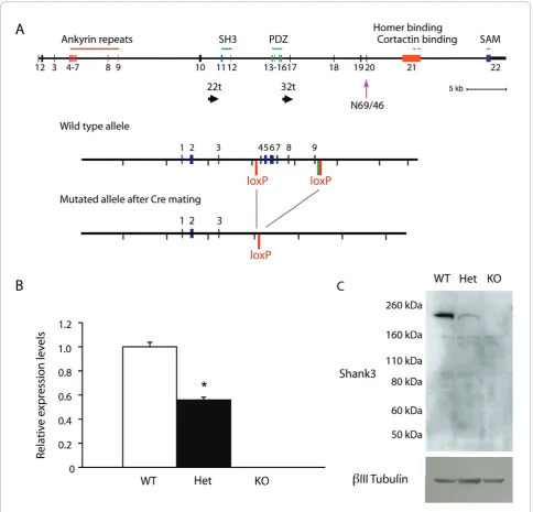
Figure 1Reduced expression of Shank3 inShank3-deficient mice.(A)Targeting strategy.Top: The genomic structure ofShank3is shown. The position of the 22t and 32t transcripts are also shown, as is the epitope for antibody N69/46.Bottom: The location of the loxP sites introduced for targeting are shown before and after the activity of the recombinase. The green bar in the wild-type allele indicates the FRT site remaining after deletion of the selection cassette.(B)Expression of Shank3 mRNA. Brain-derived mRNA from wild-type, heterozygous and knockout mice were subjected to quantitative polymerase chain reaction (qPCR) assay for Shank3 mRNA using probes across exons 6 and 7 with normalization against reference genes.(C)Expression of Shank3 in postsynaptic density (PSD) fractions. PSD fractions from wild-type,
SHANK3 in synaptic function and plasticity and impli-cate specific pathways as potential therapeutic targets forSHANK3-haploinsufficiency syndromes.
Methods
Generation of mice with a disruption of theShank3gene
Animal procedures were approved by the Mount Sinai School of Medicine and the National Institute of Mental Health Animal Care and Use committees. We made use of gene targeting in Bruce4 C57BL/6 embryonic stem (ES) cells [32] to generate a mouse line that has loxP sites inserted before exon 4 and after exon 9 (encoding the ankyrin repeats), with the selection cassette (flanked by FRT sites) excised by FLP recombinase. This floxed strategy was chosen to allow us the option of doing con-ditional (region-specific) knockouts if needed. C57BL/6 was used as the chosen embryonic stem cell line because of the more robust social and cognitive abilities in this line as compared to many of the 129-derived ES lines. For all studies reported here, the floxed allele was first excised by crossing with a CMV-Cre transgenic line (again on a C57BL/6 background) that has ubiquitous Cre expression and a line maintained with a deletion of exons 4 through 9. This Shank3-deficient line was car-ried forward by crossing heterozygotes with the C57BL/ 6 strain to maintain a pure C57 background suitable for electrophysiology and behavioral analyses.
Quantitative polymerase chain reaction
RNA was extracted from brain cortex using the RNeasy Mini Kit (Qiagen, Valencia, CA, USA) according to the manufacturer’s instructions. cDNA was synthesized with the High Capacity cDNA Reverse Transcription Kit (Applied Biosystems, Carlsbad, CA, USA). The universal probe library (UPL) system (Roche, Indianapolis, IN, USA) was used to perform quantitative polymerase chain reaction (qPCR). Primers located in exons 6 and 7 of Shank3 (NM_021423) were designed using ProbeFinder Software (Roche). Three reference genes (Actb,Gapdand Rpl13a) were used for normalization, and relative expres-sion levels were calculated using qBase software [33], now available from Biogazelle (Ghent, Belgium). Primer sequences and UPL probe numbers were Shank3, for-ward tggttggcaagagatccat, reverse ttggccccatagaacaaaag, #1; Actb, forward ggatgcagaaggagattactgc, reverse ccacc-gatccacacagagta, #63; Gapd, Fw gccaaaagggtcatcatctc, reverse cacacccatcacaaacatgg, #29; Rpl 13a, forward tccctgctgctctcaagg, reverse gccccaggtaagcaaactt, #41. Unpairedt-tests were used for group comparisons.
Immunoblotting
PSD fractions were prepared as follows. Hemibrains of wild-type, heterozygous and homozygousShank3 mice were homogenized in
2-[4-(2-hydroxyethyl)piperazin-1-yl]ethanesulfonic acid (HEPES)-A containing 4 mM HEPES, pH 7.4, 0.32 M sucrose, Protease Inhibitor Cocktail and PhoSTOP Phosphatase Inhibitor Cocktail (both from Roche). Nuclear fractions were precipitated by centrifuging twice at 700 g for 15 min, and the resulting supernatants were further centrifuged at 21,000 gfor 15 min. The precipitates were resuspended in HEPES-B containing 4 mM HEPES, pH 7.4, Protease Inhibitor Cocktail and PhoSTOP Phosphatase Inhibitor Cocktail, homogenized and rotated at 4°C for 1 hour. The lysates were centrifuged at 32,000gfor 20 min and washed twice with HEPES-C containing 50 mM HEPES, pH 7.4, 0.5% Triton X-100, Protease Inhibitor Cocktail and PhoSTOP Phosphatase Inhibitor Cocktail. Finally, postsynaptic density fractions were resuspended in HEPES-C containing 1.8% sodium dodecyl sulfate (SDS) and 2.5 M urea. Fifty-twoμg of each PSD fraction were loaded to 4-12% SDS-polyacrylamide gel electrophoresis (PAGE gel (Invitrogen, Carlsbad, CA, USA), transferred to polyvinylidene fluoride membrane and immuno-blotted with either the N69/46 anti-Shank3 antibody directed against an epitope downstream of the PDZ domain (UC Davis/NIH NeuroMab Facility, Davis, CA) or the anti-ProSAP2 anti-Shank3 antibody directed against the last 100 amino acids of Shank3 (Millipore, Billerica, MA, USA). For bIII tubulin, the membrane was stripped and immunoblotted with an anti-bIII tubu-lin antibody (Abcam, Cambridge, MA, USA).
Hippocampal slice electrophysiology
Whole cell recording, two-photon time-lapse imaging and analysis
in excitation [36]. Spine volume in theθ-burst stimulation (TBS) experiments was also calculated using the Rayburst algorithm in NeuronStudio software (available from the Computational Neurobiology and Imaging Center, Mount Sinai School of Medicine, New York, NY, USA) following deconvolution of the data [37-39], and we obtained similar results using either approach.
Extracellular recordings
Hippocampal slices (350μm thick) were prepared from 4- to 6-week-old heterozygous mice and their wild-type littermate controls. Slices were perfused with Ringer’s solution containing (in mM): NaCl, 125.0; KCl, 2.5; MgSO4, 1.3; NaH2PO4, 1.0; NaHCO3, 26.2; CaCl2, 2.5; and
glucose, 11.0. The Ringer’s solution was bubbled with 95% O2and 5% CO2 at 32°C during extracellular recordings
(electrode solution: 3 M NaCl). Slices were maintained for 1 hour prior to establishment of a baseline of field excita-tory postsynaptic potentials (fEPSPs) recorded from stra-tum radiastra-tum in area CA1, evoked by stimulation of the Schaffer collateral-commissural afferents (100-μs pulses every 30 s) with bipolar tungsten electrodes placed into area CA3 [40]. Test stimulus intensity was adjusted to obtain fEPSPs with amplitudes that were one-half of the maximal response. The EPSP initial slope (mV/ms) was determined from the average waveform of four consecu-tive responses. Input-output (I/O) curves were generated by plotting the fEPSP slope versus fiber volley amplitude in low-Mg2+(0.1 mM) solution. AMPA receptor-mediated and NMDA receptor-mediated I/O relationships were measured in the presence of 2-amino-5-phosphonopenta-noic acid (APV; 50μM) and 6-cyano-7-nitroquinoxaline-2,3-dione (CNQX; 100μM), respectively (Sigma, St. Louis, MO, USA).
Paired-pulse responses were measured with an inter-stimulus interval (ISI) of 50 ms and are expressed as the ratio of the average responses to the second stimulation pulse (FP2) to the first stimulation pulse (FP1). LTP was induced by either a high-frequency stimulus (four trains of 100-Hz, 1-s stimulations separated by 5 min) or TBS (15 bursts of four pulses at 100 Hz separated by 200 ms). To induce long-term depression (LTD), Schaffer collaterals were stimulated by low-frequency stimulation (LFS; 900 pulses at 1 Hz, 15 min) or by a paired-pulse low-frequency stimulation (PP-LFS; 1 Hz for 20 min, 50-ms interstimulus interval [41]) to induce mGlu receptor-dependent LTD. Data were expressed as means ± SD, and statistical analyses were performed using ana-lysis of variance (ANOVA) or Student’st-test, with sig-nificance set at analevel of 0.05.
Measurement of GluR1-immunoreactive puncta Immunohistochemistry
Three-month-old animals were anesthetized with 250μl of 15% chloral hydrate and perfused transcardially with
1% paraformaldehyde for 1 min followed by 4% parafor-maldehyde for a total of 13 min. The brains were then removed, hemisected, cut in 50-μm-thick sections using a Leica VT1000S Vibratome (Vibratome, Bannockburn, IL, USA) and subsequently stored in phosphate-buffered saline (PBS) until use. Sections were incubated at 37°C for 5 min, followed by incubation in acidified pepsin (1 ml in a 0.2 N HCl solution) for 6.5 min. The tissue was then washed at room temperature in PBS-B (3 × 20 min) and incubated in a 0.3% Triton X-100, 0.5% bovine serum albumin (BSA), 5% normal goat serum for 1 h on an orbital shaker. The blocking step was followed by overnight incubation in the primary antibody (rabbit polyclonal antiglutamate receptor 1 AB1504; Millipore, Billerica, MA, USA), which was made in blocking solu-tion at the appropriate dilusolu-tion (1 μg/ml). The tissue sections were then washed in PBS-B (5 × 5 min) and incubated in secondary antibody (goat antirabbit Alexa Fluor 488, Invitrogen) in a 2% BSA and 0.3% Triton PBS-B solution at the appropriate dilution (1:400) for 1 h at room temperature on an orbital shaker. Finally, the tissue was washed in PBS-B (3 × 5 min), stained with 4”,6"-diamino-2-phenylindole-2HCl (DAPI) and mounted on charged Aqua ColorFrost slides using Vec-tashield mounting medium (Vector Laboratories, Burlin-game, CA, USA).
Tissue sampling
To quantify puncta, we used a systematic random sam-pling approach whereby a 1:6 series of sections were stained, the stratum radiatum of the CA1 was contoured using SteroInvestigator (MBF Bioscience, Williston, VT, USA) and the sampling sites were determined using an optical fractionator with the size of the grid set at 18μm2 (the dimension of the confocal image stacks to be later sampled with a ×100 lens objective), at a digital zoom of 5 on a Zeiss LSM510 META confocal microscope (Zeiss, Oberkochen, Germany). The trace of the contoured area with an optical fractionator sampling grid placed on it was used as a guide to obtain confocal image stacks of the above-mentioned dimensions that were 100μm apart from each other (i.e., the size of the probe).
Confocal imaging and puncta quantification
The resultant stacks were then deconvolved using AutoDeblur 1.4.1 (Media Cybernetics, Bethesda, MD, USA), using an adaptive point-spread function (PSF) deconvolution method with a theoretical PSF, and then analyzed with custom Vamp2D software [42] that reli-ably calculates the size of individual puncta on the basis of three-dimensional estimates and circumvents the problem of object superimposition found with more traditional methods that collapse the stacks into two-dimensional projections. The relative density of puncta was then calculated per cubic micrometer, and the dif-ferences between groups were assessed using the non-parametric Mann-WhitneyUtest.
Behavioral analyses
Shank3wild-type and heterozygote breeding pairs were imported from Mount Sinai School of Medicine to the National Institute of Mental Health. Mice were main-tained by breeding C57BL/6 wild-type mice withShank3 heterozygotes and housed in a conventional tempera-ture- and humidity-controlled vivarium. Littermates were housed by sex in mixed genotype groups of two to four per cage on a 12:12-h circadian cycle with lights on at 0600. Behavioral experiments were conducted between 1000 and 1600 in dedicated testing rooms.
Developmental milestones were tested across postnatal days 2-14, including measures of body weight, body length, tail length, pinnea detachment, eye opening, inci-sor eruption, fur development, righting reflex, negative geotaxis, cliff avoidance, grasping reflex, auditory startle, bar holding, level screen and vertical screen as pre-viously described [43,44]. In addition, the mice were evaluated in a standard, automated three-chambered social approach task as previously described [44].
Male-female social interactions were evaluated in a 5-min test session as previously described [43,45], with the exception that subject males were group-housed and individually tested in clean cages with clean litter. Each of the 12 wild-type and 14 heterozygous male subject mice, ages 2.5-4 months, was paired with a different unfamiliar estrus C57BL/6J female. A digital closed-circuit television camera (Panasonic, Secaucus, NJ, USA) was positioned horizontally 30 cm from the cage. An ultrasonic microphone (Avisoft UltraSoundGate conden-ser microphone capsule CM15; Avisoft Bioacoustics, Berlin, Germany) was mounted 20 cm above the cage. Sampling frequency for the microphone was 250 kHz, and the resolution was 16 bits. The entire apparatus was contained in a sound-attenuating environmental cham-ber (ENV-018V; Med Associates, St. Albans, VT, USA) illuminated by a single 25-Watt red light. Videos from the male subjects were subsequently scored by an inves-tigator uninformed of the subject’s genotype on mea-sures of nose-to-nose sniffing, nose-to-anogenital
sniffing and sniffing of other body regions, using Noldus Observer software (Noldus Information Technology, Leesburg, VA, USA) as previously described. Ultrasonic vocalizations were played back and spectrograms were displayed using Avisoft software [43,45]. Ultrasonic vocalizations were identified manually by two highly trained investigators blinded to genotype information, and summary statistics were calculated using the Avisoft package. Interrater reliability was 95%. Data were ana-lyzed using an unpaired Student’st-test.
Olfactory habituation/dishabituation testing was con-ducted in male and femaleShank3wild-type and hetero-zygous mice ages 2.5-4 months using methods previously described [44,46,47]. Nonsocial and social odors were presented on a series of cotton swabs inserted into the home cage sequentially, each for 2 min, in the following order: water, water, water (dis-tilled water); almond, almond, almond (1:100 dilution almond extract); banana, banana, banana (1:100 dilution artificial banana flavoring); social 1, social 1, social 1 (swiped from the bottom of a cage housing unfamiliar sex-matched B6 mice); and social 2, social 2, social 2 (swiped from the bottom of a second cage housing a different group of unfamiliar sex-matched 129/SvImJ mice). One-way repeated measures ANOVA was per-formed within each genotype for each set of habituation events and each dishabituation event, followed by a Tukeypost hoctest.
Results
Generation and characterization of a Shank3-deficient mouse
We made use of gene targeting in Bruce4 C57BL/6 embryonic stem (ES) cells [32] to generate a mouse line that has loxP sites inserted before exon 4 and after exon 9 (Figure 1A). For all studies reported here, the floxed allele was excised and a line was maintained with a dele-tion of exons 4 through 9. This line, which completely deletes the ankyrin repeat domains of Shank3, produced wild-type (+/+), heterozygous (+/-) and knockout (-/-) animals with Mendelian frequencies from heterozygote-heterozygote crosses.
qPCR showed 50% reduction of full-length Shank3 mRNA in the heterozygotes and complete loss in knock-outs (Figure 1B). Moreover, there was no expression of full-length Shank3 protein in PSD fractions from Shank3-knockout mice and reduced expression in the heterozygotes, using antibodies which cross-react either with an epitope downstream of the PDZ domain (anti-body N69/46; see Figure 1A) (Figure 1C) or with the COOH terminal (data not shown), consistent with haploinsufficiency.
structure or hippocampal cytoarchitecture, nor were there any obvious seizures. There was evidence for subtle motor abnormalities in homozygotes, which is being further characterized in ongoing studies. Both genotypes were normal on measures of general health, developmental milestones and exploratory activity, as well as on social approach as measured in an automated three-chambered social approach task.
BecauseSHANK3haploinsufficiency is responsible for the neurobehavioral phenotype in individuals with 22q13DS orSHANK3mutations, we focused the studies reported here on Shank3heterozygous mice generated by crossing wild-type mice with heterozygotes to be most relevant to the clinical syndromes and to be most useful in ultimately assessing potential therapeutic inter-ventions in preclinical studies. A comprehensive investi-gation of the knockout mice obtained through an alternate breeding strategy (heterozygote-heterozygote matings) is now in progress in independent experiments.
Basal glutamatergic synaptic transmission is reduced in Shank3heterozygous mice
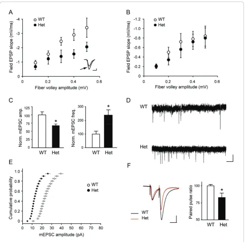
To examine the role of Shank3 in regulating synaptic glutamate receptor function, we studied glutamatergic synaptic transmission in hippocampal slices. We exam-ined the properties of basic transmission at Schaffer col-lateral-CA1 synapses in hippocampal slices from 3- to 4-week-old Shank3heterozygous mice and their litter-mates using extracellular recordings. Plotting field exci-tatory postsynaptic potential (fEPSP) slope versus stimulus intensity demonstrated a reduction in the I/O curves in the heterozygotes (data not shown), prompting us to then further examine synaptic transmission in the presence of inhibitors of specific subtypes of glutamate receptors. In the presence of the NMDA receptor antagonist APV, a decrease in AMPA receptor-mediated field potentials in the heterozygous mice was seen, reflected as a 50% decrease in the average slope of I/O function compared with wild-type mice (n= 4 mice per genotype, two to three slices per mouse; P = 0.001; Figure 2A). In contrast, when the I/O relationship was analyzed in the presence of the competitive AMPA/kai-nate receptor antagonist CNQX to measure synaptic NMDA receptor function, there was no difference between genotypes (n = 4 mice per genotype, two to three slices per mouse;P= 0.1; Figure 2B). These results indicate that there is a specific reduction in AMPA receptor-mediated basal transmission in theShank3 het-erozygous mice.
We asked whether the impairment of evoked synaptic transmission in heterozygous mice is caused by altera-tions in presynaptic and/or postsynaptic parameters. We performed whole cell patch-clamp recordings in 3-month-old littermates and monitored spontaneous
miniature postsynaptic currents in the presence of tetro-dotoxin (TTX). The amplitude of miniature excitatory postsynaptic currents (mEPSCs) from hippocampal CA1 pyramidal neurons of heterozygous mice were signifi-cantly smaller than those in control mice (n = 7 for wild-type and n= 8 for heterozygous mice, P < 0.01; Figures 2C and 2D), which was evident by the signifi-cant shift of the cumulative probability to the left (Figure 2E), again indicating a reduction in basal trans-mission. However, in heterozygous mice, the frequency of miniature excitatory postsynaptic currents was signifi-cantly higher (n = 7 for wild-type andn= 8 for hetero-zygous mice,P < 0.03; Figures 2C and 2D) and paired-pulse ratio was decreased (n= 6 for wild-type andn= 7 for heterozygous mice, P < 0.05; Figure 2F), which revealed an additional, presynaptic alteration in the het-erozygotes as well.
Long-term potentiation is impaired inShank3 heterozygous mice
We next examined long-term potentiation (LTP) with extracellular fEPSP recordings at Schaffer collateral/CA1 synapses. In the first set of experiments, LTP was induced by tetanic stimulation of the Schaffer collaterals (four trains of 100 Hz separated by 5 min). While initial expres-sion of LTP was identical across the two genotypes, the maintenance of LTP was clearly impaired in the heterozy-gous mice (average percentage of baseline 120 min after tetanus: 165.1 ± 8.8% in wild-type and 117.1 ± 9.5% in het-erozygous mice,P= 0.004; Figure 3A). In an additional set of experiments, we further tested TBS LTP (10 bursts of four pulses at 100 Hz separated by 200 ms), which also showed a significant decrease in the potentiation at 60 min after TBS in heterozygous mice (156.3 ± 9.2% of base-line in wild-type and 126.0 ± 8.9% in heterozygous mice, measured 60 min after TBS,P= 0.007; Figure 3B).
In contrast to the altered synaptic plasticity observed with LTP, long-term depression (LTD) induced by either low-frequency stimulation (LFS) (82.6 ± 1.35% of baseline in wild-type and 79.9 ± 2.5% in heterozygous mice, measured 60 min after LFS,P> 0.1; Figure 3G) or paired-pulse LFS (PP-LFS) stimulation (81.6 ± 6% of baseline in wild-type and 82.7 ± 1.9% in heterozygous mice,P > 0.1; Figure 3H) was not significantly changed in heterozygotes.
Figure 3Reduced long-term potentiation inShank3heterozygous mice.(A)Long-term potentiation (LTP) following high-frequency stimulation. Field recordings of LTP induced with high-frequency stimulation (HFS; 4 times 100 Hz, separated by 5 min) as a function of time in slices from wild-type andShank3heterozygous mice. (B) LTP followingθ-burst stimulation (TBS).(C)Long-term depression (LTD) following low-frequency stimulation (LFS), an NMDA receptor-dependent form of LTD.(D)LTD following paired-pulse low-frequency stimulation (PP-LFS), a protein synthesis-dependent form of LTD.(E)LTP recorded with whole cell patch-clamp method. LTP was induced withθ-burst pairing (TBP).(E)
increase in spine volume concurrent with an immediate increase in EPSP slope in whole cell recordings, which gradually reached a plateau by ~30 min (Figures 3C-F). However, in recordings from heterozygous mice, we found that the stabilization in synaptic potentiation and spine expansion were impaired in CA1 neurons (Figures 3C-F). In the heterozygous mice, both EPSP slope and spine volume increased immediately to values compar-able to those of control spines, but synaptic potentiation and spine expansion failed to be sustained.
GluR1 immunoreactivity is decreased inShank3 heterozygous mice
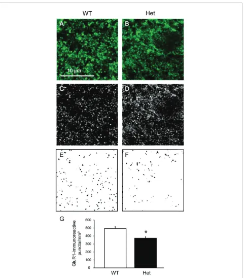
We then asked whether Shank3haploinsufficiency can lead to alterations in the numbers of AMPA receptor-positive puncta, given the results from the electrophysio-logical experiments and the relationship between synap-tic strength and AMPA receptor subunit trafficking [49]. We carried out immunolabeling for GluR1 (an AMPA receptor subunit) and quantified GluR1-immunoreactive puncta (Figures 4A-G). Neurons fromShank3 heterozy-gous mice showed significantly fewer GluR1-immunor-eactive puncta (P < 0.005) consistent with the electrophysiological data.
Behavioral analyses ofShank3heterozygotes
To more extensively study social interactions in Shank3 heterozygous mice, we examined male-female social interactions during a 5-min session of freely moving reciprocal social interactions with an estrus B6 female (Figure 5A). Cumulative duration of total social sniffing by the male test subjects was lower inShank3 heterozy-gotes than in wild-type littermates (P = 0.02). In addi-tion, fewer ultrasonic vocalizations were emitted by heterozygotes than by wild-type littermates during the male-female social interaction session (P= 0.003). Note that while the equipment used could not distinguish between calls emitted by the male subject and female partner, the preponderance of calls during male-female interactions in mice is usually emitted by the male [50].
As a control task to ensure that subject mice can detect the social pheromones that elicit approach and vocalizations, we measured olfactory abilities using the olfactory habituation/dishabituation test. Habituation and dishabituation were normal for social and nonsocial odor cues in both wild-type and Shank3 heterozygous mice (n= 8/genotype; Figure 5B). Both genotypes dis-played the expected habituation, indicated by decreased time spent in sniffing the sequence of three same odors, and the expected dishabituation, indicated by increased time sniffing the different odor (water habituation, wild-type: P < 0.001; water habituation, heterozygotes: P < 0.001; almond habituation, wild-type:P = 0.015; almond habituation, heterozygotes: P= 0.04; banana habituation,
wild-type:P < 0.001; banana habituation, heterozygotes: P = 0.033; social odor 1 habituation, wild-type: P < 0.001; social odor 1 habituation, heterozygotes: P < 0.001; social odor 2 habituation, wild-type: P < 0.001; social odor 2 habituation, heterozygotes: P < 0.001; water to almond dishabituation, wild-type: P = 0.061; water to almond dishabituation, heterozygotes: P = 0.118; almond to banana dishabituation, wild-type: P= 0.005; almond to banana dishabituation, heterozygotes: P = 0.046; banana to social odor 1 dishabituation, wild-type:P< 0.001; banana to social odor 1 dishabituation, heterozygotes:P = 0.046; social odor 1 to social odor 2 dishabituation, wild-type: P < 0.001; social odor 1 to social odor 2 dishabituation, heterozygotes:P< 0.001).
Discussion
In the present study, we summarize our results in mice with a disruption of the full-length Shank3 protein. We observed reductions in glutamatergic synaptic transmis-sion and plasticity, including deficits in AMPA receptor-mediated transmission and spine remodeling. We also identified a reduced number of GluR1-immunoreactive puncta in the striatum radiatum. Finally, we saw evi-dence of social interaction and social communication deficits in these mice. These results demonstrate the importance of Shank3 in synaptic function, ultimately leading to behavioral changes, which may be relevant to the symptoms in individuals withSHANK3 mutations.
Development of theShank3-deficient model
Figure 4Decreased density of GluR1-immunoreactive puncta inShank3heterozygous mice.(AandB)High-resolution confocal images of puncta from wild-type and heterozygous mice.(CandD)The same images from Figures 4A and 4B are shown after deconvolution.(EandF)
ankyrin repeat domain-containing form in neurodeve-lopmental disabilities. Hence, disruption of this form, as done in the current study, has direct construct validity to ASD and to neurodevelopmental disabilities. It will be of interest to determine where and to what levels the 22t and 32t forms are expressed and to assess whether neuronal and/or other phenotypes are associated with their disruption. Careful characterization of mice with a disruption of Shank3 by targeting downstream exons common to the full-length, 22t and 32t forms will be of great interest, with the caveat that such targeting may produce dominant-negative shorter products truncated at the C-terminal that, depending on the exons targeted, may or may not be relevant to known clinical mutations.
Synaptic development and function inShank3 heterozygotes
Electrophysiological studies of the mice showed a decrease in basal synaptic transmission in heterozygotes when assessed by comparing either the I/O relationship using field recordings or the amplitude of miniature excitatory postsynaptic currents (mEPSCs) using patch clamp recordings. This reduction in AMPA receptor-dependent transmission is likely mediated by a decrease in the number of synaptic AMPA receptors. Shank3
heterozygous mice also showed a decrease in the density of GluR1-immunoreactive puncta, consistent with a reduction in AMPA receptor levels. On the other hand, both increase in mEPSC frequency and decrease in paired pulse ratio in Shank3 heterozygous mice were consistent with an increase in presynaptic release.
Transfection of Shank3into aspiny cerebellar granule cells has been shown to induce an increase in the AMPA component of mEPSCs [19], and Shank3 is colo-calized with GluR1-containing AMPA receptor in typical spines [53]. In our studies, haploinsufficiency of Shank3 in vivo leads to reduced levels of AMPA receptors and GluR1-positive puncta, results which are consistent with such studies in vitro. However, while the postsynaptic alterations in the Shank3-deficient mice are consistent with studies carried out in vitro, understanding the changes in presynaptic function observed in our analyses will need further investigation. This presynaptic altera-tion could be explained by compensatory mechanisms in the Shank3heterozygous mice to maintain constant synaptic function. However, this compensatory effect falls short as evidenced by the reduction in the I/O relationship.
Selective acquisition of AMPA receptors is thought to be a crucial mechanism for synapse maturation and function, and our data confirm that Shank3 is an impor-tant mediator of AMPA receptor recruitment and of synaptic development.
Synaptic plasticity and spine remodelling inShank3 heterozygotes
We examined LTP induced by either high frequency sti-mulation or TBS. InShank3heterozygous mice, reduced LTP was apparent at 30 min after induction in either paradigm and no potentiation was observed at 120 min after induction. However, the initial expression of LTP was normal with either induction paradigm. We observed similar deficits in the spine expansion that normally accompanies LTP, such that the initial expan-sion was normal in heterozygotes but quickly decayed to baseline. The simplest explanation for these deficits is an alteration in the trafficking of AMPA receptors to synapses. AMPA receptor trafficking is involved in activ-ity-induced changes in synaptic strength associated with learning and memory [54], and synaptic insertion of GluR1-containing AMPA receptors is required [55,56]. The quantity of surface receptors controls synaptic strength [57-59]; therefore, reduction of both synaptic transmission and plasticity in theShank3heterozygotes would support a mechanism involving altered AMPA receptor trafficking. Initial LTP is likely mediated by phosphorylation of AMPA receptors, and the initial spine expansion occurs in the absence of trafficking of AMPA receptors [54]. However, AMPA receptor
Figure 5Reduced social behaviors inShank3heterozygous and wild-type littermate mice.(A)Adult male-female social
interactions.Left: Total duration of social interactions, scored as cumulative seconds spent by the male subject in sniffing the nose, anogenital and other body regions of an unfamiliar adult estrus B6 female mouse during a 5-min test session in a clean, empty mouse cage. *P= 0.02.Right: Number of ultrasonic vocalizations emitted during the social interaction test session (*P= 0.003) (n= 12 for wild-type (WT) andn= 14 for heterozygous (Het) mice).(B)
trafficking to the synapse is critical to sustain LTP and spine expansion. The transient nature of synaptic poten-tiation and spine enlargement in heterozygous mice is consistent with normal phosphorylation of existing synaptic AMPA receptors with a deficit in subsequent consolidation, the process in which both structural and functional forms of plasticity are progressively stabilized over 30 min [34].
Social behavior inShank3heterozygotes
Male Shank3 heterozygous mice displayed reduced responses to female social cues compared to their wild-type littermates on parameters of social sniffing and ultrasonic vocalizations in a male-female reciprocal social interaction context. Given that Shank3 has a cen-tral role in synaptic function, Shank3-related changes in cellular and network components that underlie social cognition could underlie the reduced social interactions in Shank3 heterozygous mice. It was interesting that social deficits were not observed in a three-chamber social approach task, implying some specificity of the social alterations. It will be of interest to better under-stand the frequency of ASD and the characteristics of social deficits inSHANK3-haploinsufficiency syndromes, as it is already evident that this is not a universal finding in these syndromes when considering 22q13DS or schizophrenia.
The social deficits observed in the mice serve as important reminders that systems outside the hippo-campus that are involved in social behaviors will need to be examined in these mice and that additional beha-viors known to be mediated by the hippocampus will be important for further investigations. Cellular and elec-trophysiological analyses in additional brain regions and large-scale behavioral studies are now underway for the Shank3-deficient mice in the authors’laboratories.
Conclusions
Haploinsufficiency of full-length Shank3resulted in a decrease in synaptic transmission, altered functional and structural plasticity of synapses and reduced social beha-viors. These findings are consistent with a model of delayed synaptic development inShank3 haploinsuffi-ciency, together with a reduction in AMPA receptor trafficking. The results highlight the importance of Shank3 in synaptic development and function and sup-port a link between deficits in synapse function and neurodevelopmental disorders. To date, pharmacological treatments for ASDs and other developmental disorders (including SHANK3-haploinsufficiency syndromes) are primarily ameliorative, focusing on managing associated systems such as anxiety, aggression, repetitive behaviors, attention deficits and epilepsy, among others (see, for example, [60]). Pharmacological treatments addressing
core diagnostic symptoms, including, in ASDs, altera-tions in reciprocal social interacaltera-tions and communica-tion, do not yet exist. Recently, the field has begun to see the evaluation of therapies targeted to etiology (that is,“personalized medicine”) using models of neurodeve-lopmental disorders including fragile X, tuberous sclero-sis, and Rett syndromes (see, for example, [61-64]). The use of model systems such as theShank3-deficient mice reported here could lead to similar advances in the case ofSHANK3-haploinsufficiency syndromes. One interest-ing outcome of the current study is that compounds that enhance glutamatergic transmission, including those that specifically enhance AMPA transmission (AMPAkines) could possibly represent therapeutic approaches in these conditions. Further analysis of this and additional models may identify targets for novel therapeutics for individuals with developmental delays arising from 22q13 deletion syndrome or SHANK3 mutations.
Acknowledgements
This work was supported by the Seaver Foundation, the Simons Foundation, the National Institute of Mental Health Intramural Research Program, and by a gift from Paulina Rychenkova, PhD, and William Gibson. OB and TS are Seaver Junior Faculty Fellows. We thank Lisa Krug and Shekhar Patil for help at early stages of the project.
Author details
1Seaver Autism Center for Research and Treatment, Mount Sinai School of
Medicine, New York, NY 10029, USA.2Department of Psychiatry, Mount Sinai
School of Medicine, New York, NY 10029, USA.3Department of
Neuroscience, Mount Sinai School of Medicine, New York, NY 10029, USA.
4Department of Neurology, Mount Sinai School of Medicine, New York, NY
10029, USA.5Department of Genetics and Genomic Sciences, Mount Sinai School of Medicine, New York, NY 10029, USA.6Laboratory of Behavioral
Neuroscience, National Institute of Mental Health, Bethesda, MD 20892-3730, USA.7Istituto Superiore di Sanità, Rome, Italy.8Genentech, South San
Francisco, CA 94080, USA.
Authors’contributions
TS and JDB generated and biochemically characterized the mice. JDB, PRH, JNC, TS, QZ and OB designed the experiments. OB, XW and QZ performed the electrophysiology experiments and analysis. Immunohistochemistry as well as confocal microscopy and analysis were conducted by DP, DLD and PRH. Behavioral experiments and analysis were conducted by MY, AMK, MLS, MJH, RS, JLS and JNC. qPCR was conducted by NT. Immunoblotting was conducted by YK. The manuscript was written by OB and JDB, and all authors reviewed the manuscript before submission.
Competing interests
OB, TS and JDB have submitted a patent on this work.
Received: 14 October 2010 Accepted: 17 December 2010 Published: 17 December 2010
References
1. Naisbitt S, Kim E, Tu JC, Xiao B, Sala C, Valtschanoff J, Weinberg RJ, Worley PF, Sheng M:Shank, a novel family of postsynaptic density proteins that binds to the NMDA receptor/PSD-95/GKAP complex and cortactin.Neuron1999,23:569-582.
2. Sheng M:Excitatory synapses. Glutamate receptors put in their place. Nature1997,386:221-223.
4. Yao I, Iida J, Nishimura W, Hata Y:Synaptic localization of SAPAP1, a synaptic membrane-associated protein.Genes Cells2003,8:121-129. 5. Zitzer H, Richter D, Kreienkamp HJ:Agonist-dependent interaction of the
rat somatostatin receptor subtype 2 with cortactin-binding protein 1.J Biol Chem1999,274:18153-18156.
6. Kreienkamp HJ, Soltau M, Richter D, Bockers T:Interaction of G-protein-coupled receptors with synaptic scaffolding proteins.Biochem Soc Trans 2002,30:464-468.
7. Tu JC, Xiao B, Naisbitt S, Yuan JP, Petralia RS, Brakeman P, Doan A, Aakalu VK, Lanahan AA, Sheng M, Worley PF:Coupling of mGluR/Homer and PSD-95 complexes by the Shank family of postsynaptic density proteins.Neuron1999,23:583-592.
8. Kim JH, Kim JH, Yang E, Park JH, Yu YS, Kim KW:Shank 2 expression coincides with neuronal differentiation in the developing retina.Exp Mol Med2009,41:236-242.
9. Qualmann B, Boeckers TM, Jeromin M, Gundelfinger ED, Kessels MM: Linkage of the actin cytoskeleton to the postsynaptic density via direct interactions of Abp1 with the ProSAP/Shank family.J Neurosci2004, 24:2481-2495.
10. Haeckel A, Ahuja R, Gundelfinger ED, Qualmann B, Kessels MM:The actin-binding protein Abp1 controls dendritic spine morphology and is important for spine head and synapse formation.J Neurosci2008, 28:10031-10044.
11. Du Y, Weed SA, Xiong WC, Marshall TD, Parsons JT:Identification of a novel cortactin SH3 domain-binding protein and its localization to growth cones of cultured neurons.Mol Cell Biol1998,18:5838-5851. 12. Cingolani LA, Goda Y:Actin in action: the interplay between the actin
cytoskeleton and synaptic efficacy.Nat Rev Neurosci2008,9:344-356. 13. Sheng M, Kim MJ:Postsynaptic signaling and plasticity mechanisms.
Science2002,298:776-780.
14. Vanderklish PW, Krushel LA, Holst BH, Gally JA, Crossin KL, Edelman GM: Marking synaptic activity in dendritic spines with a calpain substrate exhibiting fluorescence resonance energy transfer.Proc Natl Acad Sci USA 2000,97:2253-2258.
15. Lim S, Naisbitt S, Yoon J, Hwang JI, Suh PG, Sheng M, Kim E: Characterization of the Shank family of synaptic proteins. Multiple genes, alternative splicing, and differential expression in brain and development.J Biol Chem1999,274:29510-29518.
16. Sugiyama Y, Kawabata I, Sobue K, Okabe S:Determination of absolute protein numbers in single synapses by a GFP-based calibration technique.Nat Methods2005,2:677-684.
17. Sala C, Piech V, Wilson NR, Passafaro M, Liu G, Sheng M:Regulation of dendritic spine morphology and synaptic function by Shank and Homer. Neuron2001,31:115-130.
18. Hung AY, Futai K, Sala C, Valtschanoff JG, Ryu J, Woodworth MA, Kidd FL, Sung CC, Miyakawa T, Bear MF,et al:Smaller dendritic spines, weaker synaptic transmission, but enhanced spatial learning in mice lacking Shank1.J Neurosci2008,28:1697-1708.
19. Roussignol G, Ango F, Romorini S, Tu JC, Sala C, Worley PF, Bockaert J, Fagni L:Shank expression is sufficient to induce functional dendritic spine synapses in aspiny neurons.J Neurosci2005,25:3560-3570. 20. Phelan MC, Rogers RC, Saul RA, Stapleton GA, Sweet K, McDermid H,
Shaw SR, Claytor J, Willis J, Kelly DP:22q13 deletion syndrome.Am J Med Genet2001,101:91-99.
21. Luciani JJ, de Mas P, Depetris D, Mignon-Ravix C, Bottani A, Prieur M, Jonveaux P, Philippe A, Bourrouillou G, de Martinville B,et al:Telomeric 22q13 deletions resulting from rings, simple deletions, and translocations: cytogenetic, molecular, and clinical analyses of 32 new observations.J Med Genet2003,40:690-696.
22. Wilson HL, Wong AC, Shaw SR, Tse WY, Stapleton GA, Phelan MC, Hu S, Marshall J, McDermid HE:Molecular characterisation of the 22q13 deletion syndrome supports the role of haploinsufficiency of SHANK3/ PROSAP2 in the major neurological symptoms.J Med Genet2003, 40:575-584.
23. Bonaglia MC, Giorda R, Borgatti R, Felisari G, Gagliardi C, Selicorni A, Zuffardi O:Disruption of the ProSAP2 gene in a t(12;22)(q24.1;q13.3) is associated with the 22q13.3 deletion syndrome.Am J Hum Genet2001, 69:261-268.
24. Bonaglia MC, Giorda R, Mani E, Aceti G, Anderlid BM, Baroncini A, Pramparo T, Zuffardi O:Identification of a recurrent breakpoint within the
SHANK3 gene in the 22q13.3 deletion syndrome.J Med Genet2006, 43:822-828.
25. Phelan MC:Deletion 22q13.3 syndrome.Orphanet Journal of Rare Diseases 2008,3:14.
26. Durand CM, Betancur C, Boeckers TM, Bockmann J, Chaste P, Fauchereau F, Nygren G, Rastam M, Gillberg IC, Anckarsater H,et al:Mutations in the gene encoding the synaptic scaffolding protein SHANK3 are associated with autism spectrum disorders.Nat Genet2007,39:25-27.
27. Moessner R, Marshall CR, Sutcliffe JS, Skaug J, Pinto D, Vincent J, Zwaigenbaum L, Fernandez B, Roberts W, Szatmari P, Scherer SW: Contribution of SHANK3 mutations to autism spectrum disorder.Am J Hum Genet2007,81:1289-1297.
28. Gauthier J, Spiegelman D, Piton A, Lafreniere RG, Laurent S, St-Onge J, Lapointe L, Hamdan FF, Cossette P, Mottron L,et al:Novel de novo SHANK3 mutation in autistic patients.Am J Med Genet B Neuropsychiatr Genet2009,150B:421-424.
29. Gauthier J, Champagne N, Lafreniere RG, Xiong L, Spiegelman D, Brustein E, Lapointe M, Peng H, Cote M, Noreau A,et al:De novo mutations in the gene encoding the synaptic scaffolding protein SHANK3 in patients ascertained for schizophrenia.Proc Natl Acad Sci USA2010,107:7863-7868. 30. Berkel S, Marshall CR, Weiss B, Howe J, Roeth R, Moog U, Endris V,
Roberts W, Szatmari P, Pinto D,et al:Mutations in the SHANK2 synaptic scaffolding gene in autism spectrum disorder and mental retardation. Nat Genet2010,42:489-491.
31. Pinto D, Pagnamenta AT, Klei L, Anney R, Merico D, Regan R, Conroy J, Magalhaes TR, Correia C, Abrahams BS,et al:Functional impact of global rare copy number variation in autism spectrum disorders.Nature2010, 466:368-372.
32. Hughes ED, Qu YY, Genik SJ, Lyons RH, Pacheco CD, Lieberman AP, Samuelson LC, Nasonkin IO, Camper SA, Van Keuren ML, Saunders TL: Genetic variation in C57BL/6 ES cell lines and genetic instability in the Bruce4 C57BL/6 ES cell line.Mamm Genome2007,18:549-558. 33. Hellemans J, Mortier G, De Paepe A, Speleman F, Vandesompele J:qBase
relative quantification framework and software for management and automated analysis of real-time quantitative PCR data.Genome Biol2007, 8:R19.
34. Yang Y, Wang XB, Frerking M, Zhou Q:Spine expansion and stabilization associated with long-term potentiation.J Neurosci2008,28:5740-5751. 35. Bozdagi O, Wang XB, Nikitczuk JS, Anderson TR, Bloss EB, Radice GL,
Zhou Q, Benson DL, Huntley GW:Persistence of coordinated long-term potentiation and dendritic spine enlargement at mature hippocampal CA1 synapses requires N-cadherin.J Neurosci2010,30:9984-9989. 36. Holtmaat AJ, Trachtenberg JT, Wilbrecht L, Shepherd GM, Zhang X,
Knott GW, Svoboda K:Transient and persistent dendritic spines in the neocortex in vivo.Neuron2005,45:279-291.
37. Wearne SL, Rodriguez A, Ehlenberger DB, Rocher AB, Henderson SC, Hof PR: New techniques for imaging, digitization and analysis of three-dimensional neural morphology on multiple scales.Neuroscience2005, 136:661-680.
38. Rodriguez A, Ehlenberger DB, Dickstein DL, Hof PR, Wearne SL:Automated three-dimensional detection and shape classification of dendritic spines from fluorescence microscopy images.PLoS One2008,3:e1997. 39. Rodriguez A, Ehlenberger DB, Hof PR, Wearne SL:Rayburst sampling, an
algorithm for automated three-dimensional shape analysis from laser scanning microscopy images.Nat Protoc2006,1:2152-2161.
40. Bozdagi O, Rich E, Tronel S, Sadahiro M, Patterson K, Shapiro ML, Alberini CM, Huntley GW, Salton SR:The neurotrophin-inducible gene Vgf regulates hippocampal function and behavior through a brain-derived neurotrophic factor-dependent mechanism.J Neurosci2008,
28:9857-9869.
41. Huber KM, Kayser MS, Bear MF:Role for rapid dendritic protein synthesis in hippocampal mGluR-dependent long-term depression.Science2000, 288:1254-1257.
42. Dumutriu D, Hara Y, Berger S, Wadsworth S, Morrison J:Volume assisted measurement of puncta in 2 dimensions (VAMP2D): A new tool for accurate size quantification in fluorescent imaging.Society for Neurosience Annual MeetingChicago, IL: Society for Neuroscience Abstracts; 2009. 43. Scattoni ML, Gandhy SU, Ricceri L, Crawley JN:Unusual repertoire of
44. Silverman JL, Turner SM, Barkan CL, Tolu SS, Saxena R, Hung AY, Sheng M, Crawley JN:Sociability and motor functions in Shank1 mutant mice.Brain Res2010.
45. Scattoni ML, Ricceri L, Crawley JN:Unusual repertoire of vocalizations in adult BTBR T+tf/J mice during three types of social encounters.Genes Brain Behav2010.
46. Yang M, Crawley JN:Simple behavioral assessment of mouse olfaction. Curr Protoc Neurosci2009,Chapter 8(Unit 8):24.
47. Silverman JL, Yang M, Lord C, Crawley JN:Behavioural phenotyping assays for mouse models of autism.Nat Rev Neurosci2010,11:490-502. 48. Matsuzaki M, Honkura N, Ellis-Davies GC, Kasai H:Structural basis of
long-term potentiation in single dendritic spines.Nature2004,429:761-766. 49. Kopec CD, Real E, Kessels HW, Malinow R:GluR1 links structural and
functional plasticity at excitatory synapses.J Neurosci2007, 27:13706-13718.
50. Nyby J:Ultrasonic vocalizations during sex behavior of male house mice (Mus musculus): a description.Behav Neural Biol1983,39:128-134. 51. Maunakea AK, Nagarajan RP, Bilenky M, Ballinger TJ, D’Souza C, Fouse SD,
Johnson BE, Hong C, Nielsen C, Zhao Y,et al:Conserved role of intragenic DNA methylation in regulating alternative promoters.Nature2010, 466:253-257.
52. Kolevzon A, Cai G, Soorya L, Takahashi N, Grodberg D, Kajiwara Y, Willner JP, Tryfon A, Buxbaum JD:Analysis of a purported SHANK3 mutation in a boy with autism: Clinical impact of rare variant research in neurodevelopmental disabilities.Brain Res2010.
53. Uchino S, Wada H, Honda S, Nakamura Y, Ondo Y, Uchiyama T, Tsutsumi M, Suzuki E, Hirasawa T, Kohsaka S:Direct interaction of post-synaptic density-95/Dlg/ZO-1 domain-containing synaptic molecule Shank3 with GluR1 alpha-amino-3-hydroxy-5-methyl-4-isoxazole propionic acid receptor.J Neurochem2006,97:1203-1214.
54. Malinow R, Malenka RC:AMPA receptor trafficking and synaptic plasticity. Annu Rev Neurosci2002,25:103-126.
55. Shi S, Hayashi Y, Esteban JA, Malinow R:Subunit-specific rules governing AMPA receptor trafficking to synapses in hippocampal pyramidal neurons.Cell2001,105:331-343.
56. Park M, Penick EC, Edwards JG, Kauer JA, Ehlers MD:Recycling endosomes supply AMPA receptors for LTP.Science2004,305:1972-1975.
57. Ehrlich I, Malinow R:Postsynaptic density 95 controls AMPA receptor incorporation during long-term potentiation and experience-driven synaptic plasticity.J Neurosci2004,24:916-927.
58. El-Husseini AE, Schnell E, Chetkovich DM, Nicoll RA, Bredt DS:PSD-95 involvement in maturation of excitatory synapses.Science2000, 290:1364-1368.
59. Stein V, House DR, Bredt DS, Nicoll RA:Postsynaptic density-95 mimics and occludes hippocampal term potentiation and enhances long-term depression.J Neurosci2003,23:5503-5506.
60. King BH, Bostic JQ:An update on pharmacologic treatments for autism spectrum disorders.Child Adolesc Psychiatr Clin N Am2006,15:161-175. 61. Bear MF, Huber KM, Warren ST:The mGluR theory of fragile X mental
retardation.Trends Neurosci2004,27:370-377.
62. Ehninger D, Silva AJ:Genetics and neuropsychiatric disorders: treatment during adulthood.Nat Med2009,15:849-850.
63. Tropea D, Giacometti E, Wilson NR, Beard C, McCurry C, Fu DD, Flannery R, Jaenisch R, Sur M:Partial reversal of Rett Syndrome-like symptoms in MeCP2 mutant mice.Proc Natl Acad Sci USA2009,106:2029-2034. 64. Ehninger D, de Vries PJ, Silva AJ:From mTOR to cognition: molecular and
cellular mechanisms of cognitive impairments in tuberous sclerosis.J Intellect Disabil Res2009,53:838-851.
doi:10.1186/2040-2392-1-15
Cite this article as:Bozdagiet al.:Haploinsufficiency of the autism-associatedShank3gene leads to deficits in synaptic function, social interaction, and social communication.Molecular Autism20101:15.
Submit your next manuscript to BioMed Central and take full advantage of:
• Convenient online submission
• Thorough peer review
• No space constraints or color figure charges
• Immediate publication on acceptance
• Inclusion in PubMed, CAS, Scopus and Google Scholar
• Research which is freely available for redistribution