0095-1137/07/$08.00
⫹
0
doi:10.1128/JCM.00521-07
Copyright © 2007, American Society for Microbiology. All Rights Reserved.
Evaluation of the GenoType MTBDRplus Assay for Rifampin and
Isoniazid Susceptibility Testing of
Mycobacterium tuberculosis
Strains and Clinical Specimens
䌤
Doris Hillemann,* Sabine Ru
¨sch-Gerdes, and Elvira Richter
Forschungszentrum Borstel, Nationales Referenzzentrum fu
¨r Mykobakterien, D-23845 Borstel, Germany
Received 8 March 2007/Returned for modification 6 May 2007/Accepted 21 May 2007
The new GenoType MTBDRplus assay (Hain Lifescience GmbH, Nehren, Germany) was tested with 125
clinical isolates and directly with 72 smear-positive sputum specimens for its ability to detect rifampin (RMP)
and isoniazid (INH) resistance in
Mycobacterium tuberculosis
complex (MTBC) strains. In total, 106 RMP
r/
INH
r, 10 RMP
s/INH
r, and 80 RMP
s/
INH
sMTBC strains were comparatively analyzed with the new and the old
MTBDR assays. Besides the detection of mutations within the 81-bp hot spot region of
rpoB
and
katG
codon
315, the GenoType MTBDRplus assay is designed to detect mutations in the regulatory region of
inhA
. The
applicability of the new assay directly to specimens was shown, since 71 of 72 results for smear-positive sputa
and all 125 results for clinical isolates were interpretable and no discrepancies compared with the results of
real-time PCR or DNA sequencing were obtained. In comparison to conventional drug susceptibility testing,
both assays were able to identify RMP resistance correctly in 74 of 75 strains (98.7%) and 30 of 31 specimens
(96.8%). The misidentification of RMP resistance was obtained for two strains containing
rpoB
P533L
muta-tions. Compared to the old MTBDR assay, the new GenoType MTBDRplus assay enhanced the rate of detection
of INH resistance from 66 (88.0%) to 69 (92.0%) among the 75 INH-resistant strains and 36 (87.8%) to 37
(90.2%) among the 41 specimens containing INH-resistant strains. Thus, the new GenoType MTBDRplus assay
represents a reliable and upgraded tool for the detection of INH and RMP resistance in strains or directly from
smear-positive specimens.
The worldwide increase in the rates of multidrug-resistant
(MDR) tuberculosis (resistance to at least rifampin [RMP] and
isoniazid [INH]) has made the timely identification of resistant
Mycobacterium tuberculosis
complex (MTBC) strains to
achieve effective disease management and to prevent their
spread extremely important. INH and RMP are the most
im-portant first-line antituberculosis drugs, and resistance to these
drugs often results in treatment failures and fatal clinical
out-comes (6, 7).
Recently, nonradiometric fully automated systems that are
used to screen for resistance and that have technical and safety
advantages have been introduced (23). However, the time for
resistance testing still is about 7 to 10 days, beginning from the
time that a positive culture is obtained (23). The most rapid
results could be achieved by direct testing of patient specimens
by fast molecular methods (11, 25). These methods are based
on the knowledge that resistance to RMP and INH in
M.
tuberculosis
is most often attributed to mutations in the
rpoB
,
katG
, and
inhA
genes. By targeting mutations in the 81-bp
“core region” of the
rpoB
gene, more than 95% of all
RMP-resistant strains can be detected (28). On the contrary, the
mutations that cause INH resistance are located in several
genes and regions. Between 50% and 95% of INH-resistant
strains have been found to contain mutations in codon 315 of
the
katG
gene (18, 20, 28), between 20 and 35% of
INH-resistant strains have been found to contain mutations in the
inhA
regulatory region (20, 22, 28), and an additional 10 to
15% of INH-resistant strains had mutations in the
ahpC-oxyR
intergenic region (13, 22, 28), often in conjunction with
katG
mutations outside of codon 315 (26). In a recent study, the
strong statistical association between specific mutations in the
katG
,
inhA
, and
ahpC
genes and INH resistance could be
confirmed (8). The authors estimated that a simple test for five
molecular markers is able to detect 74% of INH-resistant
(INH
r) isolates; 0 to 5% of the INH
rM. tuberculosis
isolates
had mutations in the
inhA
open reading frame and 8 to 20%
had mutations in the
inhA
promoter region (8, 20, 31).
DNA strip assays targeting
rpoB
(INNO-LiPA Rif;
Innoge-netics N.V., Ghent, Belgium) or
rpoB
plus
katG
(GenoType
MTBDR; Hain Lifescience GmbH, Nehren, Germany) were
developed and evaluated for use with
M. tuberculosis
cultures
and smear-positive specimens (1, 11, 16, 17, 29). The DNA
strip assays are based on a multiplex PCR in combination with
reverse hybridization. Either the omission of a wild-type band
or the appearance of bands of DNA signals representing
spe-cific mutations indicates the existence of a resistant strain.
In order to enlarge the capacity for the detection of drug
resistance, the new GenoType MTBDRplus assay was
devel-oped. The assay has the ability to detect a broader variety of
rpoB
gene mutations and
inhA
gene mutations. By covering
mutations in the regulatory region of
inhA
, it can be expected
that additional INH-resistant strains can be detected.
The aim of the present study was to determine the sensitivity
and accuracy of the new MTBDRplus assay in comparison to
those of the MTBDR assay for the detection of INH and RMP
resistance-associated mutations in
rpoB
,
katG
, and
inhA
from
* Corresponding author. Mailing address: Forschungszentrum
Bor-stel, Nationales Referenzzentrum fu
¨r Mykobakterien, Parkallee 18,
D-23845 Borstel, Germany. Phone: 188761. Fax:
(49)-4537-188311. E-mail: [email protected].
䌤
Published ahead of print on 30 May 2007.
2635
on May 16, 2020 by guest
http://jcm.asm.org/
culture specimens and directly from smear-positive clinical
specimens.
MATERIALS AND METHODS
Culture strains.A set of 75 previously characterized MDR strains obtained
from patients living in Germany in 2001 was analyzed (10). The DNA prepara-tion method is described elsewhere (10). As controls, 50 randomly chosen and previously characterized fully susceptible MTBC strains were used.
Clinical specimens.Sputum specimens sent to the National Reference
Labo-ratory (from 2005 to 2006) were processed by the conventionalN -acetyl-L-cysteine–NaOH method (final NaOH concentration, 1%) (5). After decontam-ination, the concentrated sediment was suspended in 1.0 to 1.5 ml sterile phosphate buffer (pH 7.0), and smears were prepared by the Ziehl-Neelsen staining method (14). After inoculation of solid and liquid media for growth detection, the leftover sediment of the decontaminated sputum specimen was stored at⫺20°C. After growth of the cultures, species identification and, in cases in which MTBC strains were identified, drug susceptibility testing (DST) were performed. The DST results were used to enable the selection of a representative collection of 72 smear-positive specimens. The leftover sediment of these se-lected specimens was thawed and used for testing by the MTBDR and MTB-DRplus assays. For this, 500l of each sample was centrifuged at 13,000⫻gfor 15 min, the supernatant was discarded, and the pellet was resuspended in 100l distilled water. Subsequently, the suspension was boiled for 20 min and incubated in a sonic water bath at room temperature for 15 min.
Identification of MTBC strains from clinical specimens.For the identification and differentiation of MTBC strains from the grown cultures, the GenoType MTBC assay was performed according to the instructions of the manufacturer (Hain Lifescience GmbH).
DST.DST with INH and RMP was performed by the BACTEC MGIT 960 method (MGIT 960; Becton Dickinson Diagnostic Systems, Sparks, MD) and the proportion method on Lo¨wenstein-Jensen medium (LJ) (3). Tests were per-formed with the standard critical concentrations of INH (0.1g/ml for the MGIT 960 method and 0.25g/ml for LJ) and RMP (1g/ml for the MGIT 960 method and 40g/ml for LJ).
Genotypic characterization.All 125 strains derived from cultures were ana-lyzed by real-time PCR and/or DNA sequencing of the key regions involved in the development of resistance (rpoB,katG inhA, andahpC) (12, 24). Primers inhA 3F and inhA 4R (19), whose sequences flank the region encoding amino acid Ser94 ofinhA, were used to amplify a 517-bpinhAgene fragment. Primers katG 290F (5⬘-ACT ACG GGC CGC TGT TTA TC-3⬘) and katG 583R (5⬘-T CCTTGCCCCAATAGACCTC-3⬘), which were designed in this study, were used to amplify a 250-bp fragment ofkatGwhich included akatGregion (codons 108 to 138) with INH resistance-associated mutations (8). Direct sequencing of the PCR products was carried out with an ABI Prism 3100 capillary sequencer (Applied Biosystems) and the ABI Prism BigDye Terminator kit (version 1.1), according to the manufacturer’s instructions. For the 72 smear-positive speci-mens, sequencing of specific DNA fragments was performed only if any discrep-ancy was observed between conventional DST and the MTBDR assays. DNA sequencing analysis was also performed if a mutation was detected only by omission of a wild-type band in one of the assays.
GenoType MTBDR assays.Since for all samples the results of DST were
known, both the MTBDR assay and the new MTBDRplus assay were performed in a blinded manner.
[image:2.585.44.543.89.392.2]For all strains derived from cultures, the strip assays were performed as recommended by the manufacturer. Briefly, for amplification, 35l of a primer-nucleotide mixture (provided with the kit), amplification buffer containing 2.5 mM MgCl2, 1.25 U hot startTaqpolymerase (QIAGEN, Hilden, Germany), and
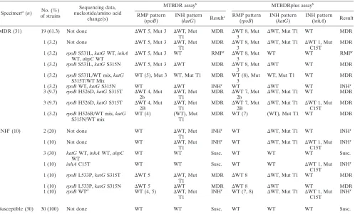
TABLE 1. GenoType MTBDRplus test results in comparison with GenoType MTBDR test results for detection of RMP and INH resistance
in 75 MDR strains
aNo. (%) of strains
Sequencing data, nucleotide/ amino acid change(s)b
MTBDR assay MTBDRplus assay
RMP pattern (rpoB)
INH pattern
(katG) Result
RMP pattern (rpoB)
INH pattern (katG)
INH pattern (inhA) Result
45 (60.0) rpoBS531L,katG315T1c ⌬WT 5, Mut 3 ⌬WT, Mut T1 MDR ⌬WT 8, Mut 3 ⌬WT, Mut T1 WT MDR 2 (2.7) rpoBS531L,katGS315T1,
inhAT8C ⌬
WT 5, Mut 3 ⌬WT, Mut T1 MDR ⌬WT 8, Mut 3 ⌬WT, Mut T1 ⌬WT1, Mut T8C
MDR
1 (1.3) rpoBS531L,katGS315T1,
inhAC15T
⌬WT 5, Mut 3 ⌬WT, Mut T1 MDR ⌬WT 8, Mut 3 ⌬WT, Mut T1 ⌬WT1, Mut C15T
MDR
2 (2.7) rpoBS531L,inhAC15T ⌬WT 5, Mut 3 WT RMPr ⌬
WT 8, Mut 3 WT ⌬WT1, Mut C15T
MDR
2 (2.7) rpoBS531L ⌬WT 5, Mut 3 WT RMPr ⌬WT 8, Mut 3 WT WT RMPr
2 (2.7) rpoBS531L,katGS315N ⌬WT 5, Mut 3 ⌬WT MDR ⌬WT 8, Mut 3 ⌬WT WT MDR 1 (1.3) rpoBS531W,katG315T1 ⌬WT 5 ⌬WT, Mut T1 MDR ⌬WT 8 ⌬WT, Mut T1 WT MDR 1 (1.3) rpoBS531W,inhAC15T ⌬WT 5 WT RMPr ⌬
WT 8 WT ⌬WT1, Mut C15T
MDR
1 (1.3) rpoBS531W ⌬WT 5 WT RMPr ⌬WT 8 WT WT RMPr
1 (1.3) rpoBS531P,katGS315T1 ⌬WT 5 ⌬WT, Mut T1 MDR ⌬WT 8 ⌬WT, Mut T1 WT MDR 1 (1.3) rpoBS531L,katGS315T1, T2 ⌬WT 5, Mut 3 ⌬WT, Mut T1,
T2
MDR ⌬WT 8, Mut 3 ⌬WT, Mut T1, Mut T2
WT MDR
1 (1.3) rpoBH526D,katG315T1 ⌬WT 4, Mut 2B ⌬WT, Mut T1 MDR ⌬WT 7, Mut 2B ⌬WT, Mut T1 WT MDR 2 (2.7) rpoBH526L,katG315T1 ⌬WT 4 ⌬WT, Mut T1 MDR ⌬WT 7 ⌬WT, Mut T1 WT MDR 2 (2.7) rpoBH526N,katG315T1 ⌬WT 4 ⌬WT, Mut T1 MDR ⌬WT 7 ⌬WT, Mut T1 WT MDR 1 (1.3) rpoBH526N,katGS315T1,
inhAC15T
⌬WT 4 ⌬WT, Mut T1 MDR ⌬WT 7 ⌬WT, Mut T1 ⌬WT1, Mut C15T
MDR
2 (2.7) rpoBH526R,katGS315T1,
inhAC15T
⌬WT 4 ⌬WT, Mut T1 MDR ⌬WT 7 ⌬WT, Mut T1 ⌬WT1, Mut C15T
MDR
1 (1.3) rpoBH526R,ahpCC52T ⌬WT 4 WT RMPr ⌬WT 7 WT WT RMPr
1 (1.3) rpoBD516V,katG315T1 ⌬WT 2, Mut 1 ⌬WT, Mut T1 MDR ⌬WT 3, 4, Mut 1 ⌬WT, Mut T1 WT MDR 1 (1.3) rpoBD516V,katGS315T1,
inhAC15T
⌬WT 2, Mut 1 ⌬WT, Mut T1 MDR ⌬WT 3, 4, Mut 1 ⌬WT, Mut T1 ⌬WT1, Mut C15T
MDR
1 (1.3) rpoBN518I,katGS315T1 ⌬WT 2, Mut 1 ⌬WT, Mut T1 MDR ⌬WT 3, 4, Mut 1 ⌬WT, Mut T1 WT MDR 1 (1.3) rpoBdel514-516,katG
S315T1 ⌬
WT 1, 2 ⌬WT, Mut T1 MDR ⌬WT 2–4 ⌬WT, Mut T1 WT MDR
1 (1.3) rpoBS522Q,katG315T1 ⌬WT 3 ⌬WT, Mut T1 MDR ⌬WT 5, 6 ⌬WT, Mut T1 WT MDR 1 (1.3) rpoBQ513P ⌬WT 1 WT RMPr ⌬
WT 2, 3 WT WT RMPr
1 (1.3) rpoBV176F,katGK152T,
ahpCG48A
WT WT Susc. WT WT WT Susc.
aResistance was determined by conventional DST.r, resistant; Susc., susceptible;⌬deletion; WT, wild type; Mut, mutation.
bAccording to previously published data (28); the GenBank accession numbers are L27989 for therpoBgene, X68081 for thekatGgene, U66801 for theinhAgene, and U16243 for theahpC-oxyRintergenic region.
cS315T1, AGC3ACC/Ser3Thr exchange; S315T2, AGC3ACA/Ser3Thr.
on May 16, 2020 by guest
http://jcm.asm.org/
5l of a preparation of chromosomal DNA in a final volume of 50l were used. The amplification protocol consisted of 15 min of denaturation at 95°C, followed by 10 cycles comprising 30 s at 95°C and 120 s at 58°C; an additional 20 cycles comprising 25 s at 95°C, 40 s at 53°C, and 40 s at 70°C; and a final extension at 70°C for 8 min. Hybridization and detection were performed in an automated washing and shaking device (Profiblot; Tekan, Maennedorf, Switzerland). The hybridization procedure was performed at 45°C for 0.5 h, followed by washing steps and the colorimetric detection of the hybridized amplicons. After a final wash, the strips were air dried and fixed on paper.
For the sputum specimens, an altered amplification protocol was applied which consisted of 15 min of denaturation at 95°C, followed by 10 cycles com-prising 30 s at 95°C and 120 s at 58°C; an additional 35 cycles comcom-prising 25 s at 95°C, 40 s at 53°C, and 40 s at 70°C; and a final extension at 70°C for 8 min. Hybridization and detection were performed as described above.
The MTBDR strip contains 17 probes, including 5 amplification and hybrid-ization controls to verify the test procedures. For the detection of RMP resis-tance, fiverpoBwild-type probes (probes WT1 to WT5) encompass the region of therpoBgene encoding amino acids 509 to 534. Four probes (probesrpoBMUT D516V,rpoBMUT H526Y,rpoBMUT H526D, andrpoBMUT S531L) specif-ically target the most common mutations. For the detection of INH resistance, one probe covers the wild-type S315 region ofkatG, while two others (probes
katGMUT T1 and MUT T2) are designed to assess the AGC-to-ACC (S315T) and the AGC-to-ACA (S315T) mutations.
The new MTBDRplus strip contains, in addition to all probes included in the MTBDR assay, three furtherrpoBwild-type probes to fill the gaps between the probes of the old MTBDR assay and to increase the detectablerpoBDNA fragment to amino acids 505 to 533. Furthermore, the promoter region of the
inhAgene is included on the new strip and encompasses the regions from positions⫺9 to⫺22 for theinhAWT1 probe and positions⫺1 to⫺12 for the
inhAWT2 probe. Four mutations (⫺15C/T,⫺16A/G,⫺8T/C, and⫺8T/A) can be targeted with theinhAMUT1, MUT2, MUT3A, and MUT3B probes. Again, either the omission of a wild-type probe or the staining of a mutant probe is an indication of a resistant strain.
RESULTS
A total of 117 drug-resistant isolates (75 from cultures
and 42 from smear-positive specimens) and 80
pansuscep-tible isolates (50 from cultures and 30 from smear-positive
specimens) characterized previously were included in the
study and tested by both assays. Overall, interpretable
re-sults were obtained for 196 isolates. The
rpoB
-specific bands
were repeatedly weak for only one smear-positive specimen
and therefore were excluded from the analysis. In general,
interpretation of the results for both test strips was easy,
but the intensities of the different hybridization bands
var-ied. The TUB control band that should be positive in all
MTBDR tests for MTBC strains was, in some cases, very
weak with smear-positive specimens in the MTBDR assay
but had good visibility in the new MTBDRplus assay.
Results of the GenoType MTBDR and MTBDRplus assays
with DNA of MDR and pansusceptible culture isolates.
A total
of 125 strains (75 MDR strains and 50 pansusceptible strains
precharacterized by conventional DST; real-time PCR; and/or
sequencing of the
rpoB
,
katG
,
inhA
, and
ahpC
genes) were
tested by both assays. Both MTBDR assays accurately detected
MTBC isolates by detection of the specific band in all (100%)
strains. The interpretation of the banding pattern was
compa-rable between the assays. Moreover, no discrepancies between
the two assays and the real-time PCR/DNA sequencing results
could be detected.
All 50 pansusceptible strains showed wild-type patterns by
both assays. For 65 (86.7%) of the 75 MDR strains, the results
of both assays were in agreement with the results of both DST
and real-time PCR/DNA sequencing (Table 1). Both assays
indicated RMP resistance in 74 strains (98.7%); only 1 strain
could not be recognized as RMP resistant by the assays since it
had a mutation outside the
rpoB
hot spot region. Both assays
correctly identified the INH resistance of 65 strains (86.7%),
comprised of 63 strains with a
katG
S315T mutation and 2
strains with an S315N mutation that was detected by the
omis-sion of the wild-type band. For one of these strains, both assays
indicated the presence of two mutations (
katG
Mut T1 and
katG
Mut T2). DNA sequencing confirmed both mutations in
this strain. Seven of the 63 strains with S315T mutations in
katG
gene had additional
inhA
mutations (5 strains with C15T
alterations and 2 strains with T8C alterations).
The MTBDR assay indicated INH susceptibility in three
FIG. 1. Representative patterns of a pansusceptible strain (rows 1)
and an MDR strain (rows 2) which had an
inhA
C15T mutation in the
regulatory region of
inhA
obtained by the new MTBDRplus assay
(B) but not the MTBDR assay (A). The positions of the
oligonucleo-tides and the marker lines are given. The targeted genes and specificity
are shown from left to right, as follows: for the MTBDR assay (A),
conjugate control (CC); amplification control (UC);
M. tuberculosis
complex-specific control (Tub);
rpoB
amplification control;
rpoB
wild-type probes WT1 to WT5 located in the hot spot region WT 1 to WT
5, respectively;
rpoB
mutant probes (probes MUT1, MUT2A, MUT2B,
and MUT3) with mutations in codons
rpoB
D516V, H526Y, H526D,
and S531L, respectively;
katG
amplification control;
katG
codon 315
wild-type probe (WT); and
katG
codon 315 mutation probes (T1 and
T2, respectively) with AGC-to-ACC (S315T1) and AGC-to-ACA
(S315T2) exchanges, respectively. The MTBDR plus assay (A) has
three additional
rpoB
wild-type probes (resulting in probes WT1 to
WT8) and targets the regulatory region of the
inhA
gene with the
inhA
amplification control (WT 1 to WT 8, respectively);
inhA
gene
wild-type probes WT1, which spans the region from positions
⫺
9 to
⫺
22,
and WT, which spans the region from positions
⫺
1 to
⫺
12; and
inhA
mutant probes MUT1, MUT2, MUT3A, and MUT3B with mutations
⫺
15C/T,
⫺
16A/G,
⫺
8T/C, and
⫺
8T/A, respectively (MUT 1, MUT
2A, MUT 2B, and MUT 3, respectively). Pansusceptible isolate 1 was
positive with the wild-type
rpoB
and
katG
probes of the MTBDR assay
and, additionally, with the
inhA
wild-type probes of the MTBDRplus
assay. MDR isolate 2 showed RMP resistance in both assays (for the
MTBDR assay, the omission of
rpoB
WT 5 and positivity for
rpoB
Mut
3; for the MTBDRplus assay, the omission of
rpoB
WT 8 and positivity
for
rpoB
Mut 3). Concerning INH resistance, isolate 2 had to be
interpreted as INH susceptible (
katG
wild-type probe) by the MTBDR
assay, whereas the MTBDRplus assay indicated INH resistance
(omis-sion of the
inhA
WT1 probe and positivity for the
inhA
MUT1 probe).
Lanes M, colored markers.
on May 16, 2020 by guest
http://jcm.asm.org/
strains (4.0%), whereas the MTBDRplus assays showed INH
resistance due to a C15T alteration in
inhA
(Fig. 1). Both
MTBDR assays failed to detect INH resistance in seven strains
(9.3%) (two of these had
ahpC
mutations).
Provided that conventional DST is the “gold standard,” the
sensitivities of the MTBDR assay were 98.7% for RMP
resis-tance detection and 88.0% for INH resisresis-tance detection. While
the sensitivity of the MTBDRplus assay was identical to that of
the MTBDR assay for the detection of RMP resistance, it was
slightly higher than that of the MTBDR assay for the detection
of INH resistance (92.0%). Both assays had specificities of
100% for the detection of INH resistance.
Results of GenoType MTBDR and MTBDRplus assays with
smear-positive sputum specimens with strains resistant to
RMP and INH or INH but not RMP and with pansusceptible
strains.
A total of 72 smear-positive specimens were chosen for
analysis, including 32 MDR strains assessed with the MGIT
960 system. Ten strains were resistant to INH but not RMP,
and 30 strains were pansusceptible (Table 2). An interpretable
result could be achieved for 71 of the 72 specimens. The
sputum positivity of the samples ranged from 1
⫹
to 4
⫹
,
ac-cording to the German guidelines (5), but no correlation
be-tween the intensities of the banding patterns and the amount
of acid-fast bacteria could be observed. Thus, each of the 71
specimens contained sufficient extractable DNA without any
inhibitors to yield this high rate of recovery efficiency.
For 29 (93.5%) of the 31 MDR strains, both MTBDR assay
results were in agreement with the results of conventional DST
and DNA sequencing (Table 2). One (3.2%) MDR strain had
the RMP wild-type banding pattern by both assays. However,
DNA sequencing was also unable to find any mutations in the
rpoB
hot spot region and an
rpoB
region further upstream. For
another MDR strain (3.2%), the MTBDR assays failed to
indicate the INH resistance of the strain. With additional DNA
sequencing of the regulatory region of
ahpC
and parts of the
katG
and
inhA
open reading frame regions, no mutation
indi-cating INH resistance was detected. In two cases the MTBDR
assays showed a mixture of banding patterns indicating both
resistance and susceptibility to RMP and INH (weak
rpoB
wild-type result with
rpoB
-specific probe MUT2 and three
mu-tation-specific bands for one strain and a weak
katG
wild-type
result with strong
katG
mutation-specific bands with probe
MUT T1 for the other; Table 2). DNA sequencing confirmed
the prevalence of both the wild-type and the mutated
se-quences.
For the group of INH
rRMP
sstrains, the rate of detection of
INH resistance was lower compared to that for the MDR
strains. For 4 of the 10 specimens, the old MTBDR test
indi-TABLE 2. GenoType MTBDRplus test results in comparison with GenoType MTBDR test results for detection of MDR, INH-resistant, and
fully susceptible strains in smear-positive sputum specimens
Specimena(n) No. (%) of strains
Sequencing data, nucleotide/amino acid
change(s)
MTBDR assayb
MTBDRplus assayb
RMP pattern (rpoB)
INH pattern (katG) Result
c RMP pattern (rpoB)
INH pattern (katG)
INH pattern (inhA) Result
MDR (31) 19 (61.3) Not done ⌬WT 5, Mut 3 ⌬WT, Mut T1
MDR ⌬WT 8, Mut
3 ⌬
WT, Mut T1 WT MDR
1 (3.2) Not done ⌬WT 5, Mut 3 ⌬WT, Mut T1
MDR ⌬WT 8, Mut
3 ⌬
WT, Mut T1 ⌬WT 1, Mut C15T
MDR
1 (3.2) rpoBS531L,katGWT,inhA
WT,ahpCWT
⌬WT 5, Mut 3 WT RMPr ⌬
WT 8, Mut 3
WT WT RMPr
1 (3.2) rpoBS531L,katGS315N ⌬WT 5, Mut 3 ⌬WT MDR ⌬WT 8, Mut 3
⌬WT WT MDR
1 (3.2) rpoBS531L/WT mix,katG
S315T/WT Mix
WT (5), Mut 3 WT, Mut T1 MDR WT (8), Mut 3
WT, Mut T1 WT MDR
1 (3.2) rpoBWT,katGS315N WT ⌬WT INHr WT ⌬WT WT INHr
3 (9.7) rpoBH526D,katGS315T ⌬WT 4, Mut 2b
⌬WT, Mut T1
MDR ⌬WT 7, Mut 2b
⌬WT, Mut T1 WT MDR
3 (9.7) rpoBH526D,katGS315T ⌬WT 4, Mut 2B
⌬WT, Mut T1
MDR ⌬WT 7, Mut 2B
⌬WT, Mut T1 ⌬WT 1, Mut C15T
MDR
1 (3.2) rpoBH526R/WT mix,katG
S315N/WT mix
WT (4) (WT), Mut T1
MDR WT (7) (WT), Mut T1 WT MDR
INHr
(10) 2 (20) Not done WT ⌬WT, Mut T1
INHr
WT ⌬WT, Mut T1 WT INHr
1 (10) Not done WT ⌬WT, Mut T1
INHr WT ⌬WT, Mut T1 ⌬WT 1, Mut
C15T
INHr
3 (30) katGWT,inhAWT,ahpC
WT
WT WT Susc. WT WT WT Susc.
1 (10) inhAC15T WT WT Susc. WT WT ⌬WT 1, Mut C15T
INHr
1 (10) rpoBL533P,katGS315T ⌬WT 5 ⌬WT, Mut T1
MDR ⌬WT 8 ⌬WT, Mut T1 WT MDR
1 (10) rpoBL533P,katGS315N ⌬WT 5 ⌬WT MDR ⌬WT 8 ⌬WT WT MDR 1 (10) rpoBWTd WT (4, 5) ⌬WT, Mut
T1
INHr WT (7, 8) ⌬WT, Mut T1 ⌬WT 1, Mut
C15T
INHr
Susceptible (30) 30 (100) Not done WT WT Susc. WT WT WT Susc.
a
Resistance was determined by conventional DST. b
WT, wild-type pattern with all respective bands visible;⌬, missing bands; weak or very weak bands are listed in parentheses. Mut, mutation. cr
, resistant; Susc., susceptible. d
This isolate contained a secondrpoBsequence derived from a mycobacterial strain not further characterized.
on May 16, 2020 by guest
http://jcm.asm.org/
[image:4.585.45.543.89.390.2]cated INH susceptibility (no detectable mutation at
katG
codon 315). However, the new MTBDRplus assay detected
INH resistance due to a C15T exchange in
inhA
in one of these
four strains. Furthermore, in two of the INH
rRMP
sstrains,
mutations were indicated by the omission of MTBDR and
MTBDRplus
rpoB
bands 5 and 8, respectively. DNA
sequenc-ing confirmed this alteration as an CTG-to-CCG exchange in
rpoB
codon 533.
Sensitivities and specificities of the GenoType MTBDR and
MTBDRplus assays.
Overall, both assays had identical
sensi-tivities of 98.1% for the detection of RMP resistance. The
sensitivities of the assays for the detection of INH resistance
differed: 87.8% for the MTBDR assay and 90.2% for the
MTBDRplus assay. The specificities of both assays were 97.8%
for the detection of RMP resistance and 100% for the
detec-tion of INH resistance.
DISCUSSION
The results of the present study have shown that the MTB
DRplus assay is easy to perform and has the capability for the
rapid detection of RMP- and INH-resistant
M. tuberculosis
. As
previously shown for other DNA strip assays (1, 11, 16, 17, 25,
29), the MTBDRplus assay has been proven to be suitable for
application both with culture isolates and directly with
smear-positive specimens.
With respect to culture isolates, the sensitivities of the
MTBDR assay for the detection of RMP resistance were
re-cently reported to be in the range of 95% to 99% (4, 10, 17).
This is in concordance with the high sensitivity of the MTB
DRplus assay measured in our study (98.7%). Both the rarity
of RMP-resistance-associated mutations in codons other than
the
rpoB
81-bp hot spot region and the rarity of silent
muta-tions in the hot spot region are responsible for the high rate of
detection of RMP resistance by investigation of this region (9,
10, 12; this study). In this study, the S531L mutation in
rpoB
was the most frequent (78 of 106 strains [73.6%]), followed by
mutations in codon 526 (16 of 106 strains [15.1%]). This is
comparable to the frequencies reported in other studies (17),
although the distribution can also differ in some settings (2).
Although the prevalence of false-resistant and
false-suscepti-ble results seems to be infrequent, some authors have
dis-cussed the relevance of mutations in codon 533 to RMP
resis-tance (15).
M. tuberculosis
isolates with mutations in codon 533
showed RMP susceptibility (15, 21) or low-level or high-level
resistance (27, 30). Our data support the suspicion that
muta-tions in codon 533 are not associated with RMP resistance and
that RMP resistance in isolates with codon 533 mutations may
be due to other mutations. The detection of a codon 533
mutation by the MTBDR and MTBDRplus assays is due to the
omission of the respective wild-type
rpoB
band (WT 5 and WT
8, respectively). The recommendation in these cases is either to
control the result by DNA sequencing or confirm the result by
the conventional DST.
Some authors discussed a main limitation of the MTBDR
test system, which is the low sensitivity for the detection of
INH resistance (16, 17). This was due to the fact that the test
targets only the
katG
S315T mutation. By using the MTBDR
plus assay, this problem is now at least partly solved by the
addition of a second target for the detection of INH resistance,
the regulatory region of the
inhA
gene. In our study we could
show that this addition led to an improvement in the rate of
detection of INH resistance of 3%, although it can be expected
that in other settings this advantage may be higher. Concerning
the distribution of mutations associated with INH resistance, a
disequilibrium between mono-INH
risolates compared to
those with resistance to more drugs was identified (8, 10). INH
resistance-associated mutations and, in particular,
katG
315
mutations were less prevalent in mono-INH
risolates than in
the other group of isolates. Although the number of samples
used in the present study is too small for a statistical analysis,
this trend can be approved.
As already reported for the MTBDR assay, the new MTB
DRplus assay can be applied directly to smear-positive
speci-mens. With a turnaround time of approximately 6 h, these
techniques save several weeks of time, which is required for
primary isolation and conventional DST. However, these
mo-lecular tests should not be applied alone and therefore cannot
totally replace culture methods for several reasons: (i) apart
from RMP and INH susceptibility testing, culture is needed for
all other drug susceptibility tests; (ii) RMP and INH
suscepti-bility must be confirmed, since the possisuscepti-bility that a strain is
resistant cannot be excluded for a strain with a wild-type
pat-tern by the MTBDR assay; and (iii) in the case of a mixed
infection with an MTBC strain and a nontuberculous
myco-bacterium, interpretation of the MTBDR assay results could
be difficult (data not shown). Furthermore, it should not be
used as a nucleic acid amplification technique for the direct
detection of MTBC strains in primary specimens.
Neverthe-less, the new MTBDRplus assay is a major improvement
among assays for the routine detection of RMP- and
INH-resistant MTBC strains, since with this rapid and reliable tool,
the therapeutic management of patients can be optimized. This
is of special importance for MDR strains and highly infectious
patients, since the prevention of transmission of resistant
strains is one of the challenges of the present and future.
ACKNOWLEDGMENTS
We thank K. Ott, B. Schlu
¨ter, and A. Witt, Forschungszentrum
Borstel, for their excellent technical assistance; S. Kretschmer,
Fors-chungszentrum Borstel, for the provision of DNA samples; and Hain
Lifescience GmbH for providing the GenoType MTBDR and MTB
DRplus assay kits.
REFERENCES
1.Bang, D., A. B. Andersen, and V. O. Thomsen.2006. Rapid genotypic
de-tection of rifampin and isoniazid-resistantMycobacterium tuberculosis di-rectly in clinical specimens. J. Clin. Microbiol.44:2605–2608.
2.Ba´rtfai, Z., A´. Somosko¨vi, C. Ko¨dmo¨n, N. Szabo´, E. Puska´s, L. Kosztola´nyi,
E. Farago´, J. Mester, L. M. Parsons, and M. Salfinger.2001. Molecular
characterization of rifampin-resistant isolates ofMycobacterium tuberculosis
from Hungary by DNA sequencing and the line probe assay. J. Clin. Micro-biol.39:3736–3739.
3.Canetti, G., W. Fox, A. Khomenko, H. T. Mahler, M. K. Menon, D. A.
Mitchison, N. Rist, and N. A. Smelov.1969. Advances in techniques of
testing mycobacterial drug sensitivity, and the use of sensitivity tests in tuberculosis control programmes. Bull. W. H. O.41:21–43.
4.Cavusoglu, C., A. Turhan, P. Akinci, and I. Soyler.2006. Evaluation of the GenoType MTBDR assay for rapid detection of rifampin and isoniazid resistance inMycobacterium tuberculosisisolates. J. Clin. Microbiol.44:2338– 2342.
5.Deutsches Institut fu¨r Normung.1986. Medical microbiology. Diagnosis of tuberculosis. Parts 3 and 32. Beuth Verlag, Berlin, Germany.
6.Espinal, M. A.2003. The global situation of MDR-TB. Tuberculosis
(Edin-burgh)83:44–51.
7.Goble, M., M. D. Iseman, L. A. Madsen, D. Waite, L. Ackerson, and C. R.
on May 16, 2020 by guest
http://jcm.asm.org/
Horsburgh, Jr.1993. Treatment of 171 patients with pulmonary tuberculosis resistant to isoniazid and rifampin. N. Engl. J. Med.328:527–532.
8.Hazbon, M. H., M. Brimacombe, M. Bobadilla del Valle, M. Cavatore, M. I.
Guerrero, M. Varma-Basil, H. Billman-Jacobe, C. Lavender, J. Fyfe, L. Garcia-Garcia, C. I. Leon, M. Bose, F. Chaves, M. Murray, K. D. Eisenach,
J. Sifuentes-Osornio, M. D. Cave, A. Ponce de Leon, and D. Alland.2006.
Population genetics study of isoniazid resistance mutations and evolution of multidrug-resistantMycobacterium tuberculosis. Antimicrob. Agents Che-mother.50:2640–2649.
9.Heep, M., B. Brandtsta¨tter, U. Rieger, N. Lehn, E. Richter, S. Ru¨sch-Gerdes,
and S. Niemann.2001. Frequency ofrpoBmutations inside and outside the
cluster I region in rifampin-resistant clinicalMycobacterium tuberculosis iso-lates. J. Clin. Microbiol.39:107–110.
10.Hillemann, D., M. Weizenegger, T. Kubica, E. Richter, and S. Niemann.
2005. Use of the GenoType MTBDR assay for rapid detection of rifampin and isoniazid resistance in Mycobacterium tuberculosis complex isolates. J. Clin. Microbiol.43:3699–3703.
11.Hillemann, D., S. Ru¨sch-Gerdes, and E. Richter.2006. Application of the GenoType MTBDR assay directly on sputum specimens. Int. J. Tuberc. Lung Dis.10:1057–1059.
12.Hillemann, D., T. Kubica, S. Ru¨sch-Gerdes, and S. Niemann.2005. Disequi-librium in distribution of resistance mutations among Beijing and non-Bei-jingMycobacterium tuberculosisstrains isolated from patients resident in Germany. Antimicrob Agents Chemother.49:1229–1231.
13.Kelley, C. L., D. A. Rouse, and S. L. Morris.1997. Analysis ofahpCgene mutations in isoniazid-resistant clinical isolates ofMycobacterium tuberculo-sis. Antimicrob. Agents Chemother.41:2057–2058.
14.Kent, P. T., and G. P. Kubica.1985. Public health mycobacteriology. A guide for a level III laboratory. Centers for Disease Control, Atlanta, GA.
15.Ma, X., H. Wang, Y. Deng, Z. Liu, Y. Xu, X. Pan, J. M. Musser, and E. A.
Graviss.2006.rpoBgene mutations and molecular characterization of
ri-fampin-resistantMycobacterium tuberculosisisolates from Shandong Prov-ince, China. J. Clin. Microbiol.44:3409–3412.
16.Ma¨kinen, J., H. J. Marttila, M. Marjamaki, M. K. Viljanen, and H. Soini.
2006. Comparison of two commercially available DNA line probe assays for detection of multidrug-resistantMycobacterium tuberculosis. J. Clin. Micro-biol.44:350–352.
17.Miotto, P., F. Piana, V. Penati, F. Canducci, G. B. Migliori, and D. M.
Cirillo.2006. Use of GenoType MTBDR assay for molecular detection of
rifampin and isoniazid resistance in Mycobacterium tuberculosis clinical strains isolated in Italy. J. Clin. Microbiol.44:2485–2491.
18.Mokrousov, I., O. Narvskaya, T. Otten, E. Limeschenko, L. Steklova, and B.
Vyshnevkiy.2002. High prevalence ofkatGSer315Thr substitution among
isoniazid-resistantMycobacterium tuberculosisclinical isolates from north-western Russia, 1996 to 2001. Antimicrob. Agents Chemother.46:1417– 1424.
19.Morlock, G. P., B. Metchock, D. Sikes, J. T. Crawford, and R. C. Cooksey.
2003.ethA,inhA, andkatGloci of ethionamide-resistant clinical Mycobacte-rium tuberculosisisolates. Antimicrob. Agents Chemother.47:3799–3805.
20.Musser, J. M., V. Kapur, D. L. Williams, B. N. Kreiswirth, D. van Soolingen,
and J. D. van Embden.1996. Characterization of the catalase-peroxidase
gene (katG) andinhAlocus in isoniazid-resistant and -susceptible strains of
Mycobacterium tuberculosisby automated DNA sequencing: restricted array of mutations associated with drug resistance. J. Infect. Dis.173:196–202.
21.Ohno, H., H. Koga, S. Kohno, T. Tashiro, and K. Hara.1996. Relationship
between rifampin MICs for andrpoBmutations ofMycobacterium tubercu-losisstrains isolated in Japan. Antimicrob. Agents Chemother.40:1053– 1056.
22.Piatek, A. S., A. Telenti, M. R. Murray, H. El-Hajj, W. R. Jacobs, Jr., F. R. Kramer, and D. Alland.2000. Genotypic analysis ofMycobacterium tubercu-losisin two distinct populations using molecular beacons: implications for rapid susceptibility testing. Antimicrob. Agents Chemother.44:103–110. 23.Piersimoni, C., A. Olivieri, L. Benacchio, and C. Scarparo.2006. Current
perspectives on drug susceptibility testing of Mycobacterium tuberculosis
complex: the automated nonradiometric systems. J. Clin. Microbiol.44:
20–28.
24.Sajduda, A., A. Brzostek, M. Poplawska, E. Augustynowicz-Kopec, Z. Zwolska, S. Niemann, J. Dziadek, and D. Hillemann.2004. Molecular characterization of rifampin- and isoniazid-resistantMycobacterium tuberculosisstrains isolated in Poland. J. Clin. Microbiol.42:2425–2431.
25.Somoskovi, A., J. Dormandy, D. Mitsani, J. Rivenburg, and M. Salfinger.
2006. Use of smear-positive samples to assess the PCR-based GenoType MTBDR assay for rapid, direct detection of theMycobacterium tuberculosis
complex as well as its resistance to isoniazid and rifampin. J. Clin. Microbiol.
44:4459–4463.
26.Sreevatsan, S., X. Pan, Y. Zhang, V. Deretic, and J. M. Musser.1997.
Analysis of theoxyR-ahpCregion in isoniazid-resistant and -susceptible My-cobacterium tuberculosiscomplex organisms recovered from diseased hu-mans and animals in diverse localities. Antimicrob. Agents Chemother.41:
600–606.
27.Taniguchi, H., H. Aramaki, Y. Nikaido, Y. Mizuguchi, M. Nakamura, T.
Koga, and S. Yoshida.1996. Rifampicin resistance and mutation of therpoB
gene inMycobacterium tuberculosis. FEMS Microbiol. Lett.144:103–108. 28.Telenti, A., N. Honore´, C. Bernasconi, J. March, A. Ortega, H. E. Takiff, and
S. T. Cole.1997. Genotyping assessment of isoniazid and rifampin resistance inMycobacterium tuberculosis: a blind study at reference laboratory level. J. Clin. Microbiol.35:719–723.
29.Traore, H., A. van Deun, I. C. Shamputa, L. Rigouts, and F. Portaels.2006. Direct detection ofMycobacterium tuberculosiscomplex DNA and rifampin resistance in clinical specimens from tuberculosis patients by line probe assay. J. Clin. Microbiol.44:4384–4388.
30.Yue, J., W. Shi, J. Xie, Y. Li, E. Zeng, and H. Wang.2003. Mutations in the
rpoBgene of multidrug-resistantMycobacterium tuberculosisisolates from China. J. Clin. Microbiol.41:2209–2212.
31.Zhang, M., J. Yue, Y. P. Yang, H. M. Zhang, J. Q. Lei, R. L. Jin, X. L. Zhang,
and H. H. Wang.2005. Detection of mutations associated with isoniazid
resistance inMycobacterium tuberculosisisolates from China. J. Clin. Micro-biol.43:5477–5482.