0095-1137/11/$12.00 doi:10.1128/JCM.02538-10
Copyright © 2011, American Society for Microbiology. All Rights Reserved.
Multiple-Locus Variable-Number Tandem-Repeat Analysis Genotyping
of Human
Brucella
Isolates from Turkey
䌤
†
Selc
¸uk Kılıc
¸,
1* Ivan N. Ivanov,
2Rıza Durmaz,
1,3Mehmet Refik Bayraktar,
4Ergin Ayas¸lıog
˘lu,
5M. Hamidullah Uyanık,
6Hikmet Alıs¸kan,
7Ekrem Yas¸ar,
8Gu
¨lc
¸in Bayramog
˘lu,
9Ahmet Arslantu
¨rk,
1,10Gilles Vergnaud,
11,12and Todor V. Kantardjiev
2Refik Saydam National Public Health Agency, Ankara, Turkey1; National Centre of Infectious and Parasitic Diseases, Sofia, Bulgaria2;
Department of Microbiology and Clinical Microbiology, Inonu University, Malatya, Turkey3; Department of Microbiology and
Clinical Microbiology, Harran University, Sanliurfa, Turkey4; Department of Infectious Diseases and Clinical Microbiology,
Kirikkale University, Kirikkale, Turkey5; Department of Microbiology and Clinical Microbiology, Ataturk University, Erzurum,
Turkey6; Department of Microbiology and Clinical Microbiology, Baskent University, Adana, Turkey7;
Microbiology Laboratory, Children’s Hospital, Diyarbakir, Turkey8; Department of Microbiology and
Clinical Microbiology, Karadeniz Technical University, Trabzon, Turkey9; Microbiology Laboratory,
State Hospital, Kutahya, Turkey10; Universite´ Paris Sud 11, CNRS, UMR 8621, Institut de
Ge´ne´tique et Microbiologie, Orsay, France11; and DGA/MRIS, Mission pour la
Recherche et l’Innovation Scientifique, Bagneux, France12
Received 15 December 2010/Returned for modification 13 February 2011/Accepted 25 April 2011
A multiple-locus variable-number tandem-repeat analysis (MLVA) was applied to investigate the
epidemi-ological relationship and genetic diversity among 162 humanBrucellaisolates collected from all geographic
regions of Turkey in an 8-year period (2001 to 2008). The isolates were genotyped by using an MLVA assay
developed in Orsay, France (MLVA-16Orsay) including eight minisatellite (panel 1) and eight microsatellite
(panel 2, subdivided into 2A and 2B) markers. Panels 1 and 2A distinguish 14 genotypes; two of these represented 85% of the strains. Panel 2B displayed a very high discriminatory power. Three loci from panel 2B
had diversity index values higher than 0.74. MLVA-16Orsayyielded 105 genotypes; 73 were represented by a
unique isolate, and 32 included two to eight isolates. The isolates from different patients within the same outbreak or from the same patient before first-line therapy and after relapse showed identical genotypes. A number of MLVA genotypes appeared to be partially restricted to some geographic areas and displayed no annual variation, possibly reflecting persistence of genotypes in certain areas for a time span of at least a
decade. This study, representing the first molecular typing results of humanBrucellaisolates from Turkey,
indicated that Turkish humanBrucella melitensisisolates were most closely related to the neighboring
coun-tries’ isolates included in the East Mediterranean group.
Brucellosis is the most common anthropozoonosis, with more than 500,000 cases annually. While the disease was erad-icated in the vast majority of industrialized regions around the world, it remains a significant public health concern, mainly in the Mediterranean littoral, the Middle East, the Arabian Pen-insula, the Indian subcontinent, Asia, Africa, and Central and South America (19, 26).
Turkey is a relatively large country in the eastern Mediter-ranean region, with a geographical surface of 783,562 km2, and
comprises seven regions. It has a population of 72 million, 70% of which lives in cities and 30% in rural areas. Brucellosis is endemic, and approximately 10,000 human brucellosis cases are reported annually. The reported incidence is 150 cases per 1 million inhabitants (24). Its prevalence varies widely from region to region due to several factors, including food habits,
milk processing methods, husbandry practices, nomadism, so-cial customs, climatic conditions, socioeconomic status, and environmental conditions. A steady increase of reported hu-man cases was observed from 1986 (3.03/100,000 population) until 2004 (25.65/100,000). Livestock vaccination, elimination of infected animals, control of animal movements, and educa-tion induced a decline in the number of annually reported human cases, from 18,563 cases in 2004 to 9,818 cases in 2008 (17).
Rapid and accurate typing procedures are crucial for epide-miologic surveillance, investigation of outbreaks, and fol-low-up of a control program. Many molecular typing methods commonly used for the subtyping of isolates of other bacterial species are not appropriate for routine typing of Brucella
strains, and none has proven to be fully satisfactory for epide-miological trace-back investigations of brucellosis (1, 9, 25). Recently, a selection of 16 variable-number tandem repeats has been proposed for fingerprintingBrucellaisolates (7, 14, 25). This multiple-locus variable-number tandem-repeat anal-ysis (MLVA) genotyping system, MLVA-16Orsay, comprised eight minisatellite markers (panel 1, Bruce06, Bruce08, Bruce11, Bruce12, Bruce42, Bruce43, Bruce45, and Bruce55) for species identification and eight complementary microsatel-lite markers (panel 2A, Bruce18, Bruce19, and Bruce21; panel
* Corresponding author. Mailing address: Refik Saydam National Public Health Agency, Department of Communicable Diseases Re-search, Bacterial Zoonoses Research and Reference Laboratory (Na-tional Brucellosis Reference Laboratory), Cemal Gursel Street 18, 06100 Sihhiye Ankara, Turkey. Phone: 00 90 312 458 21 69. Fax: 00 90 312 458 24 04. E-mail: [email protected].
† Supplemental material for this article may be found at http://jcm .asm.org/.
䌤Published ahead of print on 27 July 2011.
3276
on May 16, 2020 by guest
http://jcm.asm.org/
2B, Bruce04, Bruce07, Bruce09, Bruce16, and Bruce30) for further subspecies differentiation. The MLVA-16Orsay assay
has been shown to be an appropriate method for species iden-tification in theBrucellagenus, for discriminating isolates orig-inating from restricted geographic sources at the subspecies level, and for trace-back analyses (1, 14). This method has been reported to be highly discriminatory to distinguish strains within a local outbreak, and to some extent phylogenetically relevant (1, 11, 14, 16, 18, 23) and typing data from several hundred isolates can be queried and accessed via the Internet (http://mlva.u-psud.fr). The genetic diversity ofBrucellastrains isolated from human and animal infection has not yet been investigated in Turkey. In the present study, the MLVA-16Orsay assay was applied to investigate epidemiological
rela-tionships among human brucellosis isolates collected from all regions of Turkey and to determine the most common geno-types amongBrucellastrains in Turkey.
(This study was presented in part at the 3rd Eurasia Con-gress of Infectious Diseases [formerly ICCAID], Baku, Azer-beijan, 2009.)
MATERIALS AND METHODS
Brucellastrains.A total of 162 presumptiveBrucellaisolates from 159 patients (isolates BRU-S001 to BRU-S162) submitted to the Refik Saydam National Public Health Agency for a precise identification at the species and biovar levels were enrolled in this study. Two patients experienced relapse episodes, and two isolates were obtained from each of these patients. A total of 160 of these isolates were collected over an 8-year period (from 2001 to 2008) at various tertiary care centers in Turkey. One isolate of cerebrospinal fluid in 1998 (BRU-S131) and one from the blood culture of a preterm baby with congenital brucellosis in 2009
(BRU-S130) were also included in the study. The number ofBrucellaisolates
analyzed from each region (64 isolates from eastern Anatolia, 26 from south-eastern Anatolia, 24 from central Anatolia, 17 from the Mediterranean, 14 from the Black Sea, 11 from the Aegean, 6 from Marmara) was roughly in proportion
to brucellosis incidence.Brucellastrains isolated in early years were recovered
from freeze-dried stocks, whereas more recently isolated strains were stored at
⫺80°C in 10% skim milk.
The isolates were identified at the genus level by conventional microbiological
methods and biotyped as previously described based on requirement of CO2for
growth, urease activity, H2S production, sensitivity to the fuchsin and thionin
dyes (20 and 40g/ml), lysis by Tbilisi phage, and agglutination with
monospe-cific antiserum for A and M antigens (2).
MLVA-16Orsaygenotyping. (i) DNA sample preparation.BrucellaDNA
sam-ples were prepared by a simple thermolysate procedure. A loop of bacterial
colony was suspended into 200l TE buffer (10 mM Tris [pH 8.0], 1 mM
EDTA). Turbidity was adjusted to a McFarland standard of approximately 0.5. Bacterial suspensions were heated at 100°C for 10 min and then centrifuged at
13,000⫻gfor 10 min to obtain a clear nucleic acid-containing supernatant. Two
microliters of the supernatant was used as the template in the PCR assays.
(ii) PCR amplification.The PCR was performed as previously described (1, 14) with a slight modification. In brief, panel 1 primers were combined and run into four duplex PCR (dPCR). The primer multiplexing was arranged in a manner that avoids overlapping of the resulting PCR fragments according to published allele size ranges (14) and ensures unambiguous interpretation. Primer
concentrations were adjusted as follows: dPCR1, 0.2M Bruce06, 0.45M
Bruce12; dPCR4, 0.35M Bruce45, 0.4M Bruce55; dPCR2, 0.4M Bruce08,
0.4M Bruce42; and dPCR3, 0.4M Bruce11, 0.4M Bruce43. PCR
amplifi-cation was performed in a total volume of 25l containing 1⫻Gold buffer, 0.25
mM deoxynucleoside triphosphate (dNTP) mix, 2 mM MgCl2, 0.16 mg/ml bovine
serum albumin, 2.5% dimethyl sulfoxide, 1 U AmpliTaq Gold (Applied
Biosys-tems), appropriate concentrations of each flanking primer, and 2l of
thermo-lysate solution. The amplification was run in a QB-96 cycler (Quanta Biotech Ltd., United Kingdom). The initial denaturation step (96°C for 5 min) was followed by 30 cycles of denaturation at 96°C for 30 s, primer annealing at 60°C for 30 s, and extension at 70°C for 30 s, with a final extension step at 70°C for 5 min. Electrophoretic separation was performed by applying the M500 method of the QIAxcel capillary electrophoresis system coupled with a high-resolution
cartridge (Qiagen, Germany). ThreeB. melitensisreference strains (biovar
[bv] 1, 16 M, ATCC 23456; bv. 2, 63/9, ATCC 23457; bv. 3, ether, ATCC
23458) andBrucella abortusbv. 1 reference strain (544; ATCC 23448) as well
as Brucella melitensis Rev-1 vaccine strain (BRU-S163) were included as control strains.
Data analysis.Analysis of electrophoresis patterns was carried out with the BioCalculator software version 3.0.05 (Qiagen, Germany), and fragment sizes converted to repeat unit numbers were imported into BioNumerics (Applied Maths, Belgium) as a character data set. The cluster analysis was performed using the UPGMA (unweighted pair group method with arithmetic mean) algo-rithm and the categorical (or Hamming’s) distance. Genetic diversity (Hunter-Gaston diversity index [HGDI]) and confidence intervals were calcu-lated using online tools at www.hpa-bioinformatics.org.uk/cgi-bin/DICI/DICI.pl. Values of the HGDI can range from 0 (no polymorphism) to 1 (all samples are
different). The MLVA-16Orsaygenotypes ofB. melitensisisolates were compared
to the corresponding data obtained for the reference strains and field isolates investigated previously (15). Chi-square analysis was used to correlate patient characteristics with genotype.
RESULTS
Patient characteristics.The mean age of the 159 patients
was 34.2 years (range, 0 to 85 years), and the ratio of males (n⫽86) to females (n⫽73) was 1.17. Female patients (mean age, 38.6 years; range, 2 to 79 years) were slightly older than male patients (mean age, 30.6 years; range, 0 to 85 years). Most patients (n⫽147; 92.5%) presented with acute brucellosis (⬍2 months of illness), and five (3.1%) had subacute, four (2.5%) had chronic, and two (1.3%) had relapse brucellosis. One (0.6%) case had acute and subacute phases of a single illness episode.
Brucellaisolates.A total of 162Brucellaisolates were iden-tified asB. melitensisbv. 3 (161 isolates) andB. abortusbv. 3 (one isolate). The seasonal distribution of isolates in the present collection was in agreement with the global epide-miology of Brucellain Turkey, i.e., an increased incidence from late spring (May) to midsummer (peak in June or July), with a gradual decrease in autumn and winter. These characteristics confirm the representativity of the present strain collection.
MLVA-16Orsaygenotyping results.PCR amplification
prod-ucts were obtained for all 162 isolates. Panels 1 and 2A showed limited diversity, whereas panel 2B displayed a very high discriminatory power. The HGDI in panel 1 was highest (0.329) at Bruce42. Loci Bruce06, Bruce11, and Bruce45 showed only one allele (HGDI ⫽ 0). The HGDI values ranged from 0.0 to 0.182 in panel 2A and from 0.165 to 0.788 in panel 2B. In panel 2B, Bruce04, Bruce16, and Bruce30 had the highest variability (0.778, 0.778, and 0.740, respec-tively; see Table 1).
Clustering analysis with previously published typing data from more than 500 isolates (15) is shown in Fig. S1 in the supplemental material and confirms the species identification deduced from biotyping. Panel 1 loci gave 10 different geno-types among 161B. melitensisstrains. Three of those were new genotypes (numbers 83, 84, and 85) observed in a single strain. The seven others (numbers 42, 43, 44, 57, 61, 62, 63) were previously observed. In particular, panel 1 genotypes 42 (27 strains) and 43 (109 strains) are the most common genotypes (see Table S1 in the supplemental material).
MLVA-11Orsay (composed of the eight panel 1 and three panel 2A loci) discriminated 14 genotypes, 6 of which (num-bers 100, 102, 109, 113, 119, and 121) were not previously observed. Eighty-five percent of the isolates belong to
VOL. 49, 2011 BRUCELLA MLVA GENOTYPES FROM TURKEY 3277
on May 16, 2020 by guest
http://jcm.asm.org/
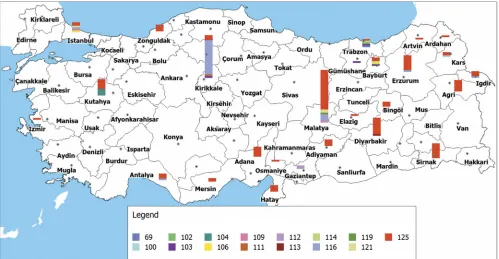
MLVA-11 genotypes 116 (27 isolates) or 125 (109 isolates). The genotype ofB. abortusstrain BRU-S093 was not identical to the previously describedB. abortusgenotypes (see Fig. S1 in the supplemental material). Distribution of MLVA-11Orsay ge-notypes showed variation in different geographical regions. MLVA-11 genotype 125 is observed all over the country, whereas genotype 116 was isolated mainly in the central Ana-tolia region. The isolates with the genotypes 103 and 104 were primarily observed in the Black Sea region, and the genotype 104 was essentially found in patients from the Aegean region (Fig. 1).
The distribution of the main genotypes was not associated with a specific period of time. Genotypes 116 and 125 were isolated during, respectively, six and eight (i.e., throughout the study period) years. The relative frequencies of the two most frequent genotypes were essentially identical in male and fe-male patients (54.1% versus 45.9%; chi-square⫽ 0.002; and
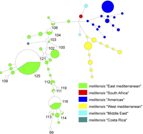
P⫽0.96). There was also no significant difference between the spectrum of genotypes isolated from children and those iso-lated from adults. The isolates in the largest MLVA-11Orsay genotype group (genotype 125) were isolated in all age groups. However, the isolates in the second largest genotype group (genotype 116) were not isolated from adults between the ages of 20 and 30 (weak significance; chi-square⫽4.14;P⫽0.24). In the minimum spanning tree clustering using previously pub-lishedB. melitensistyping data, all strains analyzed clustered within the Eastern Mediterranean group (Fig. 2).
Cluster analysis for Turkish B. melitensis genotypes.
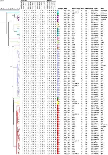
[image:3.585.41.284.90.365.2]MLVA-16Orsayyielded a total of 105 genotypes, 73 of which were represented by a unique strain. The remaining 32 geno-types included the 88 clustered strains (clustering rate was
TABLE 1. Numbers of alleles and HGDI values of 161
B. melitensisisolates from Turkey
Locus No. of
alleles
Tandem repeat copy
no. HGDI
a Confidence
interval
Panel 1
Bruce06 1 1 0.000 0.000–0.036
Bruce08 3 3, 4, 5 0.049 0.018–0.096
Bruce11 1 3 0.000 0.000–0.000
Bruce12 3 12, 13, 14 0.084 0.026–0.142
Bruce42 3 1, 2, 3 0.329 0.2522–0.406
Bruce43 3 2, 3, 4 0.049 0.0019–0.096
Bruce45 1 3 0.000 0.000–0.044
Bruce55 2 1, 2 0.012 0.000–0.037
Total 10 0.437
Panel 2A
Bruce18 3 4, 5, 6 0.161 0.0768–0.222
Bruce19 4 18, 20, 21, 23 0.182 0.115–0.270
Bruce21 1 8 0.000 0.000–0.000
Total (panels 1 and 2A)
14 0.522
Panel 2B
Bruce04 7 3, 4, 5, 6, 7, 8, 9 0.778 0.7513–0.806 Bruce07 6 3, 4, 5, 6, 7, 12 0.465 0.369–0.544 Bruce09 8 3, 4, 5, 6, 7, 8, 9, 14 0.165 0.097–0.255 Bruce16 8 3, 4, 5, 6, 7, 8, 9, 11 0.778 0.743–0.814 Bruce30 6 3, 4, 5, 6, 7, 8 0.740 0.702–0.777
MLVA-16Orsay 105 0.991
a
HGDI, Hunter-Gaston diversity index.
FIG. 1. Geographic distribution of panel 1 and 2A genotypes (genotype 69,B. abortusbv. 3). The bar size is proportional to the number of isolates. Each color corresponds to a different MLVA-11Orsaygenotype. (Adapted from a map available at www.basarsoft.com.tr with permission
of Basarsoft Ltd.)
on May 16, 2020 by guest
http://jcm.asm.org/
[image:3.585.43.542.434.693.2]54.6%) (Fig. 3). The most frequently observed genotype com-prised eight isolates obtained from five separate provinces in three geographic regions over 7 years (from 2002 to 2009). The seven strains in the second most frequent genotype were iso-lated from five separate provinces in two geographic regions between 2004 and 2008.
Relevance of MLVA-16Orsaygenotyping for patient
manage-ment and source identification.The MLVA-16Orsaygenotypes
of the isolates obtained from two patients (genotype 24 and genotype 27) during the acute phase and after relapse were identical. Additionally, two isolates cultured from one patient’s blood samples during the acute and subacute phases of a single illness episode and one isolate corresponding to a laboratory-acquired infection from this patient’s isolates showed identical MLVA-16Orsay patterns (Fig. 3, genotype 35, BRU-S157, BRU-S158, BRU-S159).
The MLVA-16Orsaytyping assay also showed very high con-cordance with available epidemiological data. For example, genotypes 22 (BRU-S080 and BRU-S081), 29 (BRU-S066, BRU-S075, BRU-S078), 43 (BRU-S094 to BRU-S097), 54 (S126 and S127), and 100 (S145 and BRU-S146) were each recovered from patients of the same family that contracted brucellosis from unpasteurized dairy products. The genotype 71 patients (isolates BRU-S058 and BRU-S059) have a common history of occupational exposure to infected animals.
Application of the MLVA panel to 11 isolates from an epidemiologically linked B. melitensis outbreak observed in June 2003 in a small village in the Kirikkale province from the central Anatolia region yielded six genotypes. Genotypes 22, 23, and 25 each comprised two isolates, and genotype 29 was shared by three isolates. The outbreak genotypes typically comprised family members and patients who were presumed to have contracted brucellosis from a common point source (con-sumption of homemade cheese). Two isolates (genotype 21, BRU-S082; genotype 30, BRU-S083) obtained from family members who did not share the same MLVA-16Orsaygenotype may either represent persistent circulating strains causing spo-radic infections or be the result of mutation events in the course of the outbreak. Genotypes 21 to 25, 29, and 30 differ only by⫾1 repeat unit at one or two of the most variable loci, Bruce04, Bruce16, or Bruce30 (Fig. 3).
DISCUSSION
In the present study, a total of 162 humanBrucellaisolates collected from different parts of Turkey during an 8-year pe-riod was evaluated by bacteriological, epidemiological, and molecular typing characteristics. All isolates but one wereB. melitensis (bv. 3). Previous studies conducted in different regions of Turkey found that human brucellosis was almost exclusively caused byB. melitensis, accounting for 99% of the total cases, andB. melitensis bv. 3 was the biovar most frequently isolated in humans (4–6, 8, 12, 13, 22). The data obtained in Turkey are consistent with the results obtained in the Mediterranean region (1, 16, 21, 26). These results reveal that human brucellosis in Turkey seems to be related more to ovicaprine than to cattle infection, which may be partly attributed to the virulence of the organism. In addi-tion, brucellosis control measures, such as the financial com-pensation of owners of slaughtered seropositive cattle, may play a significant role. No such measure exists for sheep or goats.
MLVA-16Orsay yielded a total of 105 genotypes. Panel 2B
markers in MLVA-16Orsayloci displayed very high discrimina-tory power, while panels 1 and 2A showed limited diversity. MLVA genotypes did not show significant differences among gender or different age groups. The frequency of different MLVA genotypes varied among the seven geographical re-gions. There was good correlation between molecular typing results and epidemiological data, and epidemiologically related isolates were of identical or very closely related genotypes.
MLVA-11Orsay(combined panel 1 and 2A markers) yielded 14 genotypes, whereas the added panel 2B increased the num-ber of genotypes to 105. These findings showed that the geno-typic variation of Turkish isolates was mostly associated with the highly variable panel 2B loci and to a much lesser extent panel 2A (locus Bruce19) and panel 1 (loci Bruce01, -42, and -55) loci. This may reflect microevolution via a stepwise mu-tational event of the most variable loci from a very limited number of ancestors. In agreement with previous molecular studies (1, 11), in the setting of a local outbreak investigation, the highly polymorphic panel 2B might be sufficient for a rapid and low-cost result.
Although the discrimination power of MLVA-8Orsay and MLVA-11Orsayis very low for evaluation of the
cross-transmis-FIG. 2. Minimum spanning tree analysis of publishedB. meliten-sisisolates using the MLVA-11Orsay data. Color codes are
associ-ated with the mainB. melitensisMLVA clusters. The published data forB. melitensis strains were recovered from the compilation by Maquart et al. (15). The 161 TurkishB. melitensisisolates, which were representing 14 MLVA-11Orsaygenotypes, are associated with
the green East MediterraneanB. melitensisstrains. Turkish isolates are shown in white. The numbers represent the 14 MLVA-11Orsay
genotypes found in this study. The size of the shapes indicates the number of strains described in the genotype. Each of the circles showing white and green colors included the Turkish genotype (white) and the genotypes found in Eastern Mediterranean B. melitensisisolates (green).
VOL. 49, 2011 BRUCELLA MLVA GENOTYPES FROM TURKEY 3279
on May 16, 2020 by guest
http://jcm.asm.org/
[image:4.585.43.284.70.296.2]FIG. 3. Cluster analysis for 162 human isolates ofBrucellaand Rev1 vaccine strain based on the data set of MLVA-16Orsay. In the columns, the
following data are indicated: genotype, strain, strain ID, MLVA-8Orsay(panel 1), and MLVA-11Orsay(panels 1 and 2A) genotypes corresponding
to each isolate in the database for each set of loci; isolation date (year), geographic region, and the specimen source. Under panels 1, 2A, and 2B are shown the individual MLVA-16Orsayloci and the numbers of tandem-repeat units for each isolate. A total of 105 genotypes were observed. The
color code reflects the MLVA-11Orsaygenotype and is identical to the Fig. 1 color code.
3280
on May 16, 2020 by guest
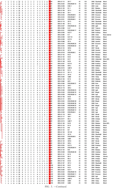
FIG. 3. —Continued.
VOL. 49, 2011 BRUCELLA MLVA GENOTYPES FROM TURKEY 3281
on May 16, 2020 by guest
http://jcm.asm.org/
sion among the cases, the results of these panels provide useful information about distribution of the genotypes among coun-tries. With regard to MLVA-8Orsay genotypes, the most com-mon genotypes (42 and 43) found in the current study were also observed in other parts of the world (1, 11, 18, 23). In contrast, those of the typical West Mediterranean family, in-cluding MLVA-8Orsaygenotypes 49 and 51, were not detected in this study. These data indicate that humanBrucellaisolates in Turkey form a highly homogeneous group belonging to the East Mediterranean group.
The proportion of strains being in clusters suggests that a significant proportion of brucellosis in Turkey is due to multi-ple contaminations from a single source. The large clusters included strains from different provinces and different regions. For instance, the largest cluster included eight isolates col-lected over an 8-year period (from 2002 to 2009) from five separate provinces in three geographic regions. The seven strains in the second most frequent genotype were isolated from five separate provinces in two geographic regions be-tween 2004 and 2008. These data show that ongoing transmis-sion of human brucellosis has continued for a long period not only in a specific region but also among the regions in Turkey. Additionally, the 73 isolates showing distinct genotypes re-flected that more than 45% of the brucellosis in Turkey had epidemiologically unrelated sporadic characteristics.
In agreement with the previous investigations (1, 11, 18), the MLVA-16Orsaygenotyping results showed good correlation with the epidemiological data. The present findings also confirmed relapses, laboratory-acquired brucellosis, and intrafamiliary bru-cellosis resulting from food sharing. The isolates of two patients from the acute and relapse stages showed identical MLVA-16Orsaygenotypes. In the current study, MLVA-16Orsay typing
enabled us to identify the source of laboratory-acquired brucel-losis in a laboratory worker who was exposed toBrucellawhile processing a blood culture specimen. MLVA genotype also con-firmed intrafamilial brucellosis in many cases, in whom brucellosis most probably resulted from traditional food habits, including the consumption of homemade cheese and cream, which are partic-ularly common in rural areas or farmland people residing in the southeastern, eastern, and central Anatolia regions of Turkey. These traditional food habits also lead to a higher incidence of brucellosis in these regions than in other parts of Turkey (3).
We detailed a small outbreak in a village where a major part of the population was occupied with agriculture and/or live-stock farming. It was supposed that this outbreak originated from a single source from sharing improperly processed milk products among households and relatives. MLVA-16Orsay di-vided the 11 cases investigated here into six genotypes (geno-types 21, 22, 23, 25, 29, 30). These six geno(geno-types were very closely related and differ by single repeat unit differences at one or two of the most variable loci. One hypothesis is that independent contamination occurred from different sources contaminated by historically very closely related strains. A more precise investigation of the strains circulating in the an-imal reservoir in this village will be required to answer this question. For such purposes, we have planned a project to characterize the genotypes circulating in livestock. We will then be able to compare then with the genotypes observed in human isolates as described in the present study.
Conversely, some isolates recovered from separate
re-gions and with no known direct epidemiological links dis-played identical MLVA-16Orsayprofiles (genotypes 7, 13, 15,
16, 24, 55, 85, 86, and 91). This observation might reflect homoplasy and convergent evolution. Alternatively, some of the associated isolates may result from either the lack of control of animal movements between regions or the circu-lation of improperly processed milk products or household products in the market.
In agreement with its location, the most prevalent MLVA genotypes found in Turkey are typically from the East Medi-terranean region. Molecular typing confirmed that more than half of the human brucellosis cases resulted from either very close cross-transmission in a location or persistent and ongoing transmission among the different regions. MLVA-16Orsay
proved to be highly discriminatory among related human Bru-cellaisolates that could not be differentiated by conventional microbiological methods. Hence, MLVA can significantly con-tribute to epidemiological trace-back analysis ofBrucella in-fections and may advance surveillance and control of brucel-losis in Turkey. The data produced in this investigation can be queried in theBrucellaMLVA database release (starting from theBrucella2010 release) at http://mlva.u-psud.fr.
REFERENCES
1.Al Dahouk, S., et al.2007. Evaluation of Brucella MLVA typing for human
brucellosis. J. Microbiol. Methods69:137–145.
2.Alton, G. G., L. M. Jones, R. D. Angus, and J. M. Verger.1988. Techniques for the brucellosis laboratory, p. 34–36. Institut National de la Recherche Agronomique (INRA), Paris, France.
3.Arda, M., O. Akay, and O. M. Esendal.1991. Bovine brucellosis: in relation
to epidemiology and human infection, p. 67–72.InE. Tumbay, S. Hilmi, and
O. Ang (ed.), Brucella and brucellosis in man and animals. The Turkish Microbiological Society, Istanbul, Turkey.
4.Ayaslıoglu, E., et al.2008. Antimicrobial susceptibility ofBrucella melitensis
isolated from blood samples. Turk. J. Med. Sci.38:257–262.
5.Baykam, N., et al.2004. In vitro antimicrobial susceptibility of Brucella
species. Int. J. Antimicrob. Agents23:405–407.
6.Bodur, H., et al.2003. Biotypes and antimicrobial susceptibilities of Brucella
isolates. Scand. J. Infect. Dis.35:337–338.
7.Bricker, B. J., D. R. Ewalt, and S. M. Halling.2003. Brucella “HOOFPrints”: strain typing by multilocus analysis of variable number tandem repeats
(VNTRs). BMC Microbiol.3:15.
8.Dokuzoguz, B., et al.2005. Characteritics ofB. melitensisversusB. abortus
bacteraemias. J. Infect.1:41–45.
9.García-Yoldi, D., et al.2007. Comparison of multiple-locus variable-number
tandem-repeat analysis with other PCR-based methods for typingBrucella
suisisolates. J. Clin. Microbiol.45:4070–4072.
10. Reference deleted.
11.Kattar, M. M., et al.2008. Evaluation of a multilocus variable-number
tandem-repeat analysis scheme for typing humanBrucellaisolates in a region
of brucellosis endemicity. J. Clin. Microbiol.46:3935–3940.
12.Kilic, S., M. Dizbay, and H. Cabadak.2008. In vitro activity of tigecycline,
tetracycline and fluoroquinolones againstBrucella melitensis. J. Chemother.
20:33–37.
13.Kose, S., S. Kilic, and Y. Ozbel. 2005. Identification ofBrucellaspecies isolated from proven brucellosis patients in Izmir, Turkey. J. Basic
Micro-biol.45:323–327.
14.Le Fle`che, P., et al.2006. Evaluation and selection of tandem repeat loci for aBrucellaMLVA typing assay. BMC Microbiol.6:9.
15.Maquart, M., et al.2009. MLVA-16 typing of 295 marine mammal Bru-cellaisolates from different animal and geographic origins identifies 7
major groups withinBrucella cetiandBrucella pinnipedialis. BMC
Micro-biol.9:145.
16.Marianelli, C., et al.2007. Molecular epidemiological and antibiotic
suscep-tibility characterization ofBrucellaisolates from humans in Sicily, Italy.
J. Clin. Microbiol.45:2923–2928.
17.Ministry of Health, Turkey.2007. Brucellosis cases registered in Turkey 1970-2007. Primary Health Care General Directorate, Ministry of Health, Ankara, Turkey.
18.Nockler, K., et al.2009. Molecular epidemiology ofBrucellagenotypes in
patients at a major hospital in Central Peru. J. Clin. Microbiol.47:3147–
3155.
19.Pappas, G., P. Papadimitriou, and N. Akritidis.2006. The new global map
of human brucellosis. Lancet Infect. Dis.6:91–99.
on May 16, 2020 by guest
http://jcm.asm.org/
20. Reference deleted.
21.Rodriguez-Torres, A., R. Landinez, and R. Abad.1984. Species and biotypes
of human brucellosis in Spain. Dev. Biol. Stand.56:107–112.
22.Simsek, H., S. Erdenlig, B. Oral, and N. Tulek.2004. Typing-biotyping of
Brucellaisolates of human origin and their epidemiologic evaluation. Klimik
17:103–106.
23.Smits, H. L., et al.2009.BrucellaMLVA genotyping of humanBrucella
isolates from Peru. Trans. R. Soc. Trop. Med. Hyg.103:399–402.
24.Turkish Statistical Institute.2008. Address-based population register sys-tem, December 31, 2008 census. Turkish Statistical Institute, Ankara, Tur-key. www.turkstat.gov.tr.
25.Whatmore, A. M., et al.2006. Identification and characterization of
variable-number tandem-repeat markers for typing ofBrucellaspp. J. Clin. Microbiol.
44:1982–1993.
26.WHO.2006. Brucellosis in human and animals. WHO/CDS/EPR/2006.7.
World Health Organization, Geneva, Switzerland.
VOL. 49, 2011 BRUCELLA MLVA GENOTYPES FROM TURKEY 3283