Rapid Identification of Patients at High Risk for Carriage of
Vancomycin-Resistant Enterococci
N. Esther Babady,aKathleen Gilhuley,aDiane Cianciminio-Bordelon,aand Yi-Wei Tanga,b
Clinical Microbiology Service, Department of Laboratory Medicine,a
and Infectious Disease Service, Department of Medicine,b
Memorial Sloan-Kettering Cancer Center, New York, New York, USA
We compared the performance characteristics of culture and the Cepheid XpertvanAassay for routine surveillance of vancomy-cin-resistant enterococci (VRE) from rectal swabs in patients at high risk for VRE carriage. The Cepheid XpertvanAassay had a limit of detection of 100 CFU/ml and correctly detected 101 well-characterized clinical VRE isolates with no cross-reactivity in 27 non-VRE and related culture isolates. The clinical sensitivity, specificity, positive predictive value, and negative predictive value of the XpertvanAPCR assay were 100%, 96.9%, 91.3%, and 100%, respectively, when tested on 300 consecutively collected rectal swabs. This assay provides excellent predictive values for prompt identification of VRE-colonized patients in hospitals with rela-tively high rates of VRE carriage.
V
ancomycin-resistant enterococci (VRE) are recognized as nosocomial pathogens alongside methicillin-resistant Staphylococcus aureus(MRSA) andClostridium difficile. Van-comycin resistance in enterococci species is conferred mainly by the presence of thevanAorvanBgene, although the pres-ence of other genes, includingvanC,vanD,vanE, andvanG, can also result in a resistant phenotype (7,25). In North America, the vanAgene is the most prevalent resistance marker in en-terococci species, followed by the vanB gene, which can be found in bacteria other than enterococci (4,10). Both thevanA and vanB genes are carried on transposable plasmids, and transfer of these plasmids to other enterococci andS. aureus has been shown bothin vitroandin vivo(7).Several reports have shown that in allogeneic hematopoietic stem cell transplant recipients, VRE colonization, prior to stem cell transplantation, is a significant risk factor for the develop-ment of VRE bacteremia, which is associated with poor clinical outcomes (3,14,22,24). In order to decrease the spread of VRE in hospital settings, the Hospital Infection Control Practices Advisory Committee (HICPAC) recommends a multipronged approach that includes rapid identification and reporting of VRE-positive stools or rectal swabs by the microbiology labo-ratory in order to ensure prompt isolation of colonized pa-tients (2).
Currently, VRE surveillance is performed at our institution using traditional culture. This procedure requires 48 to 96 h to obtain a final result and involves multiple media and incubation steps. Recently, the Food and Drug Administration (FDA) ap-proved a rapid molecular assay, the XpertvanA(Cepheid, Sunny-vale, CA), for the detection of VRE directly from rectal swab spec-imens only. The assay is a real-time, one-step PCR assay performed on the GeneXpert instrument and provides results in less than 1 h, compared to 48 to 96 h with culture. In addition to providing rapid results for timely isolation of colonized patients, rapid and more sensitive detection of VRE may also result in the timely identification of patients at risk for the development of VRE bacteremia. The objective of the present study was to evalu-ate the performance characteristics of this novel PCR assay
com-pared to those of traditional culture for the detection of VRE from rectal swabs. To our knowledge, this is the first reported evalua-tion of the XpertvanAassay in a patient population at high risk for VRE colonization.
(This study was presented in part at the 52nd Interscience Con-ference on Antimicrobial Agents and Chemotherapy.)
MATERIALS AND METHODS
Isolates and patient specimens.One hundred and twenty-eight archived, previously characterized clinical isolates of enterococci (including both
vancomycin-resistant [n⫽101] and
vancomycin-susceptible/intermedi-ate [n⫽7] isolates) and other nonenterococci isolates (n⫽20) were
tested to determine the analytical sensitivity and specificity of the Xpert
vanAPCR assay (Table 1). Additionally, 300 consecutive rectal swabs
(BBLCulturette; BD Diagnostics, Sparks, MD) from 162 patients that were submitted during a 4-week period to the laboratory for VRE surveil-lance culture were tested to determine the clinical sensitivity and
specific-ity of the XpertvanAPCR assay. The study was approved by the
Memo-rial-Sloan Kettering Cancer Center (MSKCC) institutional review board.
Surveillance culture.VRE surveillance culture was performed by streaking a rectal swab onto a Campy agar plate containing cefoperazone, vancomycin, and amphotericin B (CVA) (BD Diagnostics, Sparks, MD),
followed by incubation at 37°C in 5 to 10% CO2for 24 to 48 h. Suspicious
colonies were Gram stained and tested for the presence of pyrrolidonyl arylamidase activity and the lack of catalase activity. Any isolates
consis-tent withEnterococcusspecies were tested for vancomycin susceptibility by
the Kirby-Bauer method using a 30-g vancomycin disk according to
Clinical and Laboratory Standards Institute (CLSI) guidelines (6). The
final species identification was generated using the MicroScan dried Gram-positive identification (ID) type 3 panel on the automated Mi-croScan instrument (Siemens, West Sacramento, CA).
Received6 July 2012 Returned for modification6 August 2012 Accepted1 September 2012
Published ahead of print12 September 2012
Address correspondence to N. Esther Babady, [email protected].
Copyright © 2012, American Society for Microbiology. All Rights Reserved.
doi:10.1128/JCM.01776-12
on May 16, 2020 by guest
http://jcm.asm.org/
XpertvanAPCR.A PCR assay was performed according to the man-ufacturer’s instructions using rectal swabs collected for VRE surveillance culture. The limit of detection (LOD) of the assay was determined by
testing a dilution series (0 CFU/ml to 107CFU/ml) of a
vancomycin-resistantEnterococcus faeciumisolate (identification confirmed by
cul-ture) in 2 to 5 replicates.
Additional assays.The vancomycin-teicoplanin Etest (AB Biodisk North America, Inc., Culver City, CA) was used to determine the
pheno-type of any VRE culture isolates that were negative by the XpertvanA
assay. VRE isolates with a vancomycin MIC of⬎32g/ml and a
teicopla-nin MIC of⬎32g/ml were consideredvanApositive, and any VRE
isolates with a vancomycin MIC of⬎32g/ml and a teicoplanin MIC of
⬍32g/ml were consideredvanBpositive. Enriched broth culture was
also used on discordant results and performed by inoculating the rectal swab in Trypticase soy broth for 5 days, followed by subculture and fur-ther testing as described above for surveillance culture.
vanAandvanBreal-time PCRs.Additional real-time PCR assays were
developed to confirm the presence of thevanAorvanBgene in XpertvanA
PCR-positive, culture-negative specimens. Primers (vanAPCR forward
primer, GGCTGTTTCGGGCTGTGA-3=;vanAPCR reverse primer, 5=-A
CTAACGCGGCACTGTTTCC-3=;vanBPCR forward primer, 5=-GGGA
ACGAGGATGATTTGATTG-3=;vanBPCR reverse primer, 5=-CGTGGC
TCAGCCGGATT-3=) were designed using the Applied Biosystems
Primer Express software version 3.0 (Life Technology Corp., Carlsbad, CA). The analytical specificity was determined by performing a Basic Lo-cal Alignment Search Tool (BLAST) search of each primer and the entire amplicon sequence using the National Center for Biotechnology
Informa-tion website (http://www.ncbi.nlm.nih.gov) and testing of isolates listed
inTable 1. The analytical sensitivity of this laboratory-developed PCR
assay was determined by performing serial dilution ofvanA/vanB-positive
VRE. Detection of the amplified product was performed using Fast SYBR green master mix (Life Technology Corp., Carlsbad, CA) on the 7500 real-time PCR system (Life Technology Corp., Carlsbad, CA) in a final
volume of 20l with the following thermal cycler profile: 1 cycle of 95°C
for 2 min, 40 cycles at 95°C for 5 s, 60°C for 10 s, and 75°C for 35 s, and a dissociation step of 95°C for 15 s, 60°C for 1 min, and 95°C for 15 s. The amplified sequences were run on a 2% gel (E-gel; Life Technology Corp., Carlsbad, CA) to confirm their correct size. Known positive and negative VRE isolates were included in each run and on each gel. To test culture
isolates, 2 to 3 colonies of each isolate were diluted in 500l of
nuclease-free water (Roche Applied Sciences, Indianapolis, IN), vortexed for 10 s at high speed, and boiled for 10 min at 95°C. Five microliters of the
super-natant was used for amplification. To test rectal swab specimens, 5l of
the remaining sample reagent buffer used for the XpertvanAPCR was
used for the real-time PCR.
Discordant result analysis.The reference standard used to determine true-positive and false-negative results was a combination standard (i.e., a true-positive sample was a specimen that was positive by at least two methods). Any specimen with a discordant result was further analyzed by (i) a review of medical records to determine if the patient had a recent positive VRE culture (within 4 weeks) and/or (ii) additional testing of rectal swabs by enriched broth culture. Discordant test results were con-sidered true positive only if the enriched broth culture and/or chart review confirmed the presence of VRE and true negative if neither the broth culture nor the chart review confirmed the presence of VRE.
Statistical analysis. The sensitivity, specificity, positive predictive value (PPV), and negative predictive value (NPV) were calculated for both
the XpertvanAassay and direct culture using the reference standard
de-scribed above. The significance of the observed difference was determined using Fisher’s test for sensitivity and specificity and the 1-way analysis of variance (ANOVA) test to compare the median semiquantitative culture
results to the corresponding median cycle threshold (CT) values of the
XpertvanAPCR. APvalue ofⱕ0.05 was considered significant. Statistical
analysis was performed using the GraphPad Prism software (GraphPad Software, Inc., La Jolla, CA).
RESULTS
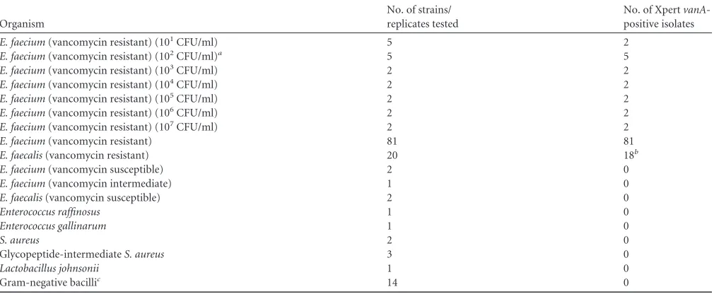
[image:2.585.41.546.78.286.2]Out of 128 well-characterized isolates, the XpertvanAPCR cor-rectly identified 101 clinical VRE isolates with no cross-reactivity with 27 non-VRE isolates (Table 1). Three VRE isolates originally tested negative by XpertvanAPCR. Additional testing of these isolates with the vancomycin-teicoplanin Etest strips (AB Biodisk North America, Inc., Culver City, CA) identified two isolates as vancomycin-susceptibleE. faecium(vancomycin MICs of 1.0 and 1.5g/ml and teicoplanin MICs of 1.5 and 2.0g/ml) and one isolate as vancomycin-resistantE. faecaliswith avanBphenotype (a vancomycin MIC of⬎32g/ml and a teicoplanin MIC of 2 TABLE 1Analytical sensitivity and specificity of the XpertvanAPCR
Organism
No. of strains/ replicates tested
No. of XpertvanA
-positive isolates
E. faecium(vancomycin resistant) (101CFU/ml) 5 2
E. faecium(vancomycin resistant) (102CFU/ml)a 5 5
E. faecium(vancomycin resistant) (103CFU/ml) 2 2
E. faecium(vancomycin resistant) (104CFU/ml) 2 2
E. faecium(vancomycin resistant) (105CFU/ml) 2 2
E. faecium(vancomycin resistant) (106CFU/ml) 2 2
E. faecium(vancomycin resistant) (107CFU/ml) 2 2
E. faecium(vancomycin resistant) 81 81
E. faecalis(vancomycin resistant) 20 18b
E. faecium(vancomycin susceptible) 2 0
E. faecium(vancomycin intermediate) 1 0
E. faecalis(vancomycin susceptible) 2 0
Enterococcus raffinosus 1 0
Enterococcus gallinarum 1 0
S. aureus 2 0
Glycopeptide-intermediateS. aureus 3 0
Lactobacillus johnsonii 1 0
Gram-negative bacillic 14 0
aThe lower limit of detection was 100 CFU/ml.
b
Two isolates wereE. faecalis vanBpositive. cIncludes severalEnterobacteriaceaeisolates.
on May 16, 2020 by guest
http://jcm.asm.org/
g/ml). Testing of this isolate using thevanBPCR described above confirmed the presence of thevanBgene (data not shown). Fol-lowing resolution of discordant results, both the analytical sensi-tivity and specificity of the XpertvanAassay were 100%.
A total of 300 specimens from 162 patients were tested by both XpertvanAPCR and direct culture. ThevanAgene was detected in 81 specimens from 60 patients (37.0% of patients), while VRE isolates were recovered in 56 specimens from 46 patients (28.4% of patients). The lower limit of detection of the assay, as deter-mined by 10-fold serial dilutions of VRE, was 100 CFU/ml (Table 1). The median CT value was compared to the corresponding semiquantitative result of the surveillance culture (Fig. 1) to fur-ther compare the sensitivity of the PCR assay to the results ob-tained by culture. The medianCTvalue for PCR-positive, culture-negative specimens was 34.1, which was approximately one dilution away from the medianCT value (30.1) for 1⫹(1 to 9 colonies) positive cultures (Fig. 1). This suggests that the observed discrepancy might be due to a bacterial load below the sensitivity of direct culture, although the difference inCT values between these two groups was not statistically significant (P⬎0.05).
Among the 25 PCR-positive, culture-negative swabs, 13 (52%) had a positive culture within 3 weeks (range, 1 day to 21 days; median, 7 days) of the PCR results and were considered true pos-itive. The remaining 12 discordant swabs were incubated for 5 days in Trypticase soy broth, and VRE was detected in 5/12 swabs for a total of 18/25 (72%) true-positive PCR results. Although only 5/12 swabs became positive by enriched broth culture, 11/12 swabs tested by a second, laboratory-developed real-time PCR
(LOD, 100 CFU/ml; specificity, 100%; data not shown) were pos-itive for thevanAgene.
Following resolution of discordant results, the clinical sensitiv-ity, specificsensitiv-ity, positive predictive value (PPV), and negative pre-dictive value (NPV) of the XpertvanA PCR assay were 100%, 96.9%, 91.4%, and 100%, respectively, while those of direct cul-ture were 75.7%, 100%, 100%, and 92.6%, respectively (Table 2). The difference between XpertvanAand direct culture results was statistically significant (P⬍0.001).
DISCUSSION
We report our evaluation of the FDA-approved Cepheid Xpert vanAassay for the detection of VRE from rectal swabs com-pared to that by direct culture. Phenotypic identification of VRE isolates by culture is based on a MIC ofⱖ32g/ml (CLSI). Using the Campy agar plate, which contains 10g/ml of van-comycin, as our primary plating medium for VRE surveillance culture, both intermediate (8 to 16g/ml) and resistant (⬎32 g/ml) enterococci can be isolated. Enterococci isolates grow-ing on the Campy agar plate are then further tested usgrow-ing a 30-g vancomycin disk to identify resistant strains. The Xpert vanAassay identifies VRE based solely on the presence of the vanAgene, which confers a high level of inducible resistance to both vancomycin and teicoplanin (7).
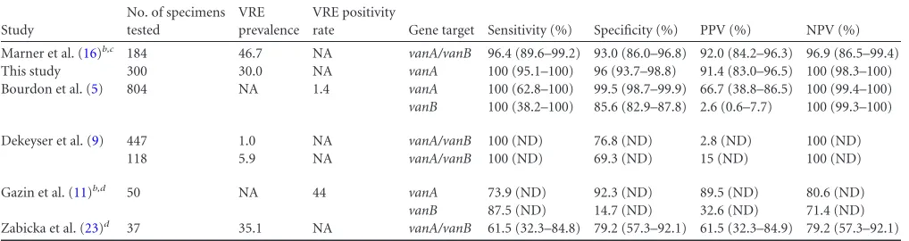
Similar studies have evaluated the performance of a Confor-mité Européenne (CE)-marked version of this assay that detects the presence of both thevanAandvanBgenes (XpertvanA/vanB assay) in rectal swabs, perianal swabs, and stool specimens (5,9,
11,16,23) (Table 3). Bourdon et al. (5) tested 804 rectal swabs and detected 127 swabs positive forvanAorvanBby XpertvanA/vanB assay, with 11 swabs positive by both Xpert PCR and culture. The high sensitivity (100%) and low positive predictive value (8.7%) of the assay in the Bourdon et al. study were attributed mainly to the high detection of thevanBgene (n⫽115), which was consid-ered a false-positive result due to the lack of enterococcus species growth in culture. The PPV of the assay for thevanAgene alone, although better, was also relatively low at 66.7%. Consequently, the authors recommended that all Xpert positive results be con-firmed by culture, which negates, in part, the value of this rapid test. However, in a setting in which the prevalence of VRE is low, such an approach might be beneficial. Dekeyser et al. tested 565 rectal swabs during and following an outbreak of VRE in their hospital (9). VRE prevalences during and after the outbreak were 5.9% and 1%, respectively. However, the PPV of the assay re-mained low (15% during the outbreak versus 2.8% after the out-break), primarily due to detection of thevanBgene. The poor PPV of PCR forvanBVRE has also been reported for other PCR assays, including the BD GeneOhmVanR assay (BD GeneOhm, San Di-FIG 1XpertvanA CTvalues versus those of semiquantitative VRE culture.
The horizontal line in each floating box represents the medianCTvalue, and
the length of each box reflects the range (minimum to maximum) ofCTvalues
for each semiquantitative result. *,Pvalue of⬍0.0001 compared to
culture-negative VRE (CNVRE). VRE, vancomycin-resistant enterococcus species.
1⫹, 1 to 9 colonies; 2⫹, 1 to 49 colonies; 3⫹, 5 to 300 colonies; 4⫹,⬎300
[image:3.585.87.240.65.196.2]colonies.
TABLE 2Comparison of VRE culture to XpertvanAPCRa
Test
No. of isolates with each set of resultsb
Sensitivity (%) Specificity (%) PPV (%) NPV (%)
Reference⫹,
test⫹
Reference⫹,
test⫺
Reference⫺,
test⫹
Reference⫺,
test⫺
VRE direct culture 56 18 0 226 75.7 (64.3–84.9) 100 (98.4–100) 100 (93.6–100) 92.6 (88.6–95.6)
XpertvanAPCR 74 0 7 219 100 (95.1–100) 96.9 (93.7–98.8) 91.3 (83.0–96.5) 100 (98.3–100)
aValues in parentheses are the 95% confidence interval.
b
Reference, direct culture and results of enriched broth culture and/or chart review for discordant specimens; reference⫹, positive result for the reference; test⫹, positive result for the indicated test.
on May 16, 2020 by guest
http://jcm.asm.org/
[image:3.585.38.545.635.697.2]ego, CA) and the Roche LightCycler analyte-specific reagents (ASRs) (15,17,21). Different from Bourdon et al., our findings showed a higher PPV (91%, versus 66.7% in the study by Bourdon et al.) after resolution of discrepant results for thevanAgene.
The prevalence of VRE for patients screened in our hospital was calculated for the year 2011 at 30.0%, which is close to the prevalence during our study period. The higher prevalence of VRE-colonized patients in our population may explain the marked difference in the PPV between the two studies (Table 3). Screening of our VRE isolate library revealed a low incidence of vanBVRE isolates; only one culture isolate negative by XpertvanA was determined to be positive for thevanBgene by a vancomycin-teicoplanin Etest and a vanB PCR assay. Similar to our data, Stamper et al. detectedvanB-positive enterococcus species by the BD GeneOhm VanR assay (BD GeneOhm, San Diego, CA) in only 3/147 specimens positive by culture (21). In their evaluation of the XpertvanA/vanBassay, Marner and colleagues also detected a low number (5/88) of perianal swabs positive only for thevanBgene, with only 1 confirmed by culture (16). These results confirm the lower prevalence of thevanBgene inEnterococcusspecies in North America compared to the prevalence of thevanBgene in Entero-coccusspecies in Europe, as previously reported by the SENTRY antimicrobial surveillance program (10). Unlike Marner et al., our evaluation was performed (i) on consecutive rectal swabs rather than on a selected set of perianal swabs, (ii) using a different ref-erence method as the gold standard, and (iii) targeting only the vanAgene. These differences may explain the variations in the observed sensitivity, specificity, PPV, and NPV between the two studies. Two additional studies evaluating the performance of the XpertvanAshowed remarkably lower sensitivity by the assay than by culture (11, 23). The limited number of specimens tested (ⱕ50), as well as the use of stool specimens rather than rectal swabs, might explain the suboptimal performance of the Xpert vanAassay in those studies.
Although culture is the most common method used for sur-veillance screening, a higher bacterial burden is necessary to ob-tain a positive result. D’Agata et al. (8) showed that the sensitivity of a rectal swab culture varied from 0% when VRE density was ⱕ4.5 log10CFU/g of stool to 100% when the VRE density was
ⱖ7.5 log10CFU/g of stool (average of 58% sensitive). In our study,
the sensitivity of the surveillance culture was 75.7% (Table 2).
Additionally, there are variations in the sensitivity and specificity of different culture methods, including chromogenic agars, which, as previously reported, result in a range of sensitivities (1,
13, 20). The lower sensitivity of a culture, therefore, should be taken into consideration when following the HICPAC recom-mendation to terminate isolation following three negative cul-tures (2). Multiple studies have also shown that spontaneous de-colonization occurs only in a limited number of patients; however, reappearance of the VRE within a few weeks of decolo-nization is common (12,18,19). All XpertvanA-positive discor-dant results were tested by a second independent laboratory-de-veloped real-time PCR to confirm that the false-positive results were truly due to the presence of thevanAgene. Additional VRE isolates were also detected when broth-enriched culture, which we do not perform routinely, was used to analyze discrepant results. Further review of our XpertvanA-positive, cultunegative re-sults suggested that 68% of our discrepant rere-sults can be attributed to low bacterial load because the PCR results preceded or followed a recent positive culture result. The significance of a low bacterial VRE load detected by PCR only and its impact on nosocomial transmission of VRE are unknown and will have to be studied further, especially as it applies to discontinuation of contact pcautions for PCR-positive patients. Since a VRE PCR-positive re-sult with aCT value of⬎34 (range, 16.5 to 38.8) often corre-sponded to a negative culture, a quantitative or semiquantitative PCR assay rather than a qualitative assay might be more relevant for infection control purposes, although this remains to be deter-mined.
Our study has some limitations. First, the XpertvanAPCR was performed using the same swab used to set up the culture. Al-though this algorithm did not affect the sensitivity of the assay, more specimens might have been positive if the swabs were tested directly as opposed to following culture inoculation. Second, it is possible that false-negative results occurred due to the presence of vanBVRE, which are not detected by this assay. We did not have any rectal swabs that were culture positive and XpertvanA nega-tive, although, as described earlier, the sensitivity of the culture is not optimal.
[image:4.585.40.549.78.213.2]At MSKCC, active surveillance for VRE is performed in units with high-risk patients, including those in intensive care and bone marrow transplant units. Implementation of the XpertvanAPCR TABLE 3Comparison of published XpertvanA/vanBtest characteristicsa
Study
No. of specimens tested
VRE prevalence
VRE positivity
rate Gene target Sensitivity (%) Specificity (%) PPV (%) NPV (%)
Marner et al. (16)b,c 184 46.7 NA vanA/vanB 96.4 (89.6–99.2) 93.0 (86.0–96.8) 92.0 (84.2–96.3) 96.9 (86.5–99.4)
This study 300 30.0 NA vanA 100 (95.1–100) 96 (93.7–98.8) 91.4 (83.0–96.5) 100 (98.3–100)
Bourdon et al. (5) 804 NA 1.4 vanA 100 (62.8–100) 99.5 (98.7–99.9) 66.7 (38.8–86.5) 100 (99.4–100)
vanB 100 (38.2–100) 85.6 (82.9–87.8) 2.6 (0.6–7.7) 100 (99.3–100)
Dekeyser et al. (9) 447 1.0 NA vanA/vanB 100 (ND) 76.8 (ND) 2.8 (ND) 100 (ND)
118 5.9 NA vanA/vanB 100 (ND) 69.3 (ND) 15 (ND) 100 (ND)
Gazin et al. (11)b,d 50 NA 44 vanA 73.9 (ND) 92.3 (ND) 89.5 (ND) 80.6 (ND)
vanB 87.5 (ND) 14.7 (ND) 32.6 (ND) 71.4 (ND)
Zabicka et al. (23)d 37 35.1 NA vanA/vanB 61.5 (32.3–84.8) 79.2 (57.3–92.1) 61.5 (32.3–84.9) 79.2 (57.3–92.1)
aPPV, positive predictive value; NPV, negative predictive value; ND, not determined; NA, not available. Values in parentheses are the 95% confidence interval.
b
Study not done on consecutive specimens. When prevalence was not available, the positivity rate obtained with the culture method is listed. cPerianal swab specimens.
d
Stool specimens.
on May 16, 2020 by guest
http://jcm.asm.org/
would provide several advantages, including rapid identification and prompt reporting of VRE-colonized patients for immediate isolation, identification of patients at high-risk for developing VRE bacteremia, and decreased labor and turnaround time asso-ciated with traditional culture. In theory, identification and isola-tion of VRE-colonized patients should result in a decreased rate of VRE infections as well as a decreased rate of nosocomial cases. However, rapid identification and isolation of colonized patients is only part of the equation; other factors, such as prudent use of vancomycin and hospital staff knowledge and adherence to isola-tion precauisola-tions, also contribute to the overall VRE hospital rate (2). Practical issues associated with the use of PCR for VRE iden-tification include the inability to save VRE culture isolates (neces-sary for epidemiological studies in case of outbreak) and the in-ability to differentiate between E. faecium and Enterococcus faecalis. Furthermore, similar to other commercial PCR assays, the cost of the XpertvanAPCR is significantly higher than that of culture. Therefore, depending on the rate of VRE colonization and VRE infection in a hospital, the implementation of a more sensi-tive, albeit more costly, test might be justified for efficient infec-tion control and rapid identificainfec-tion of patients at increased risk for VRE infections.
In conclusion, the excellent sensitivity and specificity and rapid turnaround time of the Cepheid XpertvanA assay make it an attractive option for routine surveillance of VRE from rectal swabs. This assay will significantly reduce the labor and time as-sociated with the traditional surveillance culture method.
ACKNOWLEDGMENTS
We thank Steve Tulumba and Darlene Reid in the Bacteriology
Labora-tory for assistance with testing of rectal swabs by XpertvanAPCR.
REFERENCES
1.Adler H, Oezcan S, Frei R.2010. Vancomycin-resistant enterococci of
vanBgenotype may pose problems for screening with highly selective
me-dia. J. Clin. Microbiol.48:2323.
2.Anonymous.1995. Recommendations for preventing the spread of van-comycin resistance. Recommendations of the Hospital Infection Control Practices Advisory Committee (HICPAC). MMWR Recommend. Rep.
44(RR-12):1–13.
3.Avery R, et al. 2005. Early vancomycin-resistant enterococcus (VRE) bacteremia after allogeneic bone marrow transplantation is associated
with a rapidly deteriorating clinical course. Bone Marrow Transplant.35:
497– 499.
4.Ballard SA, Grabsch EA, Johnson PD, Grayson ML.2005. Comparison
of three PCR primer sets for identification ofvanBgene carriage in feces
and correlation with carriage of vancomycin-resistant enterococci:
inter-ference byvanB-containing anaerobic bacilli. Antimicrob. Agents
Che-mother.49:77– 81.
5.Bourdon N, et al.2010. Rapid detection of vancomycin-resistant
entero-cocci from rectal swabs by the Cepheid XpertvanA/vanBassay. Diagn.
Microbiol. Infect. Dis.67:291–293.
6.CLSI.2010. Performance standards for antimicrobial susceptibility test-ing; 20th informational supplement. CLSI M100-S20. Clinical and Labo-ratory Standards Institute, Wayne, PA.
7.Courvalin P.2006. Vancomycin resistance in gram-positive cocci. Clin.
Infect. Dis.42(Suppl 1):S25–S34.
8.D’Agata EM, Gautam S, Green WK, Tang YW. 2002. High rate of false-negative results of the rectal swab culture method in detection of
gastrointestinal colonization with vancomycin-resistant enterococci.
Clin. Infect. Dis.34:167–172.
9.Dekeyser S, Beclin E, Descamps D.2011. Implementation of vanA and vanB genes by PCR technique research interest in system (Xpert vanA/ vanB CepheidR) closed in a laboratory of microbiology in managing an
outbreak toEnterococcus faeciumresistant glycopeptide (EfRG). Pathol.
Biol.59:73–78. (In French.)
10. Deshpande LM, Fritsche TR, Moet GJ, Biedenbach DJ, Jones RN.2007. Antimicrobial resistance and molecular epidemiology of vancomycin-resistant enterococci from North America and Europe: a report from the SENTRY antimicrobial surveillance program. Diagn. Microbiol. Infect.
Dis.58:163–170.
11. Gazin M, Lammens C, Goossens H, Malhotra-Kumar S.2012.
Evalua-tion of GeneOhm VanR and XpertvanA/vanBmolecular assays for the
rapid detection of vancomycin-resistant enterococci. Eur. J. Clin.
Micro-biol. Infect. Dis.31:273–276.
12. Huckabee CM, Huskins WC, Murray PR.2009. Predicting clearance of colonization with vancomycin-resistant enterococci and
methicillin-resistantStaphylococcus aureusby use of weekly surveillance cultures. J.
Clin. Microbiol.47:1229 –1230.
13. Jenkins SG, Raskoshina L, Schuetz AN.2011. Comparison of perfor-mance of the novel chromogenic Spectra VRE agar to that of bile esculin
azide andCampylobacteragars for detection of vancomycin-resistant
en-terococci in fecal samples. J. Clin. Microbiol.49:3947–3949.
14. Kamboj M, et al. 2010. The changing epidemiology of vancomycin-resistant enterococcus (VRE) bacteremia in allogeneic hematopoietic stem cell transplant (HSCT) recipients. Biol. Blood Marrow Transplant.
16:1576 –1581.
15. Mak A, Miller MA, Chong G, Monczak Y.2009. Comparison of PCR and culture for screening of vancomycin-resistant enterococci: highly
dispa-rate results forvanAandvanB.J. Clin. Microbiol.47:4136 – 4137.
16. Marner ES, et al.2011. Diagnostic accuracy of the Cepheid GeneXpert
vanA/vanBassay ver. 1.0 to detect thevanAandvanBvancomycin resis-tance genes in Enterococcus from perianal specimens. Diagn. Microbiol.
Infect. Dis.69:382–389.
17. Mehta MS, et al.2008. Optimization of a laboratory-developed test
uti-lizing Roche analyte-specific reagents for detection ofStaphylococcus
au-reus, methicillin-resistantS. aureus, and vancomycin-resistant
Enterococ-cusspecies. J. Clin. Microbiol.46:2377–2380.
18. Park I, et al.2011. Rectal culture screening for vancomycin-resistant enterococcus in chronic haemodialysis patients: false-negative rates and
duration of colonisation. J. Hosp. Infect.79:147–150.
19. Patel R, et al.2001. Natural history of vancomycin-resistant entero-coccal colonization in liver and kidney transplant recipients. Liver
Transpl.7:27–31.
20. Sloan LM, et al.2004. Comparison of the Roche LightCyclervanA/vanB
detection assay and culture for detection of vancomycin-resistant
entero-cocci from perianal swabs. J. Clin. Microbiol.42:2636 –2643.
21. Stamper PD, Cai M, Lema C, Eskey K, Carroll KC.2007. Comparison of the BD GeneOhm VanR assay to culture for identification of vancomy-cin-resistant enterococci in rectal and stool specimens. J. Clin. Microbiol.
45:3360 –3365.
22. Weinstock DM, et al.2007. Colonization, bloodstream infection, and mortality caused by vancomycin-resistant enterococcus early after alloge-neic hematopoietic stem cell transplant. Biol. Blood Marrow Transplant.
13:615– 621.
23. Zabicka D, et al.2012. Efficiency of the Cepheid XpertvanA/vanBassay for screening of colonization with vancomycin-resistant enterococci
dur-ing hospital outbreak. Antonie Van Leeuwenhoek101:671– 675.
24. Zirakzadeh A, et al.2008. Vancomycin-resistant enterococcal coloniza-tion appears associated with increased mortality among allogeneic
hema-topoietic stem cell transplant recipients. Bone Marrow Transplant.41:
385–392.
25. Zirakzadeh A, Patel R.2006. Vancomycin-resistant enterococci:
coloni-zation, infection, detection, and treatment. Mayo Clin. Proc.81:529 –536.