ORIGINAL RESEARCH ARTICLE
NEUROPHYSIOLOGICAL EVALUATION OF BLADDER DYSFUNCTION IN LOWER MOTOR
NEURON LESIONS
*1
Hamdy A. Khallaf,
1Mervat I. Hussein,
2Ibrahim Khalil Ibrahim,
1Ali E. El Dib,
1Samah
H. El Medany,
3Osama E. El Dib and
4Manal S. Awadh
1
Physical Medicine & Rehabilitation Department, Tanta University, Egypt
2Physical Medicine & Rehabilitation Department, Alexandria University, Egypt
3
Neurology Department, Menofiya University, Egypt
4
Physical Medicine & Rehabilitation Department, Ain Shams University, Egypt
ARTICLE INFO ABSTRACT
I
Innttrroodduuccttiioonn::Urinary stress incontinence is almost an exclusively female phenomenon in middle age, may be due to obstetric injury. Patients with bladder dysfunction should undergo neurophysiological examination when it is suspected to happen due to central or peripheral factors Aim of The work: This work was aimed to assess the electrodiagnostic value of sphincteric electromyography, pudendal nerve terminal motor latency, sacral reflexes, and pudendal nerve somatosensory-evoked potentials in bladder dysfunction. Patients and Methods:
This study included 20 patients suffering from bladder dysfunction, with Lower motor neuron lesions selected from Tanta and Alexandria University Hospitals. Diagnosis of the patients based on a full history including a detailed urinary sheet followed by a complete physical and neurological examination. Followed by the electrodiagnostic of sphincteric electromyography, pudendal nerve terminal motor latency, sacral reflexes, and pudendal nerve somatosensory-evoked potentials. Results: The sensitivity of motor unit action potential of external anal sphincter parameters in diagnosis of neuropathic lesion include: sensitivity of amplitude 40% , duration 10 %, area 100%, , denervation 100%.The sensitivity of other parameters in diagnosis of neuropathic lesion as following: bulbocavernosus 100%, urethral Sphincter 100%, puborectalis 100%, gracilis 15%, pudendal nerve terminal motor latencies right &left 15%, reflex The bulbocavernosus 10%, Reflex Pudendo-anal 30%, Somatosensory evoked potentials of pudendal nerve 100%. Conclusion: This study results address the significance of using combined motor unit action potential parameters of different muscles to increase sensitivity for the detection of neuropathic changes in bladder dysfunction.
Copyright © 2019, Hamdy A. Khallaf et al. This is an open access article distributed under the Creative Commons Attribution License, which permits unrestricted use, distribution, and reproduction in any medium, provided the original work is properly cited.
INTRODUCTION
Urinary incontinence is a serious health issue that negatively impacts patients' daily activities and their quality of life (Joanna, 2017).The most of bladder dysfunction patients were female in middle age claiming that primary reason is childbirth, despite the fact these women mainly show symptoms after menopause only (Snooks, 1990). The etiology of stress urinary incontinence (SUI) is suggested to be caused due to neuromuscular impairment, fascial trauma and/or alteration in motor control (Vodušek, 2005).As a result, the pelvic floor muscles fail to resist the intra-abdominal pressure However, the real cause behind it is not very well understood.
Neurophysiological investigations of bladder dysfunction originated over 60 years ago, and have evolved with the developments in general clinical neurophysiology. The data from these investigations can assist clinicians in diagnosing neurological disease or injury of the uro-genital-anal organs (Vodušek, 2005). In recent years, external anal sphincter (EAS) electromyography (EMG) has gained popularity among neurologists and clinical neurophysiologists, as the muscle is innervated by the lower sacral segments. The EAS is readily accessible and examined without too much discomfort, and it is easy to compile a representative sample of its motor unit action potentials (MUPs) (Franksson, 1953). After describing studies related to motor conduction by electrical stimulation
ISSN: 2230-9926
International Journal of Development Research
Vol. 09, Issue, 07, pp. 28711-28718, July, 2019
Article History: Received 29th April, 2019 Received in revised form 11th May, 2019
Accepted 03rd June, 2019 Published online 28th July, 2019 Key Words:
Bladder dysfunction, Lower motor neuron lesions, Evoked potentials, Motor unit.
*Corresponding author: Hamdy A. Khallaf
Citation: Hamdy A. Khallaf, Mervat I. Hussein, Ibrahim Khalil Ibrahim, Ali El Deeb, Samah H. El Medany, Osama E. El Dib and Manal S. Awadh, 2019.
“Neurophysiological evaluation of bladder dysfunction in lower motor neuron lesions”, International Journal of Development Research, 09, (07), 28711-28718.
and after reporting repetitive stimulation potentials of the pudendal nerves, latency recordings of the bulbo-cavernosus reflex were specified (Podnar, 1999). Somatosensory-evoked potentials (SEP) are used to test perineum sensory deficit. It can allow to measure the impact of neuropathy on bladder and bowel functioning, especially there is no other method to detect the action of anal sensory nerve (Austin, 2016). Such information may assist in diagnosing the disease before starting patients' treatment, but also can be used during follow-up periods (Austin, 2016).
MATERIALS AND METHODS
This study included twenty (20) patients, age ranged between 29-58 yrs old, with bladder dysfunction who suffering from urinary incontinence without bowel dysfunction due to cauda equina syndrome, sacral plexus lesions or pudendal nerve neuropathy selected from Tanta and Alexandria University Hospitals Patients with upper motor neuron lesions (stroke, multiple sclerosis, multiple system atrophy, Parkinsonism, spondylotic myelopathy, transverse myelitis and spinal cord injury) excluded from the study. Diagnosis of the patients based on a full history followed by a complete physical and neurological examination, MRI lumbo-sacral, urine analysis and culture supported by the results of the electrophysiological testing Urinary dysfunction questionnaires (Vodušek, 2005).
Urinary incontinence
1. Quantity:
underwear/pads damp underwear/pads wet outer clothes wet
urine runs down legs or into floor
Quantity of usual urine leak scored: 0 = nil, 1 =underwear pads damp 2 = underwear/pads wet, 3 = outer clothes wet, 4 = urine runs down legs or onto floor
2. Frequency of incontinence: Scored: 0 = never, 1= 1/week, 2 = 2-3/week, 3 = 1/day, 4 = >1/day, 5 = constant
3. post void urine leak
Overactive bladder:
1. Urgency
The number of urgency episodes
Scored: 0 = never, 1 = <1/3, 2 = >1/3, <2/3, 3 = >2/3, 4 = constant
2. Frequency of micturition
Scored: 0= ≤ 4 hr, 1 =3 hr, 2 = 2 hr, 3 =1 hr.
3. Nocturia
Scored: 0 = none, 1 =once, 2 = twice, 3 =3 times, 4= ≤4 times
Disturbed bladder emptying
1. Stream strength
Scored: 0 = not reduced, 1 = a little reduced, 2 = quite reduced, 3 = much reduced, 4 = no stream
2. Incomplete emptying 3. Interruption of micturition 1. Electromyography(EMG)
It is a machine that consists of two EMG parts: concentric needle (50 mm long; No. 22583, Oxford Medical, Old Woking, UK) and a system (Neuropack2 11-NEM-7102 A/K.O the Nihon Kohden) with standard filter settings (5 Hz-10 kHz), gain (200 mV/division, changed as needed) and sweep speed (10 ms/division) (Podnar, 1999). During the examination, patient's bladder must be empty. A. External anal sphincter (EAS); few millimeters under the mucosa (3±6 mm) of the external anal sphincter muscle and 1 cm away from the orifice of the anus, the needle was inserted subcutaneously. The needle was also inserted into the anal orifice with a 30°- 50° angle to the anal canal axis to measure potentials at "deeper" parts (1±3 cm) (Podnar, 1999). B. Bulbocavernosus muscle; to measure the potentials at female's bulbocavernosus muscle, needle was inserted either transmucosally in a medial way to the labia minora or horizontally to the labia majora through skin (Franksson, 1953).
C. Urethral Sphincter; the potentials at this area for females were measured by placing an electrode 5 mm from the external urethral meatus and at 12 o'clock position. [3]
D. Levator ani (Puborectalis part); the concentric needle was inserted 3 to 5 cm deep into the posterior half of the external anal sphincter (Podnar, 1999).
E. Gracilis; concentric needle was iserted in the distal third of the medial thigh; this muscle is just anterior to the medial hamstring tendon (Vodušek, 2005).
2. Pudendal nerve terminal motor latencies, by 0.1 ms single stimuli, latencies were measured at the tip of the figure using St. Mark’s Hospital Electrode, London, UK. The electrode was placed 8 cm at the base of the figure and at the level of the external sphincter to record compound motor action potentials
[4]………
Sacral reflexes
A. The bulbocavernosus; the dorsal nerve of the penis and clitoris in male and female, respectively, underwent a 0.1-0.5 msec duration and 1 Hz frequency single rectangular electrical pulse using a hand-held bipolar stimulating electrode with the cathode proximal. A standard concentric EMG needle electrode was inserted into bulbocavernosus muscles from both right and left sides to record all responses. (Joanna, 2017).
B. Pudendo-anal, Clitoro-anal; the dorsal nerve of the penis and clitoris (pudendal nerve) were electrically stimulated using surface electrode. A standard concentric EMG needle electrode was inserted into bulbocavernosus muscles from both right and left sides to record all responses (Joanna, 2017).
4. Somatosensory evoked potentials (SEP) of pudendal nerve; International 10–20 EEG system was used to record SEP from the middle region, where the electrode was placed 2 cm before the central vertex [128]. The stimulation was performed on the penis and clitoris using a surface electrode with 0.1 msec duration, 2 Hz frequency singe pulses. The intensity exceeded the sensory threshold by 3 folds.
III moderate partial EAS lesions), (Category IV severe partial EAS lesions), and (Category V: bilateral complete EAS muscles denervation) (Podnar, 1999).
Statistics: Statistical presentation and analysis of the present study was conducted, using the mean, standard error, unpaired student t-test, The Wilcoxon tests, linear correlation coefficient, Analysis of variance [ANOVA] tests Paired t-test and chisquare by SPSS V17. Chi-square the hypothesis that the row and column variables are independent, without indicating strength or direction of the relationship. Pearson chi-square and likelihood-ratio chi-square. Fisher's exact test and Yates' corrected chi-square are computed for 2x2 tables.
Student t-test [Unpaired]:
2
SE
2
SE
X
-X
2 1
2 1
t
Paired t-test:
SE
d
X
d
t
Sensitivity/ true positive rate/ recall rate allows measuring the actual positives proportion of the correctly identified ones. Sensitivity= true positives/(true positive + false negative)
Specificity/ true negative rate allows measuring negatives proportion of the correctly identified ones. Specificity=true negatives/(true negative + false
positives)
False negative - negative test result when the disease is really present
False positive - positive test result when the disease is really absent (Joanna, 2018).
RESULTS
Twenty patients (100% female) were included in this study. Neurological examination of both lower limbs was performed in all patients. 100 % of patients had completely normal findings on clinical examination. Also, MRI lumbo-sacral was without significant changes. According to the bladder questionnaire used in this study the bladder dysfunction score revealed perfect urinary continence in 0 % patients, mild urinary incontinence (score: 1–4) in 50 % patients, moderate urinary incontinence(score: 5–8) in 40 % patients, and severe urinary incontinence (score: 9–12) in 10 % patients.
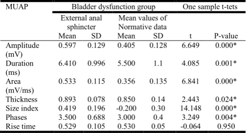
Table 1. Motor unit action potential analysis of external anal sphincter (EAS) in 20 patients with bladder dysfunction
MUAP Bladder dysfunction group One sample t-tets External anal
sphincter
Mean values of Normative data
Mean SD Mean SD t P-value Amplitude
(mV)
0.597 0.129 0.405 0.128 6.649 0.000* Duration
(ms)
6.410 0.996 5.500 1.1 4.085 0.001* Area
(mV/ms)
0.533 0.115 0.356 0.135 6.841 0.000* Thickness 0.893 0.078 0.850 0.14 2.443 0.024* Size index 0.419 0.196 -0.200 0.30 14.148 0.000* Phases 3.500 0.688 3.000 0.4 3.249 0.004* Rise time 0.529 0.105 0.530 0.05 -0.064 0.950
This table demonstrated that the potentials of the EAS motor unit action in bladder dysfunction patients were significantly larger than motor unit potentials of normal external anal sphincter in the following parameters (amplitude, duration, area, thickness, size index and phases), (normal value) (Podnar, 1999).
Table 3. The sensitivity and specificity of the amplitude as one Motor unit action potential (MUAP) parameter in diagnosis of
EAS neuropathic lesion
Sensitivity% 40
Specificity% 100
Positive predictive value% 100 Negative predictive value% 63.6 Overall agreement% 47.4
Table 4. The sensitivity and specificity of the duration as one MUAP parameter in diagnosis of EAS neuropathic lesion
Sensitivity% 10
Specificity% 100
Positive predictive value% 100 Negative predictive value% 53.8 Overall agreement% 55.6
Table 5. The sensitivity and specificity of thickness as one MUAP parameter in diagnosis of EAS neuropathic lesion
Sensitivity% 0
Specificity% 92.9
Positive predictive value% 0 Negative predictive value% 50 Overall agreement% 48.1
Table 6. The sensitivity and specificity of size index as one MUAP parameter in diagnosis of EAS neuropathic lesion
Sensitivity% 90
Specificity% 100
Positive predictive value% 100 Negative predictive value% 93.3 Overall agreement% 96.3
Table 7. The sensitivity and specificity of phases as one MUAP parameter in diagnosis of EAS neuropathic lesion
Sensitivity% 55
Specificity% 71.4
[image:3.595.36.288.640.776.2]Positive predictive value% 63.6 Negative predictive value% 62.5 Overall agreement% 63
Table 8. The sensitivity and specificity of rise time as one MUAP parameter in diagnosis of EAS neuropathic lesion
Sensitivity% 10
Specificity% 42.9
Positive predictive value% 11.1 Negative predictive value% 33.3 Overall agreement% 25.9
Table 9. The sensitivity and specificity of Interference pattern (IP) of EAS in diagnosis of pudendal nerve neuropathy
Sensitivity% 100
Specificity% 100
Table 10. The sensitivity and specificity of Denervation of EAS in diagnosis of neuropathy
Group Total
Bladder Dysfunction Normal Denervation Normal Count 20 14 34
% within Dysfunction 100 100% 100%
Total Count 20 14 34
[image:4.595.123.485.174.237.2]% within Dysfunction 100.0 100.0% 100.0% All are normal
Table 11. The sensitivity and specificity bulbocavernosus muscle (BC) EMG in diagnosis of neuropathy
Group Total
Bladder Dysfunction Normal
BC EMG Normal Count 20 14 34
% within Dysfunction 100% 100% 100%
Total Count 20 14 34
% within Dysfunction 100.0 100.0% 100.0% Table 12. The sensitivity and specificity of Urotheric sphinicter (US) EMG in diagnosis of neuropathy
Group Total
Bladder Dysfunction Normal
US EMG Normal Count 20 14 34
% within Dysfunction
Total Count 20 14 34
% within Dysfunction 100.0 100.0% 100.0% Table 13. The sensitivity and specificity of Puborectalis (PR) EMG in diagnosis of neuropathy
Group Total
Bladder Dysfunction Normal
PR EMG Normal Count 20 14 34 % within Dysfunction
Total Count 20 14 34
% within Dysfunction 100.0 100.0% 100.0% All are normal
Table 14. The sensitivity and specificity of Gracilis muscle EMG in diagnosis of neuropathy
Group Total
Bladder Dysfunction Normal EMG
Category
Abnormal Count 3 0 3
% within Dysfunction 15% 0% 8.8%
Normal Count 17 14 31
% within Dysfunction 85% 100% 91.2%
Total Count 20 14 34
% within Dysfunction 100.0 100.0% 100.0%
Sensitivity% 15%
Specificity% 100%
Positive predictive value% 100% Negative predictive value% 56% Overall agreement% 59.3%
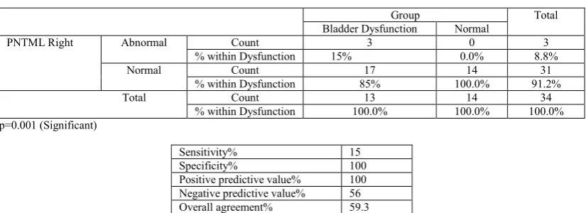
Table 15. The sensitivity and specificity of pudendal nerve terminal motor latencies (PNTML) right (RT) side in diagnosis of pudendal nerve neuropathy
Group Total
Bladder Dysfunction Normal
PNTML Right Abnormal Count 3 0 3
% within Dysfunction 15% 0.0% 8.8%
Normal Count 17 14 31
% within Dysfunction 85% 100.0% 91.2%
Total Count 13 14 34
% within Dysfunction 100.0% 100.0% 100.0% p=0.001 (Significant)
Sensitivity% 15
Specificity% 100
[image:4.595.105.501.447.594.2] [image:4.595.97.510.628.778.2]Table 16. The sensitivity and specificity of pudendal nerve terminal motor latencies (PNTML) Left (LT) side in diagnosis of pudendal nerve neuropathy
Group Total
Bladder Dysfunction Normal
PNTML left Abnormal Count 3 0 3
% within Dysfunction 15% 0.0% 8.8%
Normal Count 17 14 31
% within Dysfunction 85% 100.0% 91.2%
Total Count 20 14 34
% within Dysfunction 100.0% 100.0% 100.0% p=0.001 (Significant)
Sensitivity% 15
Specificity% 100
[image:5.595.113.494.83.240.2]Positive predictive value% 100 Negative predictive value% 56 Overall agreement% 59.3
Table 17. The sensitivity and specificity of The bulbocavernosus reflex [BCR] in diagnosis of pudendal nerve neuropathy
Group Total
Bladder Dysfunction Normal
Reflexes BCR Abnormal Count 2 0 2
% within Dysfunction 10% 0.0% 5.9%
Normal Count 18 14 32
% within Dysfunction 90% 100.0% 94.1%
Total Count 20 14 34
% within Dysfunction 100.0% 100.0% 100.0%
Sensitivity% 10
Specificity% 100
[image:5.595.111.497.264.407.2]Positive predictive value% 100 Negative predictive value% 53.8 Overall agreement% 55.6
Table 18. The sensitivity and specificity of Pudendo-anal reflex [PAR] in diagnosis of pudendal nerve neuropathy
Group Total
Bladder Dysfunction Normal
Reflexes PAR Abnormal Count 6 4 10
% within Dysfunction 30.% 28.6% 29.4%
Normal Count 14 10 24
% within Dysfunction 70% 71.4% 70.6%
Total Count 20 14 34
% within Dysfunction 100.0% 100.0% 100.0%
Sensitivity% 30
Specificity% 71.4
Positive predictive value% 50 Negative predictive value% 52.6 Overall agreement% 51.9
Table 19. The sensitivity and specificity of somatosensory evoked potentials (SSEP) of pudendal nerve
Group Total
Bladder Dysfunction Normal
Reflexes BCR Abnormal Count 2 0 2
% within Dysfunction 10% 0.0% 5.9%
Normal Count 18 14 32
% within Dysfunction 90% 100.0% 94.1%
Total Count 20 14 34
% within Dysfunction 100.0% 100.0% 100.0%
Sensitivity 10
Specificity% 100
Positive predictive value% 100 Negative predictive value% 53.8
[image:5.595.112.496.431.579.2]DISCUSSION
Since urinary incontinence impairs affected people's quality of life and their daily activities, it is considered a highly important health issue (Joanna, 2018). Patients with bladder dysfunction should be subjected to neurophysiological testing when the physician suspects it to be due to a neurogenic etiology. This is because the neurological testing helps detecting the severity of the pathological process and its corresponding mechanism, but also explains it before starting treatment (Lefaucheur, 2006 and Aldousari, 2008). Twenty patients (100% female) were included in this study. The median age was 45.1 years (range, 29-58 years -in middle age), this agreed with Snooks S.J. et al
[2]
who found in their study that most of bladder dysfunction patients were female in their middle age (female: male ratio=8:1). They suggested childbirth as the main cause to their dysfunction, knowing that the majority of the women do not show signs and symptom until after menopause (Sultan, 1993). Moreover, urinary stress incontinence is found to be exclusive to females (Aldousari, 2008).
Pudendal nerve damage during vaginal delivery may occur as a consequence of any of the following mechanisms (Fitzpatrick, 2001; Podnar, 2000 and Prather, 2007):
Direct injury to the pelvic nerves secondary to forceps delivery or compression by the fetal head;
Traction injury to the pudendal nerves during descent of the fetal head in the second stage of labour; Abnormal perineal descent which can persist for
many months following vaginal delivery and arises from stretching and disruption of the pelvic floor muscles, resulting in reduced tone.
The shape of external urethral sphincter differs between males and females. It is of horseshoe shape and thicker on the sides in females, while in males there is no distinct structure and they are much powerful compared to that of females. Urinary incontinence in females thus may be explained by the weaker sphincters and the shorter urethra, in comparison to males. (Percy, 1981 and Podnar, 2003).
EMG testing is difficult to perform/ standardize because the activity at the internal anal sphincter EMG activity vary and therefore, it is not applicable for clinical diagnosis (Daube, 1996; Lubowski, 1988; Gregory, 2008; Stalberg, 1996). It is rather based on the assumption that a proximal neural lesion affects populations of fibers that innervate all perineal and sphincter muscles to a similar extent (Petros, 2007). Table (1) demonstrated that the potentials of EAS motor unit action in bladder patients were significantly larger than motor unit potentials of normal external anal sphincter in the following parameters (area, amplitude, duration, size index and phases). These findings match the influence of nerve injury and repair. To illustrate, when it is denervated and re-innervated, the duration, the amplitude and the appearance of single motor wave become longer, larger and complex, respectively. [18,21] So, the “reduced” interference pattern when the muscle contracts is a result of force creation exerted by motor units in case of injury occurrence. In the present study, the analysis of interference pattern activity was only semi-quantitative, as many published studies (Gregory, 2008). Methods that aim to quantify single motor unit potentials' or the pattern morphology are recently proposed and they are more
complicated. However, they improve the yield of electrodiagnostic tests in neurogenic urine incontinence testing (Drake, 2008; Amarenco, 1999 and Stalberg, 1986). There was significant incomplete interference pattern affecting our patients. Podnar et al (Podnar, 2000) in their article suggested that the higher levels (exceeding the threshold) of motor units are more noticeable after reinnervation process. In the present study, the diagnostic sensitivity of mean values area (100%), size index (90%) and number of phases (55%) were the most sensitive to detect neuropathic muscles (Table 3-9). The amplitude (40%) is less sensitive, where this result is compatible with previous published studies (Sonoo, 1993). Other parameters such as thickness were less sensitive too (0 %). The duration, amplitude and area for neuropathic limb muscles, however, are more sensitive, in contrast to the above results (Nandedkar, 1988). In addition, Sonoo and Stålberg stated a higher sensitivity of thickness in their study (Sonoo, 1993) compared to this study. The short muscle fiber and the small fiber diameter in the EAS muscles could explain the shorter MUPs duration and the spike components (Malouf, 2000 and Nandedkar, 2008).
EMG has shown normal findings in category I in 85 % patients and abnormal findings in category II in 15 % of 20 patients. No activity was detected in one patient in category V due to the complete muscle denervation. EMG is useful in the early phase after neuropathic lesion and in chronic phases in patients with unclear diagnosis. This is because of the high sensitive test for motor axon damage in the first case and due to the important data provided in the second case (Drake, 2008). In order to detect a pudendal injury or in order to ascertain that this injury and/or sphincter defect cause a weak anal sphincter, the Delayed pudendal nerve terminal motor latency (PNTML) is used as an alternate marker (Drake, 2008). In this procedure, stimulating and recording electrodes are places in a glove that is wore in the index finger. The nerve around the pelvic brim is the stimulated. The examining fingers should be kept close to the nerve while measuring the shortest latency between stimulus delivery and recording. In this study Overall, sensitivity of pudendal nerve terminal motor latencies (PNTML) right (RT) side in diagnosis of pudendal nerve neuropathy was 15% for Rt side and 15 % of Lt side (Tables 15,16).
The PNTML measurements have many concerns for their usage due to several reasons (Diamant, 1999 and Hill, 2002):
Nerve latencies of the pudendal nerve could be normal and thus cannot be measured by PNTML that detects only the fastest conducting fibers;
Although as age increases, the nerve latencies increase, there was no data adjusting for the effect of age. Hence, normative data are insufficient
Unknown reproducibility among different researchers and different settings;
Inconsistent findings in the literature regarding sensitivity and specificity of the test; and
Recent studies doubt test ability to predict improvement/ no improvement status after performing defects repair surgeries.
performed systematically in all patients referred for their urine incontinence to detect lesions. In this study, prolonged bulbocavernosus reflex (BCR) latency were found in 11 of 60 patients (18.3%) and for pudendo-anal reflex (PAR) were found in 31 of 60 patients (51.7%) (Table 17), including 11 patients having abnormal findings in both conducted tests and twenty patients with only abnormal result in one side with agreement 73.3%. There was positive correlation found between bulbocavernosus reflex (BCR) and pudendo-anal reflex (PAR). Overall, sensitivity of bulbocavernosus reflex (BCR) in diagnosis of pudendal nerve neuropathy was 76.1% and for pudendo-anal reflex (PAR) was 58.7 % (Table 18). Sacral reflex testing has thus proven to provide lacking diagnostic data that are not delivered by other approaches. This finding is crucial, because during the clinical examination for perianal sensation, both sensory and motor fibers are tested, however subjectively, depending on patients' cooperation (Ertekin, 1993). Therefore, sacral reflex testing i.e.; bulbocavernosus reflex and pudendo-anal reflex, is more objective and thus has greater utility potential, that was also supported by Podnar S (Podnar, 2007).
The added value for sacral test was explained by Podnar S (Podnar, 2007). as this test is able to conduct a reflux in the sacral spinal cord that is very sensitive to de-synchronization sensory input. The test, unlike other tests, can detect minor damages in sensory nerve fibers (causing de-synchronization) and from lower regions. However, the test has a disadvantage in excluding patients with axonal lesions but with normal reflexes. A perfect example is the case of patients with genetic defect in myelination, have motor and sensory demyelinating neuropathy, have normal bladder and normal sexual function and have delayed sacral reflex responses (Amarenco, 2000). In this study, the pudendal evoked potential was abnormal in 6 patients (30%). This matches with the results of the study conducted by (Delodovici and Fowler, 1995),to look at the value of the pudendal evoked potential in 126 patients with urogenital symptoms. The study revealed that pudendal SEPs has added little to clinical examination because patients with abnormal neurological examination are the only ones who showed abnormal pudendal SEPs. Sensitivity of pudendal SEPs were found to be 30% with 100% specificity (Table 19). This in accordance with Ashraf et al (Ashraf, 2005) who found that pudendal SEPs with high specificity in bladder and bowel dysfunction. Tibial and pudendal SEPs have been found to be useful to predict bladder control recovery after spinal cord injury (Curt, 1997).
Summary
Pelvic floor nerve injury can be now objectively measured and analyzed using the multiple motor unit action potential analysis.
The study points the crucial importance of combining MUP parameters to detect neuropathic changes in EAS muscles. This combination increases the sensitivity of the test, compared to that of the individual MUP parameters that didn't exceeded 85%. Electrodiagnosis of neurogenic urine incontinence by means of Pudendal nerve terminal motor latency measurement alone is insufficient.
Sacral reflex testing is an indirect means of measurement for nerve conduction in the periphery and used as supplementary test with EMG
examination of pelvic floor muscles in patients with suspected neuropathic lesion.
No evidence that the pudendal evoked potentials of any greater value than the clinical examination in the assessment of patients with bladder dysfunction.
REFERENCE
S
Austin PF, Bauer SB, Bower W, Chase J, Franco I, Hoebeke P, Rittig S, Vande Walle J, von Gontard A, Wright A, Yang SS, Neveus T. The standardization of terminology of lower urinary tract function in children and adolescents: update report from the standardization Committee of the International Children’s Continence Society. Neurourol
Urodyn. 2016;35(4):471–481. doi: 10.1002/nau.22751.
[PubMed] [[Google Scholar].
Aldousari S. and Corcos J. Simplified anatomy of the vesico– urethral functional unit. In In: Corcos J, Schick E, (Eds). Textbook of the neurogenic bladder.; 2nd Edition. pp 13-19;2008
Amarenco, G. and Kerdraon, J. Clinical value of ipsi- and contralateral sacral reflex latency measurement: a normative data study in man. Neurourol Urodyn. 2000; 19(5): p. 565-76.
Amarenco, G. and Kerdraon, J. Pudendal nerve terminal latency: technique and normal values. J. Urol. 1999; 161, 103–106.
Ashraf V.V., Taly A.B., Nair K.P. and Rao S.S. Role of clinical neurophysiological tests in evaluation of bladder dysfunction in people with spinal cord disorders. Neurol
India.2005 ;53:32–5.
Curt, A., Rodic, B., Schurch, B. and Dietz, V. Recovery of bladder functions in patients with acute spinal cord injury: significance of ASIA scores and somatosensory evoked potentials. Spinal Cord.1997; 35, 368–373. .
Daube J.R. [1996]: Assessing the motor unit with needle electromyography ,Philadelphia.1996;pp 257–281. Delodovici, M. L. and Fowler, C. J. Clinical value of the
pudendal somatosensory evoked potential. Electroenc-ephalogr. Clin. Neuroph-ysiol.1995; 96, 509–515.
Diamant N.E., Kamm M.A., Wald A. and Whitehead WE. AGA technical review on anorectal testing techniques. Gastroenterology. 1999; 116:735–760.
Drake M. and Turner W. Physiology of the smooth muscles of the bladder and urethra. In: Corcos J, Schick E, eds. Textbook of the neurogenic bladder. 2nd Edition. 40-59;2008
Drake M. and Turner W. Physiology of the smooth muscles of the bladder and urethra. In: Corcos J, Schick E, eds. Textbook of the neurogenic bladder. 2nd Edition. 40-5;2008.
Ertekin, C. and Mungan, B. Sacral spinal cord and root potentials evoked by the stimulation of the dorsal nerve of penis and cord conduction delay for the bulbocavernosus reflex. Neurourol. Urodyn.,1993; 12, 9–22.
Fitzpatrick M. and O'Herlihy C. The effects of labour and delivery on the pelvic floor. Best Practice & Research Clinical Obstetrics & Gynaecology.2001; Vol. 15, No. 1, pp. 63±79
Gregory W.T., Lou J.S., Simmons K., et al. Quantitative anal sphincter electromyography in primiparous women with anal incontinence. Am J Obstet Gynecol.2008 ;198:550.e1-550.e6.
Gregory W.T., Lou J.S., Simmons K., et al. Quantitative anal sphincter electromyography in primiparous women with anal incontinence. Am J Obstet Gynecol. 2008; 198:550.e1-5
Henneman E., Clamann H.P., Gillies J.D., Skinner R. Rank order of motoneurons within a pool: law of combination. J Neurophysiol. 1974;37:1338-49.
Hill J., Hosker G. and Kiff E.S. Pudendal nerve terminal motor latency measurements: what they do and do not tell us. Br J Surg. 2002; 89:1268 –9.
Joanna C.C. and Anne. J.W. Dysfunctional voiding: the importance of non-invasive urodynamics in diagnosis and treatment.Pediatr Nephrol. 2018; 33(3): 381–394. Published online 2017 May 31. doi: 10.1007/s00467-017-3679-3. [PubMed] [Google Scholar].
Kafka N.J, Coller J.A., Barrett R.C., et al. Pudendal neuropathy is the only parameter differentiating leakage from solid stool incontinence. Dis Colon Rectum 1997; 40: 1220-7.
Lefaucheur J.P. Neurophysiological testing in anorectal disorders. Muscle Nerve.2006; 33:324–333.
Lose, G., Tanko, A., Colstrup, H. and Andersen, J. T. Urethral sphincter electromyography with vaginal surface electrodes: a comparison with sphincter electromyography recorded etc. J. Urol.1985; 133, 815–818.
Lubowski D.Z., Nicholls R.J., Burleigh D.E and Swash M.Internal anal sphincter in neurogenic fecal incontinence. Gastroenterology.1988; 95:997–1002. Malouf A.J., Norton C.S., Engel A.F.et al. Long term results
of overlapping anterior anal-sphincter repair for obstetric trauma. Lancet. 2000; 355:260–265.
Nandedkar, S. D., Barkhaus, P. E., Sanders, D. B. and Stålberg, E. V. Analysis of amplitude and area of concentric needle EMG motor unit action potentials. Electroencephalogr. Clin. Neurophysiol. 1988; 69, 561– 567.
Percy J.P., Neill M.E., Swash M., et al. Electrophysiological study of motor nerve supply of pelvic floor. Lancet. 1981; 1:16–17
Petros P. The female pelvic floor function, dysfunction and management according to the integral theory. Second Edition, p 4.2007
Pfeifer J., Salanga V.D., Agachan F., Weiss E.G. and Wexner S.D. Variation in pudendal nerve terminal motor latency according to disease. Dis Colon Rectum.2007; 40: 79-83. Podnar S. Saddle sensation is preserved in a few patients with
cauda equina or conus medullaris lesions. Eur J Neurol. 2007;14:48–53.
Podnar S. Electromyography of the anal sphincter: which muscle to examine. Muscle Nerve 2003;28: 377–379 Podnar S., Lukanovic A. and Vodusek D.B. Anal sphincter
electromy-ography after vaginal delivery neuropathic insufficiency or normal wear and tear. Neurourol Urodynamic. 2000;19:249-57.
Podnar S., Rodi Z., Lukanovic A., et al. Standardization of anal sphincter EMG: Technique of needle examination. Muscle Nerve.1999; 22, pp 400–403.
Podnar, S., Oblak C. and Vodušek, D.B. Sexual function in men with cauda equina lesions: a clinical and Electromyographic study. J Neurol Neurosurg Psychiatry. 2002;73(6): 715-20.
Prather H. Recognizing and treating pelvic pain and pelvic floor dysfunction. Phys Med Rehabil Clin N Am. 2007; (18) p 477–496
Snooks S.J., Swash M., Matthews S.E. & Henry M.M. Effect of vaginal delivery on the pelvic floor: a five year follow-up. British Journal of Surgery 1990; 77: 1358±1360. Sonoo, M. and Stålberg, E. The ability of MUP parameters to
discriminate between normal and neurogenic MUPs in concentric EMG: analysis of the MUP ‘thickness’ and the proposal of ‘size index’. Electroencephalogr. Clin. Neurophysiol.1993; 89, 291–303.
Stalberg E., Andreassen S., Falck B., Lang H., Rosenfalck A. and Trojaborg W. Quantitative analysis of individual motor unit potentials: a proposition for standardized terminology and criteria for measurement. J Clin Neurophysiol. 1986; 3:313– 48.
Stalberg E., Nandedkar S.D., Sanders D.B. and Falck B. Quantitative motor unit potential analysis. J Clin Neurophysiol. 1996 ;13:401-22.
Sultan A.H., Kamm M.A., Hudson C.N. and Bartram CI. Anal sphincter disruption during vaginal delivery. New England Journal of Medicine. 1993; 329: 1905±1911.
Vodušek D. B., Amarenco G., Podnar S., et al. Clinical neurophysiology. In: Abrams P, Cardozo L, Khoury S, Wein A, editors. Plymouth, UK: Health Publications; 2005. pp 675–706.
![Table 17. The sensitivity and specificity of The bulbocavernosus reflex [BCR] in diagnosis of pudendal nerve neuropathy](https://thumb-us.123doks.com/thumbv2/123dok_us/8887614.949182/5.595.112.496.431.579/table-sensitivity-specificity-bulbocavernosus-reflex-diagnosis-pudendal-neuropathy.webp)
