Nipah Virus Infection
Brenda S. P. Ang,a,b,cTchoyoson C. C. Lim,d,e Linfa Wangf,g
aDepartment of Infectious Diseases, Tan Tock Seng Hospital, Singapore
bLee Kong Chian School of Medicine, Singapore
cNational University of Singapore, Singapore
dDepartment of Neuroradiology, National Neuroscience Institute, Singapore
eDuke-NUS Medical School, Singapore
fProgramme in Emerging Infectious Disease, Duke-NUS Medical School, Singapore
gDuke Global Health Institute, Duke University, Durham, North Carolina, USA
ABSTRACT Nipah virus, a paramyxovirus related to Hendra virus, first emerged in Malaysia in 1998. Clinical presentation ranges from asymptomatic infection to fatal encephalitis. Malaysia has had no more cases since 1999, but outbreaks continue to occur in Bangladesh and India. In the Malaysia-Singapore outbreak, transmission oc-curred primarily through contact with pigs, whereas in Bangladesh and India, it is associated with ingestion of contaminated date palm sap and human-to-human transmission. Bats are the main reservoir for this virus, which can cause disease in humans and animals. There are currently no effective therapeutics, and supportive care and prevention are the mainstays of management.
KEYWORDS Nipah, encephalitis, outbreaks
T
he first cases began in late September 1998 in villages near the city of Ipoh in thestate of Perak, West Malaysia, where pig farming was a major industry. Cases continued to occur in this region until early February 1999. The second cluster occurred near Sikamat, a small town in a different state, Negri Sembilan, in December 1998 and January 1999. The third and largest cluster began near the city of Bukit Pelandok in the same state in December 1998 (1). At first, the cases were ascribed to Japanese B encephalitis (JE), which had previously caused porcine-associated outbreaks in Malay-sia, because 4 serum samples from 28 patients in this outbreak area tested positive for JE-specific IgM, and JE nucleic acids were detected in some of the patients’ sera (2). Thus, initial measures consisted of fogging to kill mosquitoes and stepping up of JE immunization (1).
However, features of this outbreak were atypical of JE (3), and most patients were adult males rather than children. A high proportion of victims had direct physical contact with pigs, unlike in the case of a mosquito-borne disease. Clustering of symptomatic cases among members of the same household was as high as 33% (1), suggesting an attack rate higher than that of JE virus, which is symptomatic in only 1 of 300 infected individuals (4). In addition, many patients had previously been immu-nized against JE, and anti-JE measures failed to stop the increase in new cases. Furthermore, there were reports of sick animals, with ill pigs developing a severe barking cough and many dying from the disease, which was also not a feature of JE (5). A look at the distribution of affected villages was striking, as there were no cases reported from the Malay villages, despite their close proximity to adjacent Chinese farms that had encephalitis cases. Malays, the largest ethnic group in Malaysia, are predominantly Muslims and are forbidden from having any close contact with pigs or pig products (6).
In early March 1999, virologists from the University of Malaya isolated a virus that,
Accepted manuscript posted online11 April 2018
CitationAng BSP, Lim TCC, Wang L. 2018. Nipah virus infection. J Clin Microbiol 56:e01875-17.
https://doi.org/10.1128/JCM.01875-17.
EditorColleen Suzanne Kraft, Emory University
Copyright© 2018 American Society for
Microbiology.All Rights Reserved.
Address correspondence to Brenda S. P. Ang, [email protected].
All authors contributed equally to this work.
crossm
on May 16, 2020 by guest
http://jcm.asm.org/
judging by its appearance, belonged to the family Paramyxoviridae, which does not include the JE virus. Further testing showed the virus reacting with antibodies to the Hendra virus, and subsequent sequencing of the viral genome at the Centers for Disease Control and Prevention showed the new virus to be about 20% different from the Hendra virus (7).
Once it was established that the cause of the outbreak was a completely new virus, and its association with pigs was recognized, completely different measures were taken. People working on pig farms were given health education and advice via radio and television on personal protection, including barrier precautions and handwashing after handling of animals, and disinfection of their environment.
Pig-culling operations were conducted in all infected pig farms in the outbreak areas. In phase I, which involved culling in areas where outbreak cases had occurred, more than 1 million pigs were culled. Phase II involved surveillance in all pig farms
throughout the country. Farms at whichⱖ3 samples had positive results of testing for
Nipah virus (NiV) were considered to be positive farms, and all pigs at the affected farms and at farms within a 500-m radius were culled. This process was carried out for 3 months (8). The last human fatality occurred on 27 May 1999. By then, 265 cases of acute NiV encephalitis with 105 deaths had been recorded in Malaysia.
In the meantime, in late February, the outbreak had spread to Singapore, which at that time, imported live pigs from Malaysia. Four cases of encephalitis admitted within a few days of each other to 3 different hospitals were noted to be abattoir workers. The Ministry of Health (MOH) was notified. Initially, importation of pigs from farms in Negri Sembilan was stopped on 3 March 1999, and on 19 March 1999, all pig importation from Malaysia was suspended and the 2 abattoirs in Singapore were closed down for investigation and thorough disinfection.
More than 500 abattoir workers were screened at the Communicable Diseases Centre (Singapore) in the following week, and those with fever and symptoms and signs of respiratory or neurological disease were admitted to hospital for investigation and management. An additional 7 identified patients, as well as the 4 index cases, were confirmed to have acute Nipah virus infection, based on raised IgM in serum. Nipah virus (NiV) was also identified by reverse transcriptase PCR in the cerebrospinal fluid (CSF) and tissue of a patient who died (9).
All 11 case patients worked at 1 of 2 Singaporean abattoirs, and the case-control study showed that significantly more case patients than control subjects had contact with live pigs. Pigs from Nipah-affected areas of Malaysia were imported and slaugh-tered 2 to 3 weeks before the development of disease in patients, which would be consistent with the expected incubation period of a paramyxovirus. This, together with the nucleotide sequences of reverse transcription-PCR (RT-PCR) products isolated from the Singaporean cases being identical to Nipah virus sequences from Malaysian cases and pigs (10), established a causal association between human Nipah virus infection in Singapore and pigs from Malaysia. The outbreak ended with the ban on importation of live pigs from Malaysia, and the ban on importation of live pigs, pork, and pork products from peninsular Malaysia is still in place to this day.
Soon after discovery of the virus, pteropodid fruit bats were identified as the natural reservoir hosts of the virus (11), and there was greater understanding of how NiV had infected pigs. Fruits partially eaten by bats may have been dropped or thrown into pigsties and subsequently infected the pigs that consumed the contaminated fruit.
An outbreak of encephalitis in Meherpur, Bangladesh, occurred in 2001, but was not investigated until 2003, when another cluster of febrile illnesses with neurologic features and eight reported deaths occurred in adjoining villages in Naogaon District,
⬇150 km from the village in Meherpur District. Similarities in the clinical manifestations
observed among patients in Naogaon and Meherpur raised the question of whether the outbreaks were caused by the same agent.
During January and February 2001, an outbreak of febrile illness associated with altered sensorium was observed in Siliguri, West Bengal, India. Laboratory investiga-tions at the time of the outbreak did not identify any known infectious agent.
on May 16, 2020 by guest
http://jcm.asm.org/
A detailed retrospective investigation for the first 2 villages in Bangladesh was conducted in March 2003 to characterize their clinical features and determine the etiologic agents, presence of asymptomatic infection, risk factors for infection and disease, and health care worker and possible animal reservoirs. Samples were sent to the CDC, tested with an immunoglobulin M (IgM) capture enzyme immunoassay (EIA) for detection of Nipah/Hendra IgM antibodies and an indirect EIA for Nipah/Hendra IgG antibodies, using the Nipah (Malaysia prototype) virus antigen. Antibodies reactive with Nipah virus antigen were found in seriously ill persons with encephalitis but were absent in asymptomatic persons or those without serious illness (12).
Because Siliguri is in close proximity to Bangladesh, where outbreaks of Nipah virus (NiV) infection had been described, clinical samples obtained during the Siliguri out-break were subsequently retrospectively analyzed and half were found to be positive for NiV infection (13).
Many of the epidemiologic features of the outbreak in Siliguri were similar to those of the NiV outbreaks in Bangladesh. Unlike in Malaysia and Singapore, pigs were not involved as an intermediary host. Bangladesh is also a predominantly Muslim country, with no pig farming, while in West Bengal, India, although there are pig farms, pig farming is not on the same scale as in Malaysia.
Transmission of NiV acquisition has been by different routes. In Bengali culture, sap harvested from the date palm tree is commonly used for fresh consumption or fermented into alcoholic drinks (toddy, tari, or palm wine). A top section of the date palm tree bark is shaved, allowing the sap to ooze overnight into collection pots
attached to the trees. A previous NiV study reported thatPteropusspp. bats frequently
feed on the shaved bark and often contaminate the sap with saliva, urine, and excreta, and investigations of NiV-associated outbreaks in Bangladesh have identified consump-tion of fresh date palm sap as the primary route of bat-to-human transmission (14).
Other risk factors have included climbing trees (probably contaminated with in-fected date palm sap) and contact with sick animals (12, 14). Fruit bats commonly drop partially eaten saliva-laden fruit, which are then eaten by domestic animals foraging for such food.
NiV outbreaks have continued to occur in Bangladesh and India on an almost annual basis since 2001 (14).
In 2014, the Philippines National Epidemiology Center received a report of human deaths in 2 villages on Mindanao, an island in the Philippines. An outbreak investigation revealed additional human deaths and nonfatal infections with concurrent neurologic disease, and sudden deaths in several horses. The case definition was met by 17 persons (11 with encephalitis, 5 with influenza-like illness, and 1 with meningitis). Testing for a range of neurotropic pathogens was negative for all agents except for henipaviruses. Neutralizing antibodies against NiV and IgM against NiV were also detected in 3 patients.
It was thought that virus transmission to humans was from direct exposure to infected horses, contact with contaminated body fluids during slaughtering of sick horses, and/or consumption of undercooked meat from infected horses. While the overall fatality rate was 53%, it was 82% for those with acute encephalitis (15).
HUMAN-TO-HUMAN TRANSMISSION
In the Malaysian outbreak, there were reports of person-to-person transmission,
especially in families of affected index cases (1). In a study of⬎300 health care workers
(HCWs) in the 3 hospitals that had looked after 80% of encephalitis patients (16), there were no reports of any serious illness, encephalitis, or hospital admissions among any HCW or pathology worker. However, 3 nurses who had cared for outbreak-related encephalitis patients had second serum samples that were positive for Nipah virus IgG antibodies. Although the authors concluded that these were false positives because they had no symptoms of encephalitis and blood samples showed no IgM response and were negative for anti-Nipah virus neutralizing antibodies, one was a staff nurse who also had magnetic resonance imaging (MRI) changes similar to those seen in acute NiV.
on May 16, 2020 by guest
http://jcm.asm.org/
Since she had cared for the infected patients but had no previous contact with pigs, it is likely that she had an asymptomatic or mild NiV infection.
The situation was very different in Bangladesh and India, where several outbreaks have resulted from person-to-person transmission. About half of the cases identified in Bangladesh between 2001 and 2007 involved human-to-human transmission (17). The clearest illustration of person-to-person transmission occurred during the Faridpur outbreak in 2004, where the chain of transmission eventually involved 5 generations and affected 34 people (18).
CLINICAL PRESENTATION
The incubation period in humans ranged from 4 days to 2 months, with more than 90% at 2 weeks or less (19). Patients presented with fever, headache, dizziness, and vomiting, which developed into a picture of severe encephalitis. Many patients had a reduced level of consciousness and prominent signs of brainstem dysfunction, includ-ing abnormal doll’s eye reflex, pupillary reflexes, vasomotor changes, seizures, and myoclonic jerks (19). Neurological involvement was diverse and multifocal, including aseptic meningitis, diffuse encephalitis, and focal brainstem involvement. Cerebellar signs were relatively common.
A unique and interesting feature of NiV infection was the development of relapse and late-onset encephalitis, some of which occurred months or years after the acute illness: In Tan’s series of 160 cases who survived the initial encephalitis, 12 (7.5%) suffered relapses (which occurred after recovery from acute encephalitis), while there were 3 (3.4%) cases who had late-onset encephalitis (where initial infection did not cause neurological manifestation) (20). The longest delay in the onset of late-onset encephalitis was 11 years (21).
In another series, a significant proportion developed psychiatric features, including depression and personality changes, while others had deficits in attention, verbal, and/or visual memory (22). There were also differences in neurological manifestations. Segmental myoclonus was prominent in the Malaysian cases, but it was not commonly seen in Bangladesh and India. A study on 22 patients who survived NiV showed that almost a third had persistent neurologic and cognitive dysfunction. Almost all of them had disabling chronic fatigue syndrome, and more than half had behavioral and neuropsychiatric changes, similar to those in the Malaysian and Singapore cases (23).
RESPIRATORY INVOLVEMENT
Though Nipah virus infection was well established as having effects on the nervous system, involvement of other organ systems was seen to various degrees. In the Malaysian series, respiratory involvement was described in 14 to 29% of cases, although it was unclear if this was part of initial presentation or was secondary to aspiration or ventilator-associated pneumonia. In Singapore, 2 out of the 11 patients had only respiratory symptoms and no encephalitis, while the remaining patients had enceph-alitis. Cases in Bangladesh and India had higher rates of respiratory involvement, comprising half to two thirds of cases, with some of them developing acute respiratory distress syndrome. This difference may be related to differences between the 2 strains, as discussed later.
NEURORADIOLOGY FINDINGS
In the Malaysian outbreak, MRI scans brain patterns revealed extensive involvement of the cortex, temporal lobe, and pons. Patients who relapsed or had late onset encephalitis also had multiple areas of patchy and confluent cortical involvement (19). In patients in the Singapore outbreak, the MRI brain pattern was different, with multiple small (less than 1 cm in maximum diameter), bilateral abnormalities within the subcortical and deep white matter (Fig. 1) and some lesions enhanced after contrast media injection; other areas involved included the cerebral cortex, brainstem, and corpus callosum. Most of these lesions were detected by diffusion-weighted (DW) MRI, a pulse sequence that has been widely used to detect ischemic stroke and cerebral
on May 16, 2020 by guest
http://jcm.asm.org/
infarction. This pattern of tiny DW abnormalities followed by T1 hyperintensities was distinctly different from the characteristic features of herpesvirus and Japanese en-cephalitis, and it may be consistent with virus-associated microangiopathy and subse-quent ischemic microinfarction.
In a follow-up MRI study in Singaporean patients, there was disappearance of lesions over time and no MRI evidence of relapse, but multiple transient T1-weighted hyper-intensities in the cerebral cortex similar in appearance to laminar cortical necrosis (25). Among a group of seropositive abattoir workers who were exposed to infection but were asymptomatic, delayed MRI revealed discrete small lesions in the brain similar to those detected in patients with symptomatic encephalitis (26).
The differences between Malaysian and Singaporean cases may have been due to the fact that with active surveillance of at-risk persons in Singapore, cases were picked up much sooner after exposure and at a much earlier stage of disease.
Fewer patients had MRI in the Bangladesh and Indian outbreaks, but MRI also showed multifocal and confluent lesions in both cortex and white matter (27).
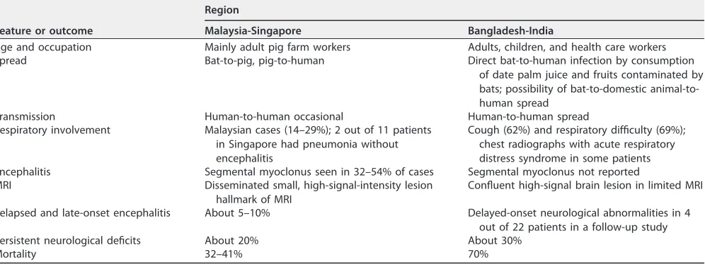
The differences in epidemiological and clinical features and outcomes of patients in the 2 large series in Malaysia-Singapore and Bangladesh-India are shown in Table 1 (6).
PATHOLOGY
In autopsies (29 full, 3 limited to the brain) performed on 32 Malaysian outbreak victims, pathological lesions were seen mainly in the brain, with disseminated
[image:5.585.87.325.71.212.2]micro-FIG 1Typical MRI pattern of multiple small white matter lesions. (A) Multiple punctate white matter lesions (arrowheads) are visible on T2-weighted MR image. (B) The largest lesion is more prominent on corresponding diffusion-weighted image (DWI). Reprinted with permission from Lim et al. (24).
TABLE 1Epidemiological and clinical features and outcomes in Nipah virus infectionsa
Feature or outcome
Region
Malaysia-Singapore Bangladesh-India
Age and occupation Mainly adult pig farm workers Adults, children, and health care workers
Spread Bat-to-pig, pig-to-human Direct bat-to-human infection by consumption
of date palm juice and fruits contaminated by bats; possibility of bat-to-domestic animal-to-human spread
Transmission Human-to-human occasional Human-to-human spread
Respiratory involvement Malaysian cases (14–29%); 2 out of 11 patients in Singapore had pneumonia without encephalitis
Cough (62%) and respiratory difficulty (69%); chest radiographs with acute respiratory distress syndrome in some patients Encephalitis Segmental myoclonus seen in 32–54% of cases Segmental myoclonus not reported
MRI Disseminated small, high-signal-intensity lesion
hallmark of MRI
Confluent high-signal brain lesion in limited MRI
Relapsed and late-onset encephalitis About 5–10% Delayed-onset neurological abnormalities in 4 out of 22 patients in a follow-up study
Persistent neurological deficits About 20% About 30%
Mortality 32–41% 70%
aAdapted from Sherrini and Tan (6).
on May 16, 2020 by guest
http://jcm.asm.org/
[image:5.585.46.544.544.731.2]infarction as a result of vasculitis-induced thrombosis and direct neuronal involvement. The respiratory tract, heart, and kidneys had similar vasculitic lesions.
All were positive for NiV (either by immunohistochemistry or serology) (28). Medium-sized and small blood vessels appeared to be the most involved by NiV, resulting in endothelial multinucleated syncytia and fibrinoid necrosis.
TREATMENT AND OUTCOME
Treatment measures were largely supportive and consisted of anticonvulsants, treatment of secondary infection, mechanical ventilation, and rehabilitation. With nothing known at the outset of the outbreak in Malaysia, empirical treatment was started with ribavirin, chosen for its broad-spectrum activity against DNA and RNA viruses and ability to cross the blood-brain barrier. Chong et al. reported a reduction in
mortality (54% in control versus 32% treatment arm,P⫽0.011) in an open-label trial
of ribavirin in 140 patients versus 54 controls (29).
In Malaysia, there were 265 cases of Nipah encephalitis and 105 deaths estimated from September 1998 to May 1999, giving a mortality rate close to 40%. The mean duration of illness from onset of symptoms to death was 16 days. Mortality was associated with positive viral culture from the CSF and severe brainstem involvement (5).
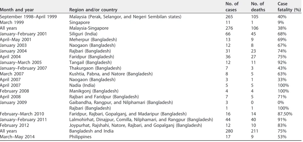
Mortality in Bangladesh and India has been much higher, approaching 70% (Table 2). This probably reflects greater involvement of the respiratory tract in the Bangladeshi-Indian outbreaks and differences in pathogenicity between the 2 viral strains, as well as less advanced health care facilities, such as intensive care units.
DISCOVERY OF THE VIRUS
In early March 1999, virologists from the University of Malaya had isolated a virus from cerebrospinal fluid of an encephalitis patient. Vero cells inoculated with cerebro-spinal fluid specimens from three fatal cases of encephalitis developed syncytia. Electron microscopic (EM) studies of the virus demonstrated features characteristic of a
virus belonging to the familyParamyxoviridae. The name, Nipah virus, was proposed
because the first isolate was made from clinical material from a fatal human case from Kampung Sungai Nipah, a village in Negeri Sembilan (7).
[image:6.585.42.546.84.320.2]Nipah virus-infected cells reacted strongly with Hendra virus antiserum, but did not react with antisera against other paramyxoviruses, including those for measles virus,
TABLE 2Morbidity and mortality of NiV in different regions
Month and year Region and/or country
No. of cases
No. of deaths
Case fatality (%)
September 1998–April 1999 Malaysia (Perak, Selangor, and Negeri Sembilan states) 265 105 40%
March 1999 Singapore 11 1 9%
All years Malaysia-Singapore 276 106 38%
January–February 2001 Siliguri (India) 66 45 68%
April–May 2001 Meherpur (Bangladesh) 13 9 69%
January 2003 Naogaon (Bangladesh) 12 8 67%
January 2004 Rajbari (Bangladesh) 31 23 74%
April 2004 Faridpur (Bangladesh) 36 27 75%
January–March 2005 Tangail (Bangladesh) 12 11 92%
January–February 2007 Thakurgaon (Bangladesh) 7 3 43%
March 2007 Kushtia, Pabna, and Natore (Bangladesh) 8 5 63%
April 2007 Naogaon (Bangladesh) 3 1 33%
April 2007 Nadia (India) 5 5 100%
February 2008 Manikgonj (Bangladesh) 4 4 100%
April 2008 Rajbari and Faridpur (Bangladesh) 7 5 71%
January 2009 Gaibandha, Rangpur, and Nilphamari (Bangladesh) 3 0 0%
Rajbari (Bangladesh) 1 1 100%
February–March 2010 Faridpur, Rajbari, Gopalganj, and Madaripur (Bangladesh) 16 14 87.50%
January–February 2011 Lalmohirhat, Dinajpur, Comilla, Nilphamari, and Rangpur (Bangladesh) 44 40 91% February 2012 Joypurhat, Rajshahi, Natore, Rajbari, and Gopalganj (Bangladesh) 12 10 83%
All years Bangladesh and India 280 211 75%
March–May 2014 Philippines 17 9 53%
Adapted from WHO data (http://www.searo.who.int/entity/emerging_diseases/links/nipah_virus_outbreaks_sear/en/).
on May 16, 2020 by guest
http://jcm.asm.org/
respiratory syncytial virus, and parainfluenzaviruses 1 and 3, as well as other viruses, including herpesvirus, enteroviruses, and JE virus, as indicated by immunofluorescence antibody assays. Cross-neutralization studies resulted in an 8- to 16-fold difference in neutralizing antibodies between Nipah and Hendra viruses, indicating that the viruses, though related, were not identical. Virus isolation or serologic testing confirmed Nipah virus infection in all cases from Singapore and in all but one of the initially identified encephalitis cases from Malaysia (7).
Classification. NiV is the second member of the genus Henipavirusin the family
Paramyxoviridae. The prototype virus of the genus is the closely related Hendra virus (HeV), discovered during an investigation of the 1994 lethal disease outbreak in horses and humans in Australia. While initially considered a potentially new member of the
genus Morbillivirus, hence tentatively named equine morbillivirus (EMV) (30),
subse-quent whole-genome analysis revealed several major molecular signatures of HeV that were not shared by any of the known morbilliviruses. Further analysis of the NiV genome sequence consolidated the notion that HeV and NiV are novel paramyxovi-ruses that did not fit into any of the existing genera in the family, and that there was a need to generate a new genus to accommodate the classification of these novel viruses (31). In 2002, the International Committee for Virus Taxonomy (ICTV) approved
the establishment of the new genusHenipavirus.
The Malaysian strain of NiV (NiV-MY) is slightly different from that of Bangladesh (NiV-BD). The outbreak in the Philippines was most likely caused by a NiV-MY strain.
Morphology. Similar to other paramyxoviruses, NiV particles are pleomorphic, spherical to filamentous, and range in size from 40 to 1,900 nm. They contain a single
layer of surface projections with an average length of 17⫾1 nm (32).
Genetic diversity.Among the NiVs known to cause disease in humans, there are two major genetic lineages, i.e., NiV Malaysia (NiV-MY) and NiV Bangladesh (NiV-BD).
Genome size and structure.The genome of the Malaysia NiV is 18,246 nucleotides (nt) in length, whereas that of the Bangladesh NiV is 18,252 nt (32). The potential role of this genome size increase in virus pathogenesis and interhost transmission is yet to be determined.
Functionally, the two strains are largely indistinguishable, but recent animal infec-tion studies suggest that the two viruses may be different in certain aspects. Infecinfec-tion studies in the African green monkey indicated that NiV-BD is more pathogenic than NiV-MY, and the window of passive antibody therapy is narrower for NiV-BD (33). In ferret infection studies, it was shown that NiV-BD infection resulted in increased oral shedding in comparison to NiV-MY (34) and a more rapid onset of productive infection and higher levels of virus replication in the respiratory tract (35). These differences may explain why more cases in Bangladesh and India have shorter incubation periods, more respiratory symptoms, greater human-to-human transmission, and higher case fatality rates.
EPIDEMIOLOGY IN ANIMALS
Reservoir host.Fruit bats (commonly known as flying foxes) in the genusPteropus,
familyPteropodidae, are main reservoir hosts of both NiV and HeV. Neither virus appears
to cause clinical disease in bats, regardless of whether they are infected naturally or experimentally (32).
Host range.Paramyxoviruses are traditionally known to have a limited host range, and interspecies transmission is rare (36). In contrast, NiV displays a very broad species tropism. In addition to multiple species of bats, NiV naturally infects pigs, horses, dogs, cats, and humans (32). NiV has also been shown to experimentally infect guinea pigs, hamsters, ferrets, squirrel monkeys, and African green monkeys. This wide range of species tropism is in part due to the fact that NiV uses ephrinB2/B3 molecules as their entry receptors, which are highly conserved among all mammals (37, 38).
It has been postulated that initial transmission of NiV from bats to pigs in Malaysia occurred in late 1997/early 1998 through contamination of pig swill by bat excretions, as a result of migration of these forest fruit bats to cultivated orchards and pig farms
on May 16, 2020 by guest
http://jcm.asm.org/
in Malaysia from Indonesia, which experienced El Nino-related drought and fires in 1997 to 1998 (39). Studies using satellite telemetry have shown that Malaysian flying foxes are highly mobile, traveling hundreds of kilometers between roosting sites within a year and occupying home ranges that extend beyond Malaysia to include Indonesia and Thailand (40). Additionally, Sendow and colleagues showed that Nipah virus circulates in populations of flying foxes in Indonesia and showed that the virus was
indistinguishable from the strains detected inPteropus vampyrusin peninsular Malaysia
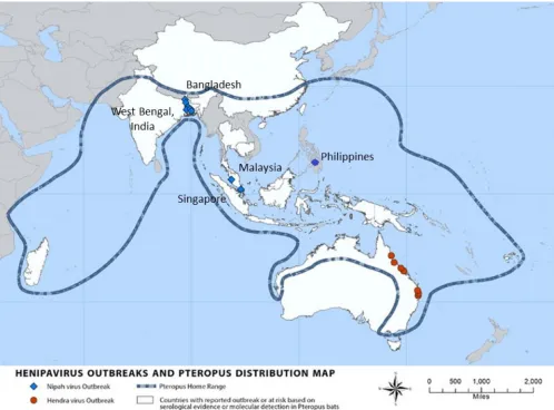
(41). The map (Fig. 2) shows the distribution ofPteropusbats and the countries where
outbreaks of Hendra virus and Nipah virus infections have occurred (41).
BIOSAFETY ISSUES OF NIPAH VIRUS
For those who have to work in the field or on farms where Nipah infection is suspected, personal protection, such as masks, goggles, gloves, gowns, and boots, is advocated, together with hand washing and disinfection of equipment (8, 42).
With its high virulence, animal-to-human and human-to-human spread, significant morbidity and mortality, and resultant fear and panic and tremendous economic losses caused, NiV fulfils some criteria to be considered a potential agent for bioterrorism (43, 44). It is thus listed as a category C agent on a list of bioterrorism agents by the Centers for Disease Control and Prevention (45), and any handling has to be done in biosafety level (BSL) 4 facilities.
FIG 2Map of henipavirus outbreaks and distribution ofPteropusbats. Adapted from Nipah virus distribution map, Centers for Disease Control and Prevention (www.cdc.gov/vhf/nipah/outbreaks/distribution-map.html).
on May 16, 2020 by guest
http://jcm.asm.org/
[image:8.585.44.542.72.440.2]PREVENTION
As treatment options are limited, focus on NiV management should be on preven-tion. Preventive strategies include interventions to prevent farm animals from acquiring NiV by eating fruit contaminated by bats. Farms should be designed to reduce overcrowding to avoid rapid spread of disease between animals and should not be near fruit trees that attract bats.
Consumption of contaminated sap should be avoided. However, efforts to reduce fresh sap consumption in general would be unpopular, as they go against social and cultural norms. Other, more acceptable methods would include physical barriers to prevent bats from accessing and contaminating sap (46).
A number of vaccine candidates have been found to be capable of complete protection against NiV disease in preclinical studies of small animal and nonhuman primate models. Candidate vaccines using a vesicular stomatitis virus vector are the most advanced, having demonstrated protection in hamsters, ferrets, and African green monkeys (47). Vaccination programs would also have to cover livestock animals, too, e.g., pigs, and perhaps horses in certain areas where NiV is endemic.
While WHO has declared NiV to be a priority pathogen, pharmaceutical companies may be reluctant to fund trials in underdeveloped countries that can ill afford medi-cations or vaccines. Fortunately, a new international coalition of governments and pharmaceutical companies called the Coalition for Epidemic Preparedness Innovations (CEPI) was formed in January 2017 to develop safe, effective, and affordable vaccines for diseases with pandemic potential, such as NiV.
CONCLUSION
NiV emerged as a new virus exactly 20 years ago, causing severe morbidity and mortality in both humans and animals and destroyed the pig-farming industry in Malaysia, and it continues to cause outbreaks in Bangladesh and India. As the reservoir hostPteropusbat is widespread, and NiV has been found in bats in various countries, the potential for outbreaks to occur in new regions remains significant.
REFERENCES
1. Tan KS, Tan CT, Goh KJ. 1999. Epidemiological aspects of Nipah virus infection. Neurol J South East Asia 4:77– 81.
2. Chua KB. 2003. Nipah virus outbreak in Malaysia. J Clin Virol 26:265–275.
https://doi.org/10.1016/S1386-6532(02)00268-8.
3. Chua KB, Goh KJ, Wong KT, Kamarulzaman A, Tan PS, Ksiazek TG, Zaki SR, Paul G, Lam SK, Tan CT. 1999. Fatal encephalitis due to Nipah virus among pig-farmers in Malaysia. Lancet 354:1257–1259.https://doi.org/ 10.1016/S0140-6736(99)04299-3.
4. Thongcharoen P. 1989. Japanese encephalitis virus encephalitis: an over-view. Southeast Asian J Trop Med Public Health 20:559 –573. 5. Looi LM, Chua KB. 2007. Lessons from the Nipah virus outbreak in
Malaysia. Malays J Pathol 29:63– 67.
6. Sherrini BA, Tan CT. 2014. Nipah encephalitis—an update. Med J Malay-sia 69(Suppl A):103–111.
7. Chua KB, Bellini WJ, Rota PA, Harcourt BH, Tamin A, Lam SK, Ksiazek TG, Rollin PE, Zaki SR, Shieh W, Goldsmith CS, Gubler DJ, Roehrig JT, Eaton B, Gould AR, Olson J, Field H, Daniels P, Ling AE, Peters CJ, Anderson LJ, Mahy BW. 2000. Nipah virus: a recently emergent deadly paramyxovirus. Science 288:1432–1435.https://doi.org/10.1126/science.288.5470.1432. 8. Lam SK, Chua KB. 2002. Nipah virus encephalitis outbreak in Malaysia.
Clin Infect Dis 34(Suppl 2):S48 –S51.https://doi.org/10.1086/338818. 9. Paton NI, Leo YS, Zaki SR, Auchus AP, Lee KE, Ling AE, Chew SK, Ang B,
Rollin PE, Umapathi T, Sng I, Lee CC, Lim E, Ksiazek TG. 1999. Outbreak of Nipah-virus infection among abattoir workers in Singapore. Lancet 354:1253–1256.https://doi.org/10.1016/S0140-6736(99)04379-2. 10. Chew MH, Arguin PM, Shay DK, Goh KT, Rollin PE, Shieh WJ, Zaki SR, Rota
PA, Ling AE, Ksiazek TG, Chew SK, Anderson LJ. 2000. Risk factors for Nipah virus infection among abattoir workers in Singapore. J Infect Dis 181:1760 –1763.https://doi.org/10.1086/315443.
11. Yob JM, Field H, Rashdi AM, Morrissy C, van der Heide B, Rota P, bin Adzhar A, White J, Daniels P, Jamaluddin A, Ksiazek T. 2001. Nipah virus
infection in bats (order Chiroptera) in peninsular Malaysia. Emerg Infect Dis 7:439 – 441.https://doi.org/10.3201/eid0703.017312.
12. Hsu VP, Hossain MJ, Parashar UD, Ali MM, Ksiazek TG, Kuzmin I, Niezgoda M, Rupprecht C, Bresee J, Breiman RF. 2004. Nipah virus encephalitis reemergence, Bangladesh. Emerg Infect Dis 10:2082–2087.https://doi .org/10.3201/eid1012.040701.
13. Chadha MS, Comer JA, Lowe L, Rota PA, Rollin PE, Bellini WJ, Ksiazek TG, Mishra A. 2006. Nipah virus-associated encephalitis outbreak, Siliguri, India. Emerg Infect Dis 12:235–240.https://doi.org/10.3201/eid1202.051247. 14. Islam MS, Sazzad HM, Satter SM, Sultana S, Hossain MJ, Hasan M, Rahman
M, Campbell S, Cannon DL, Stroher U, Daszak P, Luby SP, Gurley ES. 2016. Nipah virus transmission from bats to humans associated with drinking traditional liquor made from date palm sap, Bangladesh, 2011–2014. Emerg Infect Dis 22:664 – 670.https://doi.org/10.3201/eid2204.151747.
15. Ching PK, de los Reyes VC, Sucaldito MN, Tayag E, Columna-Vingno AB, Malbas FF, Jr, Bolo GC, Jr, Sejvar JJ, Eagles D, Playford G, Dueger E, Kaku Y, Morikawa S, Kuroda M, Marsh GA, McCullough S, Foxwell AR. 2015. Outbreak of henipavirus infection, Philippines, 2014. Emerg Infect Dis 21:328 –331.https://doi.org/10.3201/eid2102.141433.
16. Mounts AW, Kaur H, Parashar UD, Ksiazek TG, Cannon D, Arokiasamy JT, Anderson LJ, Lye MS. 2001. A cohort study of health care workers to assess nosocomial transmissibility of Nipah virus, Malaysia, 1999. J Infect Dis 183:810 – 813.https://doi.org/10.1086/318822.
17. Luby SP, Gurley ES, Hossain MJ. 2009. Transmission of human infection with Nipah virus. Clin Infect Dis 49:1743–1748.https://doi.org/10.1086/ 647951.
18. Gurley ES, Montgomery JM, Hossain MJ, Bell M, Azad AK, Islam MR, Molla MA, Carroll DS, Ksiazek TG, Rota PA, Lowe L, Comer JA, Rollin P, Czub M, Grolla A, Feldmann H, Luby SP, Woodward JL, Breiman RF. 2007. Person-to-person transmission of Nipah virus in a Bangladeshi community. Emerg Infect Dis 13:1031–1037.https://doi.org/10.3201/eid1307.061128.
on May 16, 2020 by guest
http://jcm.asm.org/
19. Goh KJ, Tan CT, Chew NK, Tan PS, Kamarulzaman A, Sarji SA, Wong KT, Abdullah BJ, Chua KB, Lam SK. 2000. Clinical features of Nipah virus encephalitis among pig farmers in Malaysia. N Engl J Med 342: 1229 –1235.https://doi.org/10.1056/NEJM200004273421701.
20. Tan CT, Goh KJ, Wong KT, Sarji SA, Chua KB, Chew NK, Murugasu P, Loh YL, Chong HT, Tan KS, Thayaparan T, Kumar S, Jusoh MR. 2002. Relapsed and late-onset Nipah encephalitis. Ann Neurol 51:703–708.https://doi .org/10.1002/ana.10212.
21. Abdullah S, Chang L-Y, Rahmat K, Goh KJ, Tan CT. 2012. Late-onset Nipah virus encephalitis 11 years after the initial outbreak: a case report. Neurol Asia 17:71–74.
22. Ng BY, Lim CC, Yeoh A, Lee WL. 2004. Neuropsychiatric sequelae of Nipah virus encephalitis. J Neuropsychiatry Clin Neurosci 16:500 –504.
https://doi.org/10.1176/jnp.16.4.500.
23. Sejvar JJ, Hossain J, Saha SK, Gurley ES, Banu S, Hamadani JD, Faiz MA, Siddiqui FM, Mohammad QD, Mollah AH, Uddin R, Alam R, Rahman R, Tan CT, Bellini W, Rota P, Breiman RF, Luby SP. 2007. Long-term neuro-logical and functional outcome in Nipah virus infection. Ann Neurol 62:235–242.https://doi.org/10.1002/ana.21178.
24. Lim CCT, Sitoh YY, Hui F, Lee KE, Ang BS, Lim E, Lim WE, Oh HM, Tambyah PA, Wong JS, Tan CB, Chee TS. 2000. Nipah viral encephalitis or Japanese encephalitis? MR findings in a new zoonotic disease. Am J Neuroradiol 21:455– 461.
25. Lim CC, Lee KE, Lee WL, Tambyah PA, Lee CC, Sitoh YY, Auchus AP, Lin BK, Hui F. 2002. Nipah virus encephalitis: serial MR study of an emerging disease. Radiology 222:219 –226.https://doi.org/10.1148/ radiol.2221010499.
26. Lim CC, Lee WL, Leo YS, Lee KE, Chan KP, Ling AE, Oh H, Auchus AP, Paton NI, Hui F, Tambyah PA. 2003. Late clinical and magnetic resonance imaging follow up of Nipah virus infection. J Neurol Neurosurg Psychi-atry 74:131–133.https://doi.org/10.1136/jnnp.74.1.131.
27. Chong HT, Hossain J, Tan CT. 2008. Differences in epidemiologic and clinical features of Nipah virus encephalitis between the Malaysian and Bangladesh outbreaks. Neurol Asia 13:23–26.
28. Wong KT, Shieh WJ, Kumar S, Norain K, Abdullah W, Guarner J, Gold-smith CS, Chua KB, Lam SK, Tan CT, Goh KJ, Chong HT, Jusoh R, Rollin PE, Ksiazek TG, Zaki SR. 2002. Nipah virus infection: pathology and patho-genesis of an emerging paramyxoviral zoonosis. Am J Pathol 161: 2153–2167.https://doi.org/10.1016/S0002-9440(10)64493-8.
29. Chong HT, Kamarulzaman A, Tan CT, Goh KJ, Thayaparan T, Kunjapan SR, Chew NK, Chua KB, Lam SK. 2001. Treatment of acute Nipah encephalitis with ribavirin. Ann Neurol 49:810 – 813.https://doi.org/ 10.1002/ana.1062.
30. Murray K, Selleck P, Hooper P, Hyatt A, Gould A, Gleeson L, Westbury H, Hiley L, Selvey L, Rodwell B. 1995. A morbillivirus that caused fatal disease in horses and humans. Science 268:94 –97.https://doi.org/10 .1126/science.7701348.
31. Wang LF, Yu M, Hansson E, Pritchard LI, Shiell B, Michalski WP, Eaton BT. 2000. The exceptionally large genome of Hendra virus: support for creation of a new genus within the family Paramyxoviridae. J Virol 74:9972–9979.https://doi.org/10.1128/JVI.74.21.9972-9979.2000. 32. Wang LF, Mackenzie JS, Broder CC. 2013. Henipaviruses, p 286 –313. In
Knipe DM, Howley PM (ed), Fields virology, 6th ed. Lippincott Williams & Wilkins, Philadelphia, PA.
33. Mire CE, Satterfield BA, Geisbert JB, Agans KN, Borisevich V, Yan L, Chan YP, Cross RW, Fenton KA, Broder CC, Geisbert TW. 2016. Pathogenic differences between Nipah virus Bangladesh and Malaysia strains in
primates: implications for antibody therapy. Sci Rep 6:30916.https://doi .org/10.1038/srep30916.
34. Clayton BA, Middleton D, Bergfeld J, Haining J, Arkinstall R, Wang L, Marsh GA. 2012. Transmission routes for Nipah virus from Malaysia and Bangladesh. Emerg Infect Dis 18:1983–1993.https://doi.org/10.3201/ eid1812.120875.
35. Clayton BA, Middleton D, Arkinstall R, Frazer L, Wang LF, Marsh GA. 2016. The nature of exposure drives transmission of Nipah viruses from Ma-laysia and Bangladesh in ferrets. PLoS Negl Trop Dis 10:e0004775.
https://doi.org/10.1371/journal.pntd.0004775.
36. Lamb RA, Parks GD. 2007.Paramyxoviridae: the viruses and their repli-cation, p 1449 –1496. In Fields BN, Knipe DN, Howley PM (ed), Fields virology, 5th ed. Lippincott Williams & Wilkins, Philadelphia, PA. 37. Negrete OA, Wolf MC, Aguilar HC, Enterlein S, Wang W, Muhlberger E, Su
SV, Bertolotti-Ciarlet A, Flick R, Lee B. 2006. Two key residues in ephrinB3 are critical for its use as an alternative receptor for Nipah virus. PLoS Pathog 2:e7.https://doi.org/10.1371/journal.ppat.0020007.
38. Bonaparte MI, Dimitrov AS, Bossart KN, Crameri G, Mungall BA, Bishop KA, Choudhry V, Dimitrov DS, Wang LF, Eaton BT, Broder CC. 2005. Ephrin-B2 ligand is a functional receptor for Hendra virus and Nipah virus. Proc Natl Acad Sci U S A 102:10652–10657. https://doi.org/10 .1073/pnas.0504887102.
39. Chua KB, Chua BH, Wang CW. 2002. Anthropogenic deforestation, El Nino and the emergence of Nipah virus in Malaysia. Malays J Pathol 24:15–21.
40. Epstein Jonathan H, Olival Kevin J, Pulliam Juliet RC, Smith C, Westrum J, Hughes T, Dobson Andrew P, Zubaid A, Rahman Sohayati A, Basir Misliah M, Field Hume E, Daszak P. 2009.Pteropus vampyrus, a hunted migratory species with a multinational home-range and a need for regional management. J Appl Ecol 46:991–1002.https://doi.org/10.1111/ j.1365-2664.2009.01699.x.
41. Sendow I, Ratnawati A, Taylor T, Adjid RMA, Saepulloh M, Barr J, Wong F, Daniels P, Field H. 2013. Nipah virus in the fruit batPteropus vampyrus
in Sumatera, Indonesia. PLoS One 8:e69544.https://doi.org/10.1371/ journal.pone.0069544.
42. Food and Agriculture Organization of the United Nations, Regional Office for Asia and the Pacific. 2002. Manual on the diagnosis of Nipah virus infection in animals. RAP publication no. 2002/01. Food and Agri-culture Organization of the United Nations, Regional Office for Asia and the Pacific, Bangkok, Thailand.
43. Balali-Mood M, Moshiri M, Etemad L. 2013. Medical aspects of bio-terrorism. Toxicon 69:131–142.https://doi.org/10.1016/j.toxicon.2013.01 .005.
44. Lam SK. 2003. Nipah virus—a potential agent of bioterrorism? Antiviral Res 57:113–119.https://doi.org/10.1016/S0166-3542(02)00204-8. 45. Centers for Disease Control and Prevention. 2018. Bioterrorism agents.
Centers for Disease Control and Prevention, Atlanta, GA.https://fas.org/ biosecurity/resource/documents/CDC_Bioterrorism_Agents.pdf. 46. Nahar N, Mondal UK, Sultana R, Hossain MJ, Khan MS, Gurley ES, Oliveras
E, Luby SP. 2013. Piloting the use of indigenous methods to prevent Nipah virus infection by interrupting bats’ access to date palm sap in Bangladesh. Health Promot Int 28:378 –386. https://doi.org/10.1093/ heapro/das020.
47. Satterfield BA, Dawes BE, Milligan GN. 2016. Status of vaccine research and development of vaccines for Nipah virus. Vaccine 34:2971–2975.
https://doi.org/10.1016/j.vaccine.2015.12.075.