Copyright © 2004, American Society for Microbiology. All Rights Reserved.
Evaluation of Performance and Potential Clinical Impact of ProSpecT
Shiga Toxin
Escherichia coli
Microplate Assay for Detection
of Shiga Toxin-Producing
E. coli
in Stool Samples
Patrick J. Gavin,
1,2Lance R. Peterson,
1,2Anna C. Pasquariello,
1Joanna Blackburn,
1Mark G. Hamming,
1Kuo J. Kuo,
3and Richard B. Thomson, Jr.
1,2*
Department of Pathology and Laboratory Medicine, Evanston Northwestern Healthcare, Evanston,1and
Northwestern University Feinberg School of Medicine,2and Enteric Laboratory,
Illinois Department of Public Health,3Chicago, Illinois
Received 9 August 2003/Returned for modification 19 October 2003/Accepted 7 January 2004
Shiga toxin-producing Escherichia coli bacteria (STEC) are emerging pathogens capable of producing sporadic and epidemic diarrhea, hemorrhagic colitis, and potentially life-threatening hemolytic-uremic syn-drome. Although the presence of E. coliO157 can be readily detected in stool by sorbitol-MacConkey agar culture (SMAC), STEC non-O157 serotypes cannot. In contrast to culture, testing for the presence of Shiga toxins 1 and 2 in stool detects both O157 and non-O157 STEC serotypes capable of causing disease. Over two consecutive summers, we evaluated the performance of the ProSpecT Shiga toxin E. coliMicroplate assay (Alexon-Trend, Ramsey, Minn.), an enzyme immunoassay for the detection of Shiga toxins 1 and 2, on all stools submitted for culture of enteric pathogens, and the potential clinical impact of Shiga toxin detection. Twenty-nine stool specimens were STEC positive by ProSpecT assay. Twenty-seven of 29 STEC-positive isolates were confirmed by SMAC and serotyping or by a second enzyme immunoassay and PCR (positive predictive value, 93%). Thirteen of 27 confirmed Shiga toxin-producing strains were serotype O157. The remaining 14 strains represented 8 other serotypes. The ProSpecT assay was 100% sensitive and specific for detection ofE. coliO157 in stool (7 of 7) compared to SMAC. In addition, the ProSpecT assay detected twice as many STEC as SMAC. Fifty-two percent of confirmed STEC-positive stools were nonbloody. Thus, in our population, screening strategies that test only visibly bloody stools for STEC would miss a majority of cases. Eleven (41%) STEC-positive patients were hospitalized, and eight (30%) developed severe disease (two developed hemolytic-uremic syndrome, and six developed hemorrhagic colitis). Prior to detection of STEC infection, seven (26%) and eight patients (30%) underwent unnecessary diagnostic procedures or received potentially deleterious empirical treatment, respectively. We propose that establishing a specific diagnosis of STEC may have prevented these potentially harmful interven-tions. We conclude that the ProSpecT assay is sensitive and specific for the detection of Shiga toxins 1 and 2 in stool and has potentially significant clinical impact for the individual patient and public health. Shiga toxin assays should be considered for routine use in settings where prevalence of STEC disease warrants testing.
Shiga toxin-producing Escherichia coli organisms (STEC) are emerging pathogens capable of producing sporadic and epidemic diarrhea, hemorrhagic colitis, and potentially life-threatening hemolytic-uremic syndrome (HUS) (4, 7, 15, 18, 25). Shiga toxin production is the defining characteristic of STEC, and Shiga toxins 1 and 2 are the virulence factors responsible for these serious complications (18, 28, 29, 32).
Escherichia coliO157:H7, the most frequently identified STEC
serotype, alone causes an estimated 60 deaths and 73,000 ill-nesses annually in the United States (27).E. coliO157 lacks the ability to rapidly ferment sorbitol and can be detected in stool with sorbitol-MacConkey agar culture (SMAC) and se-rotyping (24). In addition, approximately 100E. colinon-O157 serotypes ferment sorbitol, produce one or both Shiga toxins, and are an increasing cause of potentially serious disease (1, 9, 10, 17, 24, 27, 28; A. B. Rouse and J. M. Campos, Abstr. 100th Gen. Meet. Am. Soc. Microbiol., abstr. C-216, 2000; L. J. Nims, D. S. Horensky, L. L. Bucks, S. A. Young, J. L. Golobics, K. D.
Greene, and E. G. Sowers, Abstr. 101st Gen. Meet. Am. Soc. Microbiol., abstr. C-165, 2001).
In 1999, the Centers for Disease Control and Prevention (CDC) estimated that one-third of STEC-associated diarrhea was caused by non-O157 serotypes (27). Non-O157 STEC strains are not detected by SMAC; few microbiology laboratories test for their presence, and the true prevalence is unknown (8). However, both STEC O157 and non-O157 serotypes capable of producing disease are detected by virulence factor-based tests for the pres-ence of Shiga toxins 1 and 2 in stool. We evaluated the perfor-mance of the ProSpecT Shiga toxinEscherichia coliMicroplate assay (Alexon-Trend, Ramsay, Minn.), an enzyme immunoassay for detection of Shiga toxins 1 and 2, in stools submitted for culture of enteric pathogens, and evaluated the potential clinical impact of this diagnosis.
(This work was presented, in part, at the 103rd Annual Meeting of the American Society for Microbiology, May 2003, Washington, D.C.)
MATERIALS AND METHODS
Specimens.During the summer months of 2001 and 2002, consecutive stool specimens from outpatients and inpatients submitted to Evanston Northwestern Healthcare (ENH) for routine culture of enteric pathogens were tested prospec-tively for the presence of Shiga toxins 1 and 2 by the ProSpecT assay. Specifically, * Corresponding author. Mailing address: Department of Pathology
and Laboratory Medicine, Evanston Northwestern Healthcare, Room 1936, 2650 Ridge Ave., Evanston, IL 60201. Phone: (847) 570-2744. Fax: (847) 733-5314. E-mail: [email protected].
1652
on May 15, 2020 by guest
http://jcm.asm.org/
from June to August 2001, consecutive stool specimens were tested for Shiga toxins directly by ProSpecT assay and were tested blindly and in parallel by SMAC. Subsequently, from June to September 2002, when we were satisfied that the ProSpecT assay was sensitive and specific for detection ofE. coliO157, the initial SMAC screening plate was dropped, and stool specimens were tested by ProSpecT assay alone.
Fresh stool specimens were submitted in standard clean or sterile, leak-proof containers without preservative or in modified Cary-Blair transport medium (Remel, Lenexa, Kans.). Stools were classified as bloody when blood was visible to the technologist or reported as present by the submitting physician. Fifty microliters or a small pea-sized sample of fresh stool or stool in Cary-Blair transport medium was inoculated into 5 ml of sterile MacConkey broth (Remel) within 2 h of arrival in the laboratory. Specimens were incubated in MacConkey broth at 37°C for 18 to 24 h. Specimens that were dried, received in fixative, or held unrefrigerated without transport medium for more than 4 h were rejected. All Shiga toxin-positive specimens were screened for presence ofE. coliO157 using SMAC, and non-sorbitol-fermentingE. coliwere serotyped with anti-O157 antibody (Pro-Laboratory Diagnostics, Inc., Ontario, Canada). All STEC-posi-tive stool specimens were submitted to the Illinois Department of Public Health laboratory (the state laboratory), where SMAC was repeated. Non-sorbitol-fermenting colonies were serotyped with anti-O157 antibody (Remel) and tested by a second Shiga toxin immunoassay (Premier EHEC assay; Meridian Diag-nostics Inc., Cincinnati, Ohio). In addition, to identify sorbitol-fermenting STEC on SMAC, a colony sweep and up to five isolated sorbitol-positive colonies were tested by Premier EHEC assay at the state laboratory. All non-O157 STEC-positive strains were forwarded to the Enteric Pathogens Laboratory at the CDC, where PCR for genotype and non-O157 serotyping were performed.
ProSpecT Shiga toxinEscherichia colimicroplate assay.The ProSpecT assay uses rabbit polyclonal anti-Shiga toxin 1 and 2 capture antibodies, and a horse-radish peroxidase-labeled monoclonal mouse anti-Shiga toxin 1 and 2 conjugate. The ProSpecT assay was performed according to the manufacturer’s instructions. Briefly, 200l of specimen from MacConkey broth was added to each microplate well and the reaction mixture was incubated at room temperature for 60 min. After a series of manual washes, the enzyme conjugate was added and the mixture was incubated at room temperature for 30 min. After a further series of washes, color substrate was added, and the reaction mixtures were read spectro-photometrically at 450 nm with a dual-wavelength spectrophotometer (BIO-TEK Instruments Inc., Winooski, Vt.). Specimens were considered Shiga toxin positive when the optical density wasⱖ0.200. The materials and labor cost of testing stool specimens by ProSpecT assay was calculated to be $16 per test, based on list price for the assay and an average labor cost of $20/h.
Culture.All stool specimens were inoculated into sheep blood, MacConkey, Hecktoen enteric, andCampylobacteragars; incubated in the appropriate atmo-sphere; and examined for growth ofAeromonasspp.,Campylobacterspp., Plesi-omonasspp.,Salmonellasp.,Shigellaspp., andYersiniaspp. by standard methods. Shiga toxin-positive specimens were cultured on SMAC, and non-sorbitol-fer-menting colonies were tested with anti-O157 antibody.
Patients.Clinical and demographic data of patients with STEC-positive stools were obtained from the electronic patient record or from the physician.
RESULTS
During the study periods, stool specimens from 2,060 pa-tients were tested for presence of Shiga toxins 1 and 2 by ProSpecT assay and for enteric pathogens by culture. Stool cultures from 285 patients (14%) were positive for an enteric pathogen:Campylobacterspp., 146;Salmonellasp., 66;Shigella
spp., 36; andAeromonasspp., 10. A total of 29 of 2,060 stool specimens (1.4%) were STEC positive. No stool specimen was positive for more than one pathogen. Twenty-seven of 29 stools that were STEC positive by ProSpecT assay were con-firmed positive at the state laboratory (positive predictive value, 93%) (Tables 1 and 2). During the first summer of the study, from June to August 2001, 7 of 543 consecutive stool specimens wereE. coliO157 positive by ProSpecT assay and SMAC at ENH and at the state laboratory. No STEC-positive specimen that failed to grow sorbitol-negative colonies was subsequently serotyped asE. coliO157 (sensitivity and speci-ficity for the detection ofE. coliO157, 100%).
Overall, 14 of 27 (52%) confirmed STEC-positive stools were non-O157 serotypes. Twelve of the 14 were submitted to the Enteric Pathogen Laboratory at the CDC: 3 were serotype O126:H11 and 7 were other individual non-O157 serotypes. One sorbitol-fermenting isolate was Shiga toxin positive by both ProSpecT and Premier EHEC assays, but the presence of Shiga toxin was not confirmed by the CDC (isolate from pa-tient 26). Two non-O157 serotype isolates were not tested by the CDC (those from patients 15 and 25). Stool specimens from patients 24 and 25, who were siblings, were temporally clustered with patient 26, who attended the same day care center.
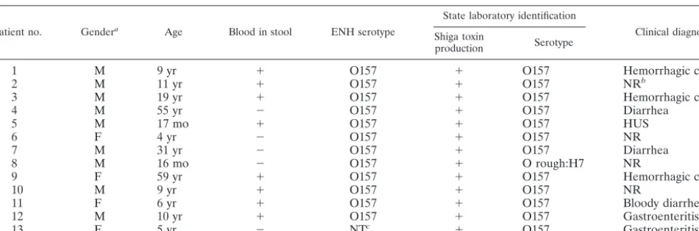
[image:2.603.50.540.80.242.2]Two of 29 stool specimens (7%) were Shiga toxin positive by the ProSpecT assay but negative by Premier EHEC assay at the state laboratory (specimens from patients 28 and 29). Both specimens were nonbloody and sorbitol fermenting. Although it is conceivable that the sweep of sorbitol-positive colonies at TABLE 1. O157 Serotype Shiga toxin-producingE. coliisolates detected in this study
Patient no. Gendera Age Blood in stool ENH serotype
State laboratory identification
Clinical diagnosis Shiga toxin
production Serotype
1 M 9 yr ⫹ O157 ⫹ O157 Hemorrhagic colitis
2 M 11 yr ⫹ O157 ⫹ O157 NRb
3 M 19 yr ⫹ O157 ⫹ O157 Hemorrhagic colitis
4 M 55 yr ⫺ O157 ⫹ O157 Diarrhea
5 M 17 mo ⫹ O157 ⫹ O157 HUS
6 F 4 yr ⫺ O157 ⫹ O157 NR
7 M 31 yr ⫺ O157 ⫹ O157 Diarrhea
8 M 16 mo ⫺ O157 ⫹ O rough:H7 NR
9 F 59 yr ⫹ O157 ⫹ O157 Hemorrhagic colitis
10 M 9 yr ⫹ O157 ⫹ O157 NR
11 F 6 yr ⫹ O157 ⫹ O157 Bloody diarrhea
12 M 10 yr ⫹ O157 ⫹ O157 Gastroenteritis
13 F 5 yr ⫺ NTc ⫹ O157 Gastroenteritis
aAbbreviations: M, male; F, female. bNR, not recorded.
cNT, not typed.
on May 15, 2020 by guest
http://jcm.asm.org/
the state laboratory may have missed Shiga toxin-producing colonies, both results were considered false positives by the ProSpecT assay. The state laboratory did have difficulty isolat-ing STEC-positive, sorbitol-negative colonies from SMAC of a stool specimen from a patient with hemorrhagic colitis (patient 27). After repeat isolation and testing of colony sweeps by ProSpecT assay at ENH, the presence of STEC was subse-quently confirmed by the state laboratory.
Of 27 confirmed STEC-positive patients, 11 (41%) were hospitalized (median stay, 4 days; range, 1 to 18 days) and 8 (30%) developed severe STEC-associated disease (2 devel-oped HUS, and 6 develdevel-oped hemorrhagic colitis).E. coliO157 and non-O157 serotypes were associated with equal numbers of cases of hemorrhagic colitis and HUS (3 and 1 each, respec-tively). Stool specimens from all patients with hemorrhagic colitis and HUS were visibly bloody. However, in our popula-tion, as in an earlier report, the majority of STEC-positive stools (14 of 27; 52%) were nonbloody (33). In addition, sim-ilar to other reports, bloody stools were more frequently asso-ciated with O157 (62%; 8 of 13) than non-O157 STEC sero-types (35%; 5 of 14) (22, 30, 31).
Eight of 27 confirmed STEC-positive patients (30%) re-ceived empirical antimicrobial (6) and/or antimotility treat-ment (4) (data not shown). Among the patients treated with antimicrobials, quinolones were the most frequently prescribed agent (5 of 6; 83%). The majority of patients with hemorrhagic colitis or HUS (5 of 8; 62%) received empirical antimicrobial (4) and/or antimotility agents (3) before diagnosis of STEC infection. All four patients treated with antimotility agents prior to the diagnosis of STEC infection were subsequently hospitalized.
Over two summers, screening 2,060 stool specimens for the presence of Shiga toxins by ProSpecT assay cost $32,960.
DISCUSSION
From June to August or September of 2001 and 2002, we prospectively evaluated the ProSpecT assay for detection of Shiga toxins 1 and 2 in stool specimens. The ProSpecT assay
was a highly sensitive and specific test forE. coliO157 in stool (seven of seven without false positives; 100%) compared to SMAC. The positive predictive value of the ProSpecT assay for the presence of O157 and non-O157 STEC serotypes com-bined was 93% (27 of 29 specimens). Performance of Pro-SpecT assay for the detection of O157 and non-O157 STEC, in our population, was similar to that observed in an evaluation by Kehl et al. (sensitivity and specificity, 99%) and comparable to studies of the Premier EHEC assay for detection of O157 STEC (sensitivity, 89 to 100%; specificity, 99.7%) and O157 and non-O157 STEC (sensitivity, 98 to 100%; specificity, 98%) (19, 20, 22, 31).
Many laboratories either fail to screen for STEC or screen only visibly bloody stools either because the local prevalence of STEC is considered to be too low or in an effort to contain laboratory costs (8). The prevalence of STEC in our popula-tion (1.3%) was almost identical to that of an earlier multi-center U.S. study, in which STEC were as prevalent asShigella
spp. (D. W. Acheson, K. Frankson, D. Willis, et al., Abstr. 98th Gen. Meet. Am. Soc. Microbiol., abstr. C-205, 1998). The prevalence ofShigellaspp. was slightly higher in our population (1.7%), so that STEC were the fourth most common enteric pathogen during the summer months. In an earlier large Bel-gian PCR-based study of more than 10,000 stool specimens, STEC were the third most prevalent enteric pathogens after
CampylobacterandSalmonella(33).
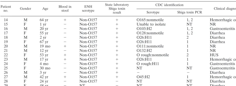
Having decided that the local prevalence of STEC warrants some form of screening, our findings emphasize again the importance of using a test that detects both O157 and O157 STEC serotypes. In the present study, as in others, non-O157 STEC were more prevalent than non-O157 STEC serotypes (D. W. Acheson et al., 98th Gen. Meet. Am. Soc. Microbiol.; L. J. Nims et al., 101st Gen. Meet. Am. Soc. Microbiol.). In the last decade, Australia, Europe, and North and South America have seen an increase in detection and reporting of non-O157 STEC as a cause of HUS (L. J. Nims et al., 101st Gen. Mtg. Am. Soc. Microbiol.) (1, 9–12, 14, 20, 33). Similarly, in our population, the majority of STEC infections and half of the cases of hemorrhagic colitis and HUS were caused by non-TABLE 2. Non-O157 serotype Shiga toxin-producingE. coliisolates detected in this studya
Patient
no. Gender Age Blood instool serotypeENH
State laboratory Shiga toxin
result
CDC identification
Clinical diagnosis Serotype Shiga toxin PCR
14 M 64 yr ⫹ Non-O157 ⫹ O165:nonmotile 1, 2 Hemorrhagic colitis
15 F 1 yr ⫺ Non-O157 ⫹ Unable to isolate NT NR
16 M 20 yr ⫺ Non-O157 ⫹ O103:H2 1, 2 Gastroenteritis
17 F 55 yr ⫺ Non-O157 ⫹ O128:nonmotile 1, 2 Diarrhea
18 M 2 yr ⫺ Non-O157 ⫹ O26:H11 2 Diarrhea
19 F 67 yr ⫺ Non-O157 ⫹ O26:H11 1 Diarrhea
20 M 19 mo ⫹ Non-O157 ⫹ O111:nonmotile 1 NR
21 M 12 yr ⫺ Non-O157 ⫹ O132:H2 1 NR
22 F 53 yr ⫹ Non-O157 ⫹ O rough:nonmotile 2 HUS
23 M 17 yr ⫹ Non-O157 ⫹ O26:H11 1 Hemorrhagic colitis
24 F 4 mo ⫺ Non-O157 ⫹ O rough:H11 1 Gastroenteritis
25 F 30 mo ⫺ Non-O157 ⫹ NT NT Gastroenteritis
26 M 3 yr ⫺ Non-O157 ⫹ ⫺ ⫺ Diarrhea
27 M 42 yr ⫹ Non-O157 ⫹ O45:H2 1 Hemorrhagic colitis
28 F 24 yr ⫺ Non-O157 ⫺ NT NT Diarrhea
29 F 48 yr ⫺ NT ⫺ NT NT Diarrhea
aAbbreviations: M, male; F, female; NR, not recorded; NT, not tested.
on May 15, 2020 by guest
http://jcm.asm.org/
[image:3.603.44.542.81.263.2]O157 STEC serotypes. As in previous studies, in which viru-lence factor-based tests led to increases of 20 to 30% in de-tection of STEC-positive isolates, the ProSpecT assay detected twice as many STEC positives as SMAC culture alone (19, 34). After deciding to screen for STEC and recognizing the in-creasing importance of non-O157 serotype STEC, the question arises as to which stool specimens to screen with a virulence-factor based test. Most U.S. laboratories that screen for STEC screen only visibly bloody stools (8). However, presence of blood in the stool is a poor predictor of the presence of STEC (19). In the present study, screening visibly bloody stools would have identified all patients with severe STEC-associated dis-ease but would have missed the majority of STEC-positive patients (14 of 27 [52%] with nonbloody stools). In addition, screening only bloody stools may miss STEC-positive patients if the presence of blood is obscured in transport medium or if a history is not conveyed to the laboratory. Specifically, in this study, screening all stools, including nonbloody specimens, by ProSpecT assay detected almost twice as a many STEC-posi-tive specimens. Previously in our laboratory, stools were screened for STEC by SMAC only if visibly bloody or upon request. Our present strategy is to screen all stools for STEC by the ProSpecT assay during the warmer summer months, when the frequency of STEC infection increases (22, 23, 30, 35). For the remainder of the year, in an effort to improve efficiency and cost-effectiveness of STEC screening, we test for STEC only if stools are bloody or upon request. For epidemi-ologic and public health purposes, all STEC-positive strains undergo SMAC and serotyping of sorbitol-negative colonies and are forwarded to the state laboratory.
Detection and identification of even a single O157 or non-O157 STEC infection have important benefits for the individ-ual and for public health (5). In searching for a cause of bloody diarrhea, physicians have performed unnecessary diagnostic imaging, colonoscopies, and surgeries on patients subsequently shown to be infected with STEC (15, 25). Similarly, seven of our STEC-positive patients (26%) had additional diagnostic procedures performed (computed tomography was performed on four patients, and colonoscopy was performed on three patients) prior to detection of STEC (data not shown). We suggest, and Park et al. (31) have recently demonstrated, that an earlier diagnosis of STEC infection would have avoided such potentially harmful and expensive investigations in a sig-nificant number of our patients. In addition, establishing a specific diagnosis may prevent potentially deleterious antimi-crobial or antimotility treatment (2, 26, 36, 37). It is a matter of concern that 30% of STEC-positive patients in the present study received empirical antimicrobials (6) and/or antimotility agents (4). The fact that quinolones were the antimicrobials prescribed in the majority of these cases is worrisome. Exper-imental evidence demonstrates that quinolones are potent in-ducers of Shiga toxin-encoding bacteriophages and toxin pro-duction in STEC (21, 39). Whereas the role of antimicrobials in the pathogenesis of HUS may be considered controversial by some, the potentially deleterious effect of antimotility treat-ment upon the evolution of STEC disease is well established (6, 16, 26). Thus, it is particularly worrisome that several pa-tients received antimotility agents prior to diagnosis of STEC infection. Our findings reinforce past data that physicians should not prescribe antimicrobials or antimotility treatment
for patients known to have STEC infection. Furthermore, at a minimum, early detection permits identification and optimal monitoring of patients at risk of progression to HUS (38). And finally, in the future, timely microbiologic diagnosis of STEC infection may permit specific anti-Shiga toxin prophylaxis to prevent severe STEC-associated disease (3, 13).
The public health benefits of diagnosing STEC infection are readily apparent in the setting of outbreaks of this potentially lethal agent (5). Detection of a STEC-positive patient requires an epidemiologic investigation to assess whether a case is truly sporadic or part of an outbreak, to determine the source of the outbreak, and to prevent further spread. Early detection, rapid notification, and timely epidemiologic investigation of a STEC outbreak in the western United States led to recall of contam-inated product and prevention of an estimated 800 additional cases of infection (5). In the present study, familial and day care center transmission of non-O157 STEC (patients 24, 25, and 26) may have gone undetected if screening was limited to bloody stools or SMAC alone was performed.
Results of the present study should be interpreted in light of certain limitations of study design. Firstly, because a reference method that detects non-O157 STEC, such as PCR, was not used, it is not possible to assess sensitivity, specificity or neg-ative predictive value for detection of non-O157 STEC sero-types. However, in the first part of the study, the ProSpecT assay demonstrated 100% sensitivity and specificity for detec-tion of O157 serotype STEC, and we expect the performance characteristics to be similar for antigenically identical toxins of non-O157 serotypes. In addition, 11 of 12 non-O157 serotypes detected by ProSpecT assay were confirmed by PCR at the CDC. The one discordant non-O157 STEC-positive specimen (positive by ProSpecT assay but negative at the CDC) was from a young child (patient 26) that was temporally clustered with non-O157 STEC-positive isolates from two siblings (patients 24 and 25) attending the same day care center. Considering that the isolate was positive by both ProSpecT and Premier EHEC assays but negative by repeat Premier EHEC assay at the CDC, difficulty isolating STEC-positive non-sorbitol-fer-menting colonies from SMAC at the state laboratory or loss of the Shiga toxin genes may account for the discordant result (17). Secondly, many laboratories may consider the cost of screening by ProSpecT assay prohibitive. Our screening strat-egy cost almost $33,000 over two consecutive summers: $1,220 per positive test; and $16 per negative test. However, notwith-standing the potential societal or public health benefits of STEC outbreak detection and prevention efforts, we believe that the additional costs of screening are justified and may, in fact, be associated with significant cost savings. Although de-tection of STEC infection, by itself, does not prevent disease in the individual patient, it may prevent potentially deleterious treatments that cause progression to HUS or hemorrhagic colitis. We speculate that cost savings from the prevention of progression of even a single case of STEC infection justify the additional expense of our screening strategy. Specifically, ad-mission and treatment of a single HUS case (patient 22) cost $97,977. And finally, our results may not be representative of populations with different prevalences of STEC infection. Al-though the frequency of STEC in our population was similar to those in previous multicenter studies, further study is required to determine if our screening strategy is applicable in other
on May 15, 2020 by guest
http://jcm.asm.org/
populations (23, 35; D. W. Acheson et al., 98th Gen. Meet. Am. Soc. Microbiol.).
In summary, the ProSpecT assay was a highly sensitive and specific test for E. coli O157 in stool (100%), with a high positive predictive value for O157 and non-O157 STEC sero-types (93%). Specifically, during the warmer summer months, screening all stools by ProSpecT assay detected twice as many STEC-positive patients as SMAC alone. In addition, testing all stools rather than only visibly bloody ones detected twice as many STEC-positive specimens. Thus, screening all stools for presence of STEC by a virulence factor-based test more accu-rately reflects the true prevalence of STEC infection, further emphasizes the contribution and pathogenic potential of non-O157 STEC, and provides important public health data. In addition, for the individual patient, tests such as ProSpecT assay may avoid unnecessary, expensive, and possibly harmful investigations and prevent potentially deleterious treatments and progression to severe disease. We conclude that virulence factor-based tests, such as the ProSpecT assay, which detect both O157 and non-O157 STEC serotypes in stool, are sensi-tive and should be more widely adopted by clinical microbiol-ogy laboratories.
REFERENCES
1. Acheson, D. W. K., and G. T. Keusch.1997. Shiga toxin-producing Esche-richia coliserotype 0X3:H21 as a cause of hemolytic-uremic syndrome. Clin. Infect. Dis.24:1280–1282.
2. Acheson, D. W. K., and C. L. Sears.2001. Dangers of empiric oral cipro-floxacin in the treatment of acute inflammatory diarrhea in children. Pediatr. Infect. Dis. J.20:817–818.
3. Armstrong, G. D., P. C. Rowe, P. Goodyear, E. Orrbine, T. P. Klassen, G. Wells, A. MacKenzie, H. Lior, C. Blanchard, F. Auclair, B. Thompson, D. J. Rafter, and P. N. McLaine.1995. A phase 1 study of chemically synthesized verotoxin (Shiga-like toxin) Pk-trisaccharide receptors attached to chro-mosorb for preventing hemolytic-uremic syndrome. J. Infect. Dis.171:1042– 1045.
4. Armstrong, G. L., J. Hollingsworth, and J. G. Morris.1996. Emerging foodborne pathogens:Escherichia coli0157:H7 as a model of entry of a new pathogen into the food supply of the developed world. Epidemiol. Rev.
18:29–51.
5. Bell, B. D., M. Goldoft, P. M. Griffin, M. A. Davis, D. C. Gordon, P. I. Tarr, C. A. Bartleson, J. A. Lewis, T. J. Barret, J. G. Wells, R. Baron, and J. Kobayashi.1994. A multistate outbreak ofEscherichia coli O157:H7-associ-ated bloody diarrhea and hemolytic syndrome from hamburgers. JAMA
272:1349–1353.
6. Bell, B. P., P. Griffin, and P. Lozano.1997. Predictors of hemolytic uremic syndrome in children during a large outbreak ofEscherichia coli0157:H7 infections. Pediatrics100:e12.
7. Besser, R. E., P. M. Griffin, and L. Slutsker.1999.Escherichia coli0157:H7 gastroenteritis and the hemolytic uremic syndrome: an emerging infectious disease. Annu. Rev. Med.50:355–367.
8. Boyce, T. G., A. G. Pemberton, J. G. Wells, and P. M. Griffin.1995. Screening forEscherichia coliO157:H7–a nationwide survey of clinical laboratories. J. Clin. Microbiol.33:3275–3277.
9. Caprioli, A., A. E., Tozzi, G. Rizzoni, and H. Karch.1997. Non-O157 shiga toxin-producingEscherichia coliinfections in Europe. Emerg. Infect. Dis.
3:578.
10. Cordovez, A., V. Prado, L. Maggi, J. Cordero, J. Martinez, A. Misraji, R. Rios, G. Soza, A. Ojeda, and M. M. Levine.1992. Enterohemorrhagic Esch-erichia coliassociated with hemolytic-uremic syndrome in Chilean children. J. Clin. Microbiol.30:2153–2157.
11. Elliott, E. J., R. M. Robins-Browne, E. V. O’Loughlin, V. Bennett-Wood, J. Bourke, P. Henning, G. G. Hogg, J. Knight, H. Powell, and D. Redmond.
2001. Nationwide study of haemorrhagic uraemic syndrome: clinical, micro-biological, and epidemiological features. Arch. Dis. Child.85:125–131. 12. Fey, P. D., R. S. Wickert, M. E. Rupp, T. J. Safranek, and S. H. Hinrichs.
2000. Prevalence of non-O157:H7 shiga toxin-producingEscherichia coliin diarrheal stool samples from Nebraska. Emerg. Infect. Dis.6:530–533. 13. Gauthier, A., and B. B. Finlay.2002. Type-III secretion system inhibitors are
potential antimicrobials. ASM News68:383–387.
14. Gerber, A., H. Karch, F. Allerberger, H. M. Verweyen, and L. B.
Zimmer-hackl.2002. Clinical course and the role of shiga toxin-producingEscherichia coliinfection in the hemolytic uremic syndrome in pediatric patients, 1997– 2000, in Germany and Austria: a prospective study. J. Infect. Dis.186:493– 500.
15. Griffin, P. M., S. Ostroff, R. Tauxe, K. Greene, J. Wells, J. Lewis, and P. Blake.1988. Illnesses associated withEscherichia coli0157:H7 infections: a broad clinical spectrum. Ann. Intern. Med.109:705–712.
16. Guerrant, R. L., T. van Gilder, T. S. Steiner, N. M. Thielman, L. Slutsker, R. V. Tauxe, T. Hennessy, P. M. Griffin, H. DuPont, R. B. Sack, P. Tarr, M. Neill, I. Nachamkin, L. B. Reller, M. T. Osterholm, M. L. Bennish, and L. K. Pickering.2001. Practice guidelines for management of infectious diarrhea. Clin. Infect. Dis.32:331–350.
17. Karch, H., T. Meyer, H. Russmann, and J. Hessemann.1992. Frequent loss of Shiga-like toxin genes in clinical isolates ofEscherichia coliupon subcul-tivation. Infect. Immun.60:3464–3467.
18. Karmali, M. A., M. Petric, C. Lim, P. C. Fleming, and B. T. Steele.1983.
Escherichia colicytotoxin, haemolytic-uraemic syndrome, and haemorrhagic colitis. Lancetii:1299–1300.
19. Kehl, K. S., P. Havens, C. E. Behnke, and D. W. K. Acheson.1997. Evalu-ation of the Premier EHEC assay for detection of Shiga toxin-producing
Escherichia coli. J. Clin. Microbiol.35:2051–2054.
20. Kehl, S. C.2002. Role of the laboratory in the diagnosis of enterohemor-rhagicEscherichia coliinfections. J. Clin. Microbiol.40:2711–2715. 21. Kimmit, P. T., C. R. Harwood, and M. R. Barer.2000. Toxin gene expression
by shiga toxin-producingEscherichia coli: the role of antibiotics and the bacterial SOS response. Emerg. Infect. Dis.6:458–465.
22. Klein, E. J., J. R. Stapp, C. R. Clausen, D. R. Boster, J. G. Wells, X. Qin, D. L. Swerdlow, and P. I. Tarr.2002. Shiga toxin-producingEscherichia coli
in children with diarrhea: a prospective point-of-care study. J. Pediatr.141:
172–177.
23. MacDonald, K., M. J. O’Leary, M. L. Cohen, P. Norris, J. G. Wells, E. Noll, J. M. Kobayashi, and P. A. Blake.1988.Escherichia coliO157:H7, an emerg-ing gastrointestinal pathogen. JAMA259:3567–3570.
24. March, S. B., and S. Ratnam.1986. Sorbitol-MacConkey medium for detec-tion ofEscherichia coliO157:H7 associated with hemorrhagic colitis. J. Clin. Microbiol.23:869–872.
25. Martin, D., K. MacDonald, K. White, J. Soler, and M. Osterholm.1990. The epidemiology and clinical aspects of the hemolytic uremic syndrome in Minnesota. N. Engl. J. Med.323:1161–1167.
26. Mead, P. S., and P. M. Griffin.1998.Escherichia coli O157:H7. Lancet
352:1207–1212.
27. Mead, P. S., L. Slutsker, V. Dietz, L. F. McCaig, J. S. Breese, C. Shapiro, P. M. Griffin, and R. V. Tauxe.1999. Food-related illness and death in the United States. Emerg. Infect. Dis.5:607–625.
28. Nataro, J. P., and J. B. Kaper.1998. DiarrheagenicEscherichia coli. Clin. Microbiol. Rev.11:142–201.
29. O’Brien, A. D., T. A. Lively, M. E. Chen, S. W. Rothman, and S. B. Formal.
1983.Escherichia coliO157:H7 strains associated with hemorrhagic colitis in the United States produce a Shigella dysenteriae 1 (shiga) like cytotoxin. Lanceti:702.
30. Pai, C. H., N. Ahmed, H. Lior, N. W. Johnson, H. V. Sims, and D. E. Woods.
1988. Epidemiology of sporadic diarrhea due to verocytotoxin-producing
Escherichia coli: a two-year prospective study. J. Infect. Dis.157:1054–1057. 31. Park, C., H. Kim, and D. Hixon.2002. Importance of testing stool specimens
for Shiga toxins. J. Clin. Microbiol.40:3542–3543.
32. Paton, J. C., and A. W. Paton.1998. Pathogenesis and diagnosis of Shiga toxin-producingEscherichia coliinfections. Clin. Microbiol. Rev.11:450–479. 33. Pierard, D., D. Stevens, L. Moriau, H. Lior, and S. Lauwers.1994. Three years PCR screening for VTEC in human stools in Brussels, p. 77–80.In
M. A. Karmali and A. G. Goglio (ed.), Recent advances in verocytotoxin producingEscherichia coliinfections. Elsevier Science B.V., Amsterdam, The Netherlands.
34. Ramotar, K., E. Henderson, R. Szumski, and T. J. Louie.1995. Impact of free verotoxin testing on epidemiology of diarrhea caused by verotoxin-producingEscherichia coli. J. Clin. Microbiol.33:1114–1120.
35. Slutsker, L., A. A. Ries, K. D. Greene, L. Hutwagner, and P. M. Griffin.1997.
Escherichia coli0157:H7 diarrhea in the United States: clinical and epide-miologic features. Ann. Intern. Med.126:505–513.
36. Tarr, P. I.1995.Escherichia coli0157:H7: clinical, diagnostic, and epidemi-ological aspects of human infection. Clin. Infect. Dis.20:1–10.
37. Wong, C. S., S. Jelacic, R. L. Habeeb, S. L. Watkins, and P. I. Tarr.2000. The risk of the hemolytic-uremic syndrome after antibiotic treatment of Esche-richia coli0157:H7 infections. N. Engl. J. Med.342:1930–1936.
38. Wood, R., M. Donaghy, and S. Dundas.2001. Monitoring patients in the community with suspected Escherichia coli 0157 infection during a large outbreak in Scotland in 1996. Epidemiol. Infect.127:413–420.
39. Zhang, X., A. D. McDaniel, L. E. Wolf, G. T. Keusch, M. K. Waldor, and D. W. K. Acheson.2000. Quinolone antibiotic induces shiga-toxin-encoding bacteriophages, toxin production and death in mice. J. Infect. Dis.181:664– 670.