Identification of the low density lipoprotein
receptor-binding site in apolipoprotein B100
and the modulation of its binding activity by the
carboxyl terminus in familial defective
apo-B100.
J Boren, … , S Taylor, T L Innerarity
J Clin Invest.
1998;
101(5)
:1084-1093.
https://doi.org/10.1172/JCI1847
.
Familial defective apolipoprotein B100 (FDB) is caused by a mutation of apo-B100
(R3500Q) that disrupts the receptor binding of low density lipoproteins (LDL), which leads to
hypercholesterolemia and premature atherosclerosis. In this study, mutant forms of human
apo-B were expressed in transgenic mice, and the resulting human recombinant LDL were
purified and tested for their receptor-binding activity. Site-directed mutagenesis and other
evidence indicated that Site B (amino acids 3,359-3,369) binds to the LDL receptor and that
arginine-3,500 is not directly involved in receptor binding. The carboxyl-terminal 20% of
apo-B100 is necessary for the R3500Q mutation to disrupt receptor binding, since removal
of the carboxyl terminus in FDB LDL results in normal receptor-binding activity. Similarly,
removal of the carboxyl terminus of apo-B100 on receptor-inactive VLDL dramatically
increases apo-B-mediated receptor-binding activity. We propose that the carboxyl terminus
normally functions to inhibit the interaction of apo-B100 VLDL with the LDL receptor, but
after the conversion of triglyceride-rich VLDL to smaller cholesterol-rich LDL, arginine-3,500
interacts with the carboxyl terminus, permitting normal interaction between LDL and its
receptor. Moreover, the loss of arginine at this site destabilizes this interaction, resulting in
receptor-binding defective LDL.
Research Article
Find the latest version:
J. Clin. Invest.
© The American Society for Clinical Investigation, Inc. 0021-9738/98/03/1084/10 $2.00
Volume 101, Number 5, March 1998, 1084–1093 http://www.jci.org
Identification of the Low Density Lipoprotein Receptor–binding Site in
Apolipoprotein B100 and the Modulation of Its Binding Activity by the Carboxyl
Terminus in Familial Defective Apo-B100
Jan Borén,*‡ Isabelle Lee,* Weimin Zhu,* Kay Arnold,* Stacy Taylor,* and Thomas L. Innerarity*‡§
*Gladstone Institute of Cardiovascular Disease, ‡Cardiovascular Research Institute, and §Department of Pathology, University of
California, San Francisco, California 94141-9100
Abstract
Familial defective apolipoprotein B100 (FDB) is caused by a mutation of apo-B100 (R3500Q) that disrupts the receptor binding of low density lipoproteins (LDL), which leads to hypercholesterolemia and premature atherosclerosis. In this study, mutant forms of human apo-B were expressed in transgenic mice, and the resulting human recombinant LDL were purified and tested for their receptor-binding activity. Site-directed mutagenesis and other evidence indicated that Site B (amino acids 3,359–3,369) binds to the LDL receptor and that arginine-3,500 is not directly involved in receptor binding. The carboxyl-terminal 20% of apo-B100 is neces-sary for the R3500Q mutation to disrupt receptor binding, since removal of the carboxyl terminus in FDB LDL results in normal receptor-binding activity. Similarly, removal of the carboxyl terminus of apo-B100 on receptor-inactive VLDL dramatically increases apo-B–mediated receptor-binding activity. We propose that the carboxyl terminus normally functions to inhibit the interaction of apo-B100 VLDL with the LDL receptor, but after the conversion of triglyceride-rich VLDL to smaller cholesterol-rich LDL, arginine-3,500 interacts with the carboxyl terminus, permit-ting normal interaction between LDL and its receptor. Moreover, the loss of arginine at this site destabilizes this in-teraction, resulting in receptor-binding defective LDL. (J. Clin. Invest. 1998. 101:1084–1093.) Key words: lipoproteins •
LDL receptor •receptor binding •transgenic mice •familial
defective apo-B100
Introduction
The interaction between LDL and the LDL receptor plays a major role in determining plasma cholesterol levels in humans and other mammalian species (1). Apo-B100 is the major pro-tein component of LDL and is responsible for the binding of these lipoproteins to the LDL receptor (2). The relevance of this catabolic pathway is best illustrated by the genetic
disor-ders familial hypercholesterolemia (FH)1 and familial
defec-tive apo-B100 (FDB), in which high levels of LDL accumulate in the circulation because mutations in the LDL receptor (FH) or in the ligand (FDB) disrupt the binding of LDL to its recep-tor (2). Many different mutations of the LDL receprecep-tor cause FH (3), but FDB is associated with a single site mutation, the substitution of glutamine (4), or, in a few cases, tryptophan (5) for the normally occurring arginine at residue 3,500 of apo-B100. With the exception of an arginine to cysteine mutation at residue 3,531 (6), which is associated with a minor decrease in LDL receptor binding, extensive searches have not found any other mutations of apo-B100 that cause defective receptor binding of LDL (6). The FDB mutation occurs at an estimated frequency of 1/500 in the normal population and is therefore one of the most common single-gene defects known to cause an inherited abnormality (2).
Much attention has focused on understanding the molecu-lar interaction between apo-B100 and the LDL receptor. The structural and functional domains of the LDL receptor have been defined in detail (3), but much less is understood about the receptor-binding domain of apo-B100, because of its large size and insolubility in aqueous buffer. Furthermore, because apo-B100 binds to the LDL receptor only after the conversion of large VLDL to smaller LDL (1), both its lipid composition and its conformation appear to be crucial to its function as an effective ligand for the LDL receptor,
Although the exact receptor-binding site has remained elu-sive, several lines of evidence have suggested that residues 3,000–3,700 are critical for mediating apo-B100 binding to the LDL receptor. Milne et al. (7) mapped the epitopes of apo-B monoclonal antibodies that inhibit the binding of LDL to its receptor to a region of apo-B100 in the vicinity of the R3500Q mutation. Another approach involved reacting apo-B100 mono-clonal antibodies with LDL that were already bound to the LDL receptor. Those antibodies with epitopes in the region of residues 3,000–3,700 bound poorly, if at all, to the receptor-bound LDL (7).
LDL isolated from patients with the genetic disorder hypo-betalipoproteinemia have also been useful in defining the apo-B100 receptor–binding site. These patients have mutations that result in premature termination of translation and the for-mation of truncated apo-B. Interestingly, LDL containing the amino-terminal 67% of apo-B100 (apo-B67) did not bind to the LDL receptor (8). Since apo-B67 contains the first 3,040 amino acids of apo-B100, the receptor-binding domain proba-bly lies carboxyl-terminal to residue 3,040. The LDL with trun-cated apo-B100 containing the protein’s amino-terminal 75%
Address correspondence to Jan Borén, M.D., Ph.D., Wallenberg Laboratory for Cardiovascular Research, Sahlgrenska University Hospital, S-413 45 Göteborg, Sweden. Phone: 46-31-60-22-10; FAX: 46-31-82-37-62; [email protected]
Received 26 September 1997 and accepted in revised form 30 De-cember 1997.
(apo-B75) or 89% (apo-B89) bound with an increased affinity to the LDL receptor (9, 10). However, these results must be evaluated with caution because SDS-polyacrylamide gels of B77 LDL indicated the presence of E, the other apo-lipoprotein ligand for the LDL receptor, which may have been responsible for the enhanced binding of this lipoprotein frac-tion (10).
Selective chemical modification of the apo-B100 of LDL demonstrated that the basic amino acids arginine and lysine were important in the interaction of LDL with its receptor (11, 12). Once apo-B100 was sequenced, several regions enriched in arginine and lysine residues became candidates for receptor binding, including Site A (residues 3,147–3,157) and Site B (residues 3,359–3,367) (13). The receptor-binding site of apo-E has been defined in detail because it is much smaller than apo-B100 (299 versus 4,536 residues) and because many different natural mutations of apo-E disrupt receptor binding (14). One of the basic sequences of apo-B100, Site B, is highly similar to the receptor-binding site of apo-E (13).
The current lack of detailed information about the confor-mation of the apo-B molecule and the mechanism of its inter-action with the LDL receptor makes it difficult to understand why only one mutation in apo-B100, the single amino acid sub-stitution in FDB, has such a dramatic effect on receptor bind-ing and why truncated forms of apo-B apparently display in-creased receptor binding. In this study, we defined the receptor-binding site (Site B) and developed a model that ex-plains how sequences and mutations carboxyl-terminal to the LDL receptor-binding site modulate the receptor-binding ac-tivity of apo-B for the LDL receptor. This model explains both the defective receptor binding seen in FDB subjects and the enhanced receptor binding and clearance of truncated forms of apo-B100. It also explains why VLDL do not bind to the LDL receptor and reveals a novel function of the carboxyl minus of apo-B100. Our results suggest that the carboxyl ter-minus of apo-B100 inhibits the interaction of VLDL with the LDL receptor and that after conversion of VLDL to smaller LDL, arginine at residue 3,500 stabilizes the carboxyl termi-nus, permitting normal interaction between LDL and its re-ceptor.
Methods
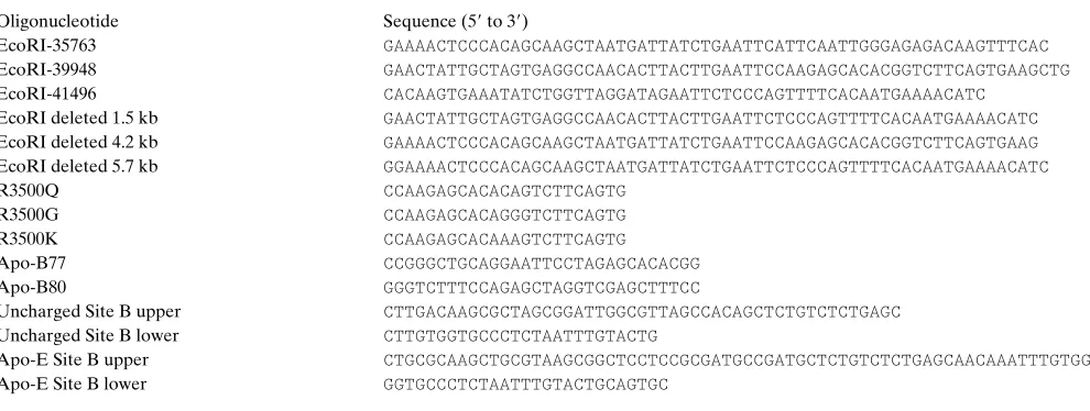
Generation of truncated P1 plasmids and isolation of DNA for mu-tagenesis. The 95-kb apo-B P1 plasmid p158 (15) was prepared and modified by RecA-assisted restriction endonuclease (RARE) cleav-age as described (16). Oligomers EcoRI-35,763 and EcoRI-41,496 were used to make a 5.7-kb–deleted P1 plasmid, and oligomers EcoRI-39,948 and EcoRI-41,496 to generate a 1.5-kb–deleted P1 plasmid and the corresponding 1.5-kb fragment, respectively (Table I). A 4.2-kb and a 5.7-4.2-kb fragment were isolated from the apo-B100 Leu-Leu plasmid with RARE cleavage using oligomers EcoRI-35,763 and EcoRI-39,948 and oligomers EcoRI-35,763 and EcoRI-41,496, re-spectively. The isolated 1.5-kb fragment and the two fragments with the Leu-Leu mutation were cloned into the pZErO-1 vector. The B100 “Leu-Leu” plasmid was used to increase the yield of apo-B100, because it contains a CAA to CTA mutation in codon 2,153 that effectively abolishes the formation of apo-B48, which is formed by an editing mechanism in mouse liver (17).
Generation of mutant P1 clones with mutations at residue 3,500 in apo-B100. The plasmid pZErO-1.5 kb was subjected to site-specific mutagenesis with the Morph System (5 Prime→3 Prime, Inc., Boul-der, CO). Three different oligomers were used to change arginine at position 3,500 in apo-B to glutamine (R3500Q), glycine (R3500G), or lysine (R3500K) (Table I). After RARE cleavage of the 1.5-kb– deleted P1 plasmid with oligonucleotide EcoRI deleted 1.5-kb, the mutated 1.5-kb fragment was ligated into the linearized and phos-phatased 5.7-kb–deleted P1 vector (16). These and all of the following constructs were sequenced over the mutated region(s) and over the 59 and 39 EcoRI sites and found to be correct.
Generation of mutant P1 clones with B77, B-80, and apo-B80 with the R3500Q mutation. Oligomers apo-B77 or apo-B80 were used to introduce stop codons at residue 3,497 (apo-B77) or residue 3,620 (apo-B80) in the plasmid pZErO-1.5 kb using the Morph Sys-tem. The latter mutation was also introduced into the pZErO-1.5 kb R3500Q plasmid. The mutated 1.5-kb fragments were isolated by EcoRI digestion and ligated into the vector pZErO-4.2 kb that had been linearized by RARE cleavage with oligomer EcoRI-39948. The resulting plasmids were subjected to RARE cleavage with oligomers EcoRI-35,763 and EcoRI-41,496, and the mutated 5.7-kb fragments were isolated and ligated into the 5.7-kb–deleted P1 plasmid (16).
Generation of mutant P1 clones with mutations in Site B. The pZ-ErO-5.7–kb plasmid was subjected to site-directed mutagenesis with the ExSite PCR System (Stratagene, Inc., La Jolla, CA). The first
mu-Table I. Oligomers Used in This Study
Oligonucleotide Sequence (59 to 39)
EcoRI-35763 GAAAACTCCCACAGCAAGCTAATGATTATCTGAATTCATTCAATTGGGAGAGACAAGTTTCAC
EcoRI-39948 GAACTATTGCTAGTGAGGCCAACACTTACTTGAATTCCAAGAGCACACGGTCTTCAGTGAAGCTG
EcoRI-41496 CACAAGTGAAATATCTGGTTAGGATAGAATTCTCCCAGTTTTCACAATGAAAACATC
EcoRI deleted 1.5 kb GAACTATTGCTAGTGAGGCCAACACTTACTTGAATTCTCCCAGTTTTCACAATGAAAACATC
EcoRI deleted 4.2 kb GAAAACTCCCACAGCAAGCTAATGATTATCTGAATTCCAAGAGCACACGGTCTTCAGTGAAG
EcoRI deleted 5.7 kb GGAAAACTCCCACAGCAAGCTAATGATTATCTGAATTCTCCCAGTTTTCACAATGAAAACATC
R3500Q CCAAGAGCACACAGTCTTCAGTG
R3500G CCAAGAGCACAGGGTCTTCAGTG
R3500K CCAAGAGCACAAAGTCTTCAGTG
Apo-B77 CCGGGCTGCAGGAATTCCTAGAGCACACGG
Apo-B80 GGGTCTTTCCAGAGCTAGGTCGAGCTTTCC
Uncharged Site B upper CTTGACAAGCGCTAGCGGATTGGCGTTAGCCACAGCTCTGTCTCTGAGC
Uncharged Site B lower CTTGTGGTGCCCTCTAATTTGTACTG
Apo-E Site B upper CTGCGCAAGCTGCGTAAGCGGCTCCTCCGCGATGCCGATGCTCTGTCTCTGAGCAACAAATTTGTGG
[image:3.612.59.554.549.736.2]tant was created with oligomers designated uncharged Site-B upper and uncharged Site-B lower. All arginines between residues 3,359 and 3,369 in apo-B were changed to serines, and all the lysines to ala-nines. The second mutant was created with oligomers designated apo-E Site-B upper and apo-E Site-B lower to substitute residues 3,358– 3,370 in apo-B with the receptor-binding sequence (residues 141–153) of apo-E. To avoid misincorporation mutations, a 708-nucleotide AvrII–EcoRI fragment of the two vectors, which had been verified by sequencing, was subcloned back into the plasmid pZErO-5.7 kb by RARE cleavage with oligomer EcoRI-39,948 followed by AvrII di-gestion. The mutated 5.7-kb fragments were isolated and ligated to the linearized 5.7-kb deleted P1 plasmid.
Transgenic mice. Transgenic mice were generated by microinjec-tion of P1 DNA into fertilized mouse eggs (C57BL/6XSJL) as de-scribed (16, 18). A human apo-B–specific radioimmunoassay of mouse plasma was used to identify transgenic mice (15). Two lines were established from each construct.
Generation of a rabbit antiserum to mouse apo-B and mouse apo-E.
Mouse apo-B was isolated from mouse LDL (d5 1.02–1.05 g/ml). Mouse apo-E was expressed in vitro and isolated. Rabbits were im-munized with 5 mg of mouse apo-B or 2.5 mg of mouse apo-E. The specificity of the antibodies was confirmed by Western analysis. No cross-reactivity against human apo-B or apo-E was detected in the antisera used.
Isolation of recombinant lipoproteins. Blood from mice fasted for 5 h was collected by cardiac puncture into tubes containing EDTA (final concentration, 1 mg/ml), and the plasma was mixed with butyl-ated hydroxytoluene (final concentration, 25 mM), phenylmethyl sul-fonylfluoride (final concentration, 1 mM), and aprotinin (final con-centration, 10 U/ml). The LDL (d 5 1.02–1.05 g/ml) were isolated by sequential ultracentrifugation (Ti 70 rotor) and dialyzed against 150 mM NaCl and 0.01% EDTA, pH 7.4, and the mouse apo-E and apo-B were removed by immunoaffinity chromatography. The d5 1.02– 1.05 g/ml fraction was mixed with an equal volume (850 ml) of Affi-Gel Hz (Bio-Rad Laboratories, Richmond, CA) and incubated for 16 h at 48C in a gently rocking tube filled with N2. The Affi-Gel Hz (100 g)
had previously been coupled with mouse apo-E or mouse apo-B rab-bit immunoglobulins from 50 ml of antiserum. Lipoproteins used for receptor-binding experiments were isolated and assayed within 1 wk. Human plasma LDL, isolated from a single blood donor, were in-cluded as a control in each experiment.
Cell culture and receptor-binding assays. Competitive receptor-bind-ing assays were performed as described (19). The amount of unla-beled lipoproteins needed to compete 50% with 125I-labeled LDL
af-ter a 3-h incubation at 48C was determined from an exponential decay curve–fitting model. Direct binding experiments were conducted with increasing concentrations of 125I-labeled lipoproteins during a 4-h
incu-bation. Surface-bound radioactivity data were linearized by Scat-chard analysis, and the Kd and maximum amount of lipoprotein
bound at receptor saturation (Bmax) were determined (19). The
re-sults shown are representative of two to four independent receptor-binding assays for each experiment.
Immunoassays of human recombinant R3500Q and control LDL. Plate immunoassays were carried out as described (20), with the following modifications. The dilution curves of the recombinant R3500Q and control LDL ranged from 15 ng/ml to 1,500 ng/ml. The monoclonal antibodies MB47 and MB3 were diluted 1:10,000 and 1:15,000, respectively, and 400,000 cpm of the 125I-labeled antibody
was added per well.
Analysis of the plasma lipoproteins. Purified lipoproteins were analyzed on Western blots of 3–15% polyacrylamide-SDS gels, using the enhanced chemiluminescense Western blotting detection re-agents (Amersham Corp., Arlington Heights, IL). Negative-staining electron microscopy was performed as described (21), and the parti-cle size distribution in each lipoprotein fraction was determined by measuring at least 1,000 particles with an Image-1/AT analysis system.
Results
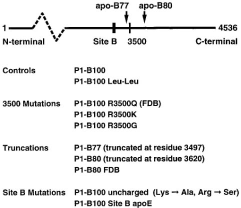
[image:4.612.57.298.453.661.2]Generation of transgenic mice expressing mutant recombinant human apo-B. Transgenic mice were generated with a human apo-B P1 plasmid in which mutations had been introduced by RARE cleavage. For this study, we created 10 lines of trans-genic mice classified into four groups (Fig. 1). The first group consisted of controls. The first control (P1-B100) was human recombinant apo-B100 without any mutation in the apo-B gene. The second control (apo-B100 Leu-Leu) was created to improve the yield of recombinant LDL by eliminating the formation of apo-B48. In the second group, codon 3,500 of the human apo-B gene was altered to understand how mutations affecting residue 3,500 cause defective receptor binding. Residue 3,500 was substituted with either glutamine (R3500Q), glycine (R3500G), or lysine (R3500K). In the third group, truncations were created to localize the receptor-binding site of apo-B100 and to clarify how mutations at residue 3,500 disrupt receptor binding. These deletion mutations were apo-B77 (truncated at residue 3,497), B80 (truncated at residue 3,620), and apo-B80 with the FDB mutation (apo-apo-B80 R3500Q). The fourth group was designed to identify the site on apo-B100 that binds to the LDL receptor. Because the basic amino acids arginine
Figure 1. Generation of transgenic mice. The mutated apo-B P1
[image:4.612.314.557.664.721.2]clones were used to generate four groups of transgenic mice express-ing recombinant LDL containexpress-ing mutated human apo-B. To improve the yield of the recombinant LDL, all lines in groups three and four and the control apo-B100 Leu-Leu contained the Leu-Leu mutation. A simplified apo-B100 diagram (above) shows the locations of the mutations.
Table II. Mutations in Site B of Apo-B100
Residues 3359 3369
Apo-B100 Arg Leu ThrArg Lys ArgGly LeuLysLeu Ala Uncharged Site B Ser Ser Ala Ser Ala
Apo-E Site B LysLeu Leu Arg
and lysine are important in receptor binding, the arginine resi-dues were converted to serines and the lysine resiresi-dues were changed to alanines in Site B of apo-B100 (Table II). Further-more, because of the homology to the receptor-binding site of apo-E, the receptor-binding region of human apo-E was sub-stituted for site-B of apo-B100.
Isolation, purification, and characterization of recombinant human LDL. Recombinant LDL were isolated from trans-genic mouse plasma by ultracentrifugation, and the endog-enous mouse apo-E– and apo-B–containing lipoproteins removed by immunoaffinity chromatography. Purified recom-binant LDLisolated from the plasma of 10 lines of transgenic mice contained nondegraded apo-B without visible contami-nation by apo-E or any other protein (Fig. 2). Western blot analysis showed that all recombinant LDL bound to the mono-clonal antibody 1D1, whose epitope is between amino acids 474 and 539 in human apo-B100 (22), and that only the unpuri-fied LDL reacted with polyclonal antibodies against mouse B and mouse E (Fig. 3), confirming that mouse apo-B100 and apo-E had been completely removed by the immu-noaffinity columns. Negative-staining electron microscopy of isolated recombinant LDL showed that the average size of re-combinant LDL containing apo-B100 was the same as that of
human plasma LDL (21.862.3 versus 20.861.9 nm, respec-tively). Recombinant LDL containing truncated forms of apo-B100 were slightly smaller (18.862.1, 19.261.9, and 19.162.4 nm for apo-B77, apo-B80, and apo-B80 R3500Q LDL, respec-tively). Thus, highly purified recombinant LDL that are struc-turally similar to plasma-derived human LDL can be isolated from transgenic mice.
Characterization of receptor-binding activity of mouse-derived human recombinant control LDL. First, we evaluated the receptor-binding activity of purified recombinant LDL de-rived from the transgenic mouse plasma and found that it was similar to that of plasma-derived human LDL (ED50 1.9 versus
1.8 mg/ml, respectively). Because the 10 transgenic mouse lines had different transgene copy numbers and different levels of expression of recombinant LDL, we were concerned that the level of expression of the recombinant apo-B might influence the receptor-binding activity of the recombinant LDL. There-fore, we isolated recombinant control LDL from one high-expressor line and one low-high-expressor line, reflected by the plasma cholesterol concentrations of the pooled blood (229 and 132 mg/dl, respectively). Competitive receptor-binding studies with 125I-LDL showed almost identical activities (ED
50
[image:5.612.316.556.62.344.2]2.1 versus 2.2 mg/ml, respectively). Thus, the receptor-binding affinity was not dependent on the degree of expression of the transgene in the different lines. Nevertheless, two transgenic mouse lines were established from each construct.
Figure 2. Coomassie staining of recombinant lipoproteins.
[image:5.612.59.298.346.659.2]Recombi-nant LDL (d 5 1.02–1.05 g/ml) from all transgenic lines were isolated by sequential ultracentrifugation and subjected to immunoaffinity chromatography to remove endogenous apo-B and apo-E. 5 mg of apo-B100 (lanes 1–8) and 2 mg of the truncated forms of apo-B (lanes 9–11) were analyzed by SDS-PAGE with 3–15% gels. Human plasma LDL (lane 1) were used as standard.
The leu-leu alteration did not influence the receptor bind-ing of the recombinant LDL (ED50 2.2 versus 2.3 mg/ml,
re-spectively, for the recombinant control LDL with and without this mutation). Thus, we could use recombinant LDL with this mutation, which increased the yield of recombinant LDL.
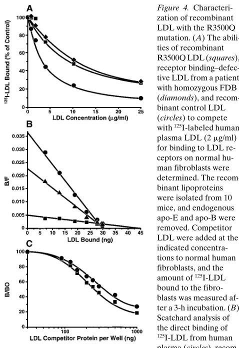
Verification of the phenotype of the R3500Q mutation. To compare recombinant R3500Q LDL with LDL from an FDB homozygote, we examined the receptor-binding affinities of recombinant R3500Q LDL, LDL from an FDB subject ho-mozygous for the R3500Q mutation, and recombinant control LDL (Fig. 4 A). The ED50 values were 9.8, 10.6, and 2.7 mg/ml,
respectively. These results were confirmed by direct binding experiments with 125I-labeled human plasma–derived LDL,
re-combinant control LDL, and rere-combinant R3500Q LDL. Scat-chard analysis (Fig. 4 B) showed that the LDL with the R3500Q mutation had a 6.4-fold reduction in affinity for the LDL receptor compared with recombinant control LDL. The Kd50 values for human plasma LDL, recombinant control
LDL, or recombinant R3500Q LDL were 0.81, 1.31, and 8.33
mg/ml, respectively.2 Thus, both competitive and direct binding
experiments demonstrated that the R3500Q mutation causes the defective receptor binding.
LDL from subjects with FDB bind with a higher affinity than control LDL to the apo-B monoclonal antibody MB47. This monoclonal antibody inhibits the binding of LDL to its receptor and has a discontinuous epitope in apo-B100 that flanks residue 3,500 (amino acids 3,429–3,453 and amino acids 3,507–3,523) (23). It has been hypothesized that the R3500Q mutation changes the apo-B100 conformation in this region, such that MB47 binds with a higher affinity to FDB LDL (20). To determine if the R3500Q mutation causes this enhanced MB47 binding we analyzed recombinant LDL with and with-out the R3500Q mutation for MB47 binding in a solid-phase radioimmunoassay (Fig. 4 C). Less LDL with the R3500Q mu-tation (ED50 285 ng) was required than recombinant control
LDL (ED50 386 ng) to displace 50% of human plasma–derived
LDL from MB47. This experiment were repeated three times with newly isolated recombinant LDL for each experiment. These experiments showed that MB47 had a 1.5-fold higher af-finity for recombinant R3500Q LDL than for recombinant control LDL (average of n 5 3). Taken together, these results prove that the R3500Q mutation enhances MB47 binding and causes the defective receptor binding seen in FDB LDL.
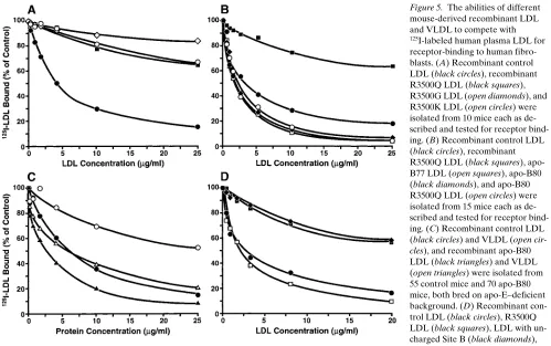
Analysis of human recombinant LDL with mutations at residue 3500. Next, we determined whether the loss of argi-nine or the loss of a positive charge at residue causes receptor binding–defective LDL. Recombinant LDL with the R3500Q, R3500K, or R3500G mutation were analyzed in a competitive in vitro receptor-binding assay (Fig. 5 A). Surprisingly,
recom-binant LDL with the R3500K mutation were as defective as the LDL with the R3500Q or the R3500G mutation (ED50.
25 mg/ml versus 5.5 mg/ml for recombinant control apo-B100 LDL). Thus, it is the presence of an arginine at residue 3,500, not simply a positive charge, that is essential for normal recep-tor binding.
Competitive receptor binding of LDL and VLDL with truncated forms of apo-B. Analysis of truncated LDL isolated from patients with familial hypobetalipoproteinemia showed that B67, containing the first 3,040 amino acids of apo-B100, did not bind to the LDL receptor (8). However, LDL containing either apo-B75, which terminates at residue 3,386, or apo-B89 apparently bound more avidly than normal LDL to the LDL receptor (9, 10). How could these truncated forms of apo-B display higher affinity for the LDL receptor than
2. The direct binding experiment showed that recombinant control LDL had slightly lower affinity for the LDL receptor than human plasma LDL. The reason for this is unclear but may be due to differ-ences in lipid composition. Recombinant human or mouse LDL over-expressed in transgenic mice have been shown to be more triglyceride rich than normal human or mouse plasma LDL (15, 31), and in-creased triglyceride content of the core of the LDL particle has been shown to alter the interaction with the LDL receptor (32, 33). Thus, it is conceivable that the slightly lower LDL receptor affinity is caused by changes in lipid composition.
Figure 4.
Characteri-zation of recombinant LDL with the R3500Q mutation. (A) The abili-ties of recombinant R3500Q LDL (squares), receptor binding–defec-tive LDL from a patient with homozygous FDB (diamonds), and recom-binant control LDL (circles) to compete with 125I-labeled human
plasma LDL (2 mg/ml) for binding to LDL re-ceptors on normal hu-man fibroblasts were determined. The recom-binant lipoproteins were isolated from 10 mice, and endogenous apo-E and apo-B were removed. Competitor LDL were added at the indicated concentra-tions to normal human fibroblasts, and the amount of 125I-LDL
bound to the fibro-blasts was measured af-ter a 3-h incubation. (B) Scatchard analysis of the direct binding of
125I-LDL from human
plasma (circles), recom-binant control LDL (triangles), or recomrecom-binant R3500Q LDL (squares). The recombinant LDL were isolated from 35 mice each and labeled with 125I after removal of endogenous apo-E and apo-B.
Nonspecific binding, obtained by the addition of a 100-fold excess of unlabeled plasma LDL, was subtracted at each concentration. Bound/Free (B/F) is the amount of LDL bound/dish divided by the amount of LDL free in the medium in each dish. The molar Kd values
[image:6.612.313.552.56.404.2]apo-B100? One explanation would be an increased apo-E/ apo-B molar ratio in truncated forms of LDL, enhancing the affinity of the apo-B75 LDL for the LDL receptor. This pos-sibility cannot be ruled out, as SDS-polyacrylamide gels of the apo-B75 LDL demonstrated the presence of apo-E, which may have been responsible for the enhanced binding of apo-B75 (10). A more intriguing hypothesis is that the carboxyl-terminal part of apo-B100 itself modulates the receptor bind-ing of LDL.
To test this hypothesis, we isolated and analyzed recombi-nant LDL containing apo-B77, apo-B80, apo-B80 R3500Q, apo-B100 R3500Q, and recombinant control LDL. Western analysis demonstrated that immunoaffinity purification re-moved all endogenous apo-B and apo-E (Fig. 1). Competitive receptor-binding assays showed repeatedly that, compared with recombinant control LDL, all LDL containing truncated forms of apo-B had slightly enhanced binding to LDL recep-tors (Fig. 5 B). The ED50 values for recombinant apo-B77,
B80, B80 R3500Q, control B100 LDL, and apo-B100 R3500Q LDL were 1.9, 1.9, 1.5, 3.7, and . 25 mg/ml, respectively. The fact that apo-B77 had increased receptor binding proves that arginine-3,500 is not directly involved in receptor binding: apo-B77 is truncated at residue 3,497 and therefore does not contain arginine-3,500. Moreover, the find-ing that apo-B80 LDL with the R3500Q mutation has en-hanced receptor binding indicates that glutamine at residue
3,500 of full-length FDB LDL interacts with residues in the carboxyl-terminal 20% of apo-B100 to disrupt receptor bind-ing or, alternatively, that arginine-3,500 in normal LDL inter-acts with residue(s) in the carboxyl tail to facilitate normal re-ceptor binding.
The observation that the carboxyl terminus of apo-B100 could inhibit LDL binding suggested that this modulation may be important in other apo-B–containing lipoproteins. For ex-ample, large VLDL bind poorly to the LDL receptor via apo-B. However, when these large particles are reduced in size by lipolysis, the apo-B100 in the smaller LDL bind with high af-finity to LDL receptors (1). To determine if the carboxyl ter-minus of apo-B100 inhibits the binding of VLDL to LDL re-ceptors, we analyzed the receptor binding of VLDL containing apo-B80 or apo-B100. In these experiments, it was essential not to have any apo-E that would interfere with the receptor-binding assay. Because apo-E is an integral component of VLDL and cannot be removed by immunoaffinity chromatog-raphy, transgenic mice expressing recombinant apo-B80 or apo-B100 were bred with gene-targeted apo-E–deficient mice. Furthermore, since endogenous B48 accumulates in apo-E–deficient mice, making complete removal of mouse apo-B difficult, the apo-B80 3 apo-E2/2 and apo-B100 3 apo-E2/2
[image:7.612.57.556.57.370.2]mice were bred onto an endogenous apo-B100–only mouse background. Apo-B100–only mice have been genetically mod-ified to produce only apo-B100 and no apo-B48 (24). VLDL
Figure 5. The abilities of different
mouse-derived recombinant LDL and VLDL to compete with
125I-labeled human plasma LDL for
receptor-binding to human fibro-blasts. (A) Recombinant control LDL (black circles), recombinant R3500Q LDL (black squares), R3500G LDL (open diamonds), and R3500K LDL (open circles) were isolated from 10 mice each as de-scribed and tested for receptor bind-ing. (B) Recombinant control LDL (black circles), recombinant R3500Q LDL (black squares), apo-B77 LDL (open squares), apo-B80 (black diamonds), and apo-B80 R3500Q LDL (open circles) were isolated from 15 mice each as de-scribed and tested for receptor bind-ing. (C) Recombinant control LDL (black circles) and VLDL (open
cir-cles), and recombinant apo-B80
(d , 1.006 g/ml) and LDL (d 5 1.02–1.05 g/ml) were isolated by ultracentrifugation, and the mouse apo-B100 was removed by immunoaffinity chromatography. Negative-staining elec-tron microscopy of the isolated lipoprotein particles showed that the apo-B100 and apo-B80 VLDL, which were the same size (35.5 and 34.9 nm, respectively), were substantially larger in diameter than the apo-B100 and apo-B80 LDL (23.5 and 21.5 nm, respectively). Further characterization of the apo-B80 and apo-B100 VLDL showed they had the same lipid composition (data not shown). In competitive binding studies with 125I-labeled
normal LDL (Fig. 5 C), apo-B80 VLDL bound with a 6.5-fold higher affinity to human fibroblasts than apo-B100 VLDL (ED50 6.0 versus 39 mg/ml, respectively). The ED50 for
apo-B100 VLDL was determined using Probit transformation (19). Apo-B80 LDL bound 2.3-fold more avidly to the LDL recep-tor than apo-B100 LDL (ED50 2.6 mg/ml versus 6.0 mg/ml,
re-spectively). These results show that the carboxyl-terminal 20% of apo-B100 inhibits the receptor binding of VLDL and con-firm that apo-B80 LDL bind more avidly than apo-B100 LDL.
The receptor-binding site of apo-B100. Studies of LDL from
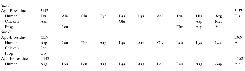
patients with hypobetalipoproteinemia have indicated that the receptor-binding region of apo-B100 probably lies between amino acids 3,040 and 3,386. Analysis of the amino acid se-quence of apo-B100 in this domain shows two regions contain-ing clusters of basic amino acids, Site A (residues 3,147–3,157) and Site B (residues 3,359–3,367), which have been proposed as the receptor-binding regions of apo-B100 (13). To identify conserved, functionally significant regions in apo-B100, we an-alyzed LDL (d 5 1.02–1.05 g/ml) isolated from Xenopus laevis and made the surprising discovery that X. laevis LDL bind to human LDL receptors with an affinity equivalent to or higher than that of human LDL (data not shown). Subsequently, we sequenced 4.5 kb of X. laevis apo-B DNA and compared this with Site A and Site B of human apo-B100 (Table III). Site B was essentially perfectly conserved despite 350 million years of evolution, strongly implicating its importance in receptor bind-ing. The fact that Site B is very similar to the receptor-binding site of apo-E also argues in favor of its significance. Moreover, Site A is poorly conserved in both chicken and frog apo-B (Ta-ble III), and receptor-binding analysis of chicken LDL (d 5 1.02–1.05 g/ml) showed that it binds with a high affinity equiva-lent to that of human LDL (data not shown). The lack of con-servation of Site A in these animals (Table III) indicated that Site A is very unlikely to be a potential site for receptor binding.
Therefore, we focused on Site B. First we changed the basic amino acids in this region to neutral amino acids (Table II), substituting serines for arginines and alanines for lysines. Sec-ond, we reasoned that if Site B in apo-B100 actually interacts with the LDL receptor, then it might be possible to substitute the receptor-binding site apo-E for Site B and retain LDL re-ceptor–binding activity. The receptor-binding ability of recom-binant LDL with the uncharged Site B or with the receptor-binding site of apo-E substituted for Site B was compared with those of recombinant control LDL and recombinant R3500Q LDL (Fig. 5 D). LDL containing the uncharged Site B had de-fective receptor binding equivalent to that of recombinant R3500Q LDL (ED50. 20 mg/ml for both). Furthermore, the
LDL with the receptor-binding region of apo-E in place of Site B and recombinant control LDL had similar receptor-binding activities (ED50 2.4 and 2.3 mg/ml, respectively). These results
prove that Site B is functionally important for LDL receptor binding.
One concern with using site-directed mutagenesis to study protein function is distinguishing between mutations that af-fect local structure and function and those that afaf-fect the fold-ing and stability of the entire protein, because both would have the same defective binding phenotype. To determine if the mutation(s) affected only the structure and function of the re-ceptor-binding site or if they had a more global effect, we used primary mouse hepatocytes to test the rate of secretion of re-combinant apo-B from all transgenic lines in vitro. No differ-ences were found (data not shown). The assembly of VLDL and LDL is a complex procedure, and if the protein is not fold-ing correctly, the apo-B100 probably will be degraded instead of being secreted (25).
Discussion
[image:8.612.55.560.576.723.2]Even though the apo-B cDNA and gene were cloned and se-quenced more than a decade ago, progress in understanding the molecular interaction between apo-B100 and the LDL re-ceptor has been slow. There are several reasons for the slow progress. Apo-B100, one of the largest proteins known (4,536 amino acids), is highly hydrophobic and aggregates when not associated with lipids. Thus, only apo-B100 present in LDL can bind to the LDL receptor. Moreover, recombinant LDL secreted by cultured hepatic cells transfected with apo-B mini-genes lacked normal receptor-binding activity (Innerarity,
Table III. Species Comparison of Site A and Site B in Apo-B100 with the Receptor-binding Site of Apo-E
Site A
Apo-B residue 3147 3157
Human Lys Ala Gln Tyr Lys Lys Asn Lys His Arg His
Chicken Asn Glu Asp Met
Frog Leu Thr Asp Val
Site B
Apo-B residue 3359 3369
Human Arg Leu Thr Arg Lys Arg Gly Leu Lys Leu Ala
Chicken Ser
Frog Gly
Apo-E3 residue 142 152
Human Arg Lys Leu Arg Lys Arg Leu Leu Arg Asp Ala
T.L., unpublished observation), and heterologous minigenes yield extremely poor expression in transgenic mice (26). To overcome these problems, we developed a new approach for studying apo-B structure: mutating a P1 bacteriophage clone, making transgenic mice, and then purifying and characterizing the mutant apo-B protein. The first step was to mutate the 95-kb P1 clone. For this purpose, we developed an efficient proto-col for performing site-directed mutagenesis on large-insert bacterial clones based on RARE cleavage, which made it pos-sible to insert subtle or complex mutations into the apo-B gene within 2 wk (16). This strategy made it feasible to generate a series of apo-B mutants to dissect the molecular interaction between apo-B100 and the LDL receptor.
Our study indicates that Site B (residues 3,359–3,369) is critical for receptor binding. Further support for this finding can be summarized as follows: LDL with apo-B truncated at amino acid 3,040 do not bind to the LDL receptor (8), but LDL with apo-B truncated at residue 3,386 (10) or 3,497 bind avidly to the LDL receptor. Site B is within the receptor-bind-ing region defined by these truncated apo-B LDLs. Further-more, Site B has clusters of basic amino acids known to be im-portant for binding to the LDL receptor and is extremely similar to the site on apo-E that binds to the LDL receptor (13, 14). Site B is highly conserved among many species (27) and essentially perfectly conserved in apo-B100 from X. laevis. This conservation over 350 million years of evolution is consis-tent with Site B’s functional importance in receptor binding. The finding that Site B peptides from apo-B100 bind heparin (28) is significant because the receptor-binding site of apo-E also binds heparin. Immunoelectron microscopy studies by Phillips et al. also support this concept (Phillips, M., and V. Schumaker, personal communication). They found that a solu-ble fragment of the LDL receptor bound immediately amino-terminal to the binding site of the monoclonal antibody MB47 on B100. Since the discontinuous MB47 epitope of apo-B100 includes amino acids 3,429–3,453 and 3,507–3,523 (23), these results place the location of binding in the vicinity of Site B (3,359–3,369). Furthermore, our receptor-binding analysis of recombinant LDL with an altered Site B indicated that Site B is functionally important for the interaction between apo-B100 and the LDL receptor. Finally, recombinant LDL maintained normal receptor binding when Site B was replaced with the re-ceptor-binding site of apo-E, indicating that Site B is
equiva-lent to apo-E’s receptor-binding site and therefore is most likely the site that binds to the LDL receptor. While it has been proposed that the receptor-binding domain is discontinu-ous and composed of several segments of apo-B100 and that Site B is only one site of several (27), our data argue against this concept. Acetylation of the remaining lysine residues of apo-B100 in the recombinant LDL with the mutated Site B did not further decrease the receptor binding (data not shown). While other interpretations are possible, it is likely that Site B is the primary or only receptor-binding site for apo-B100.
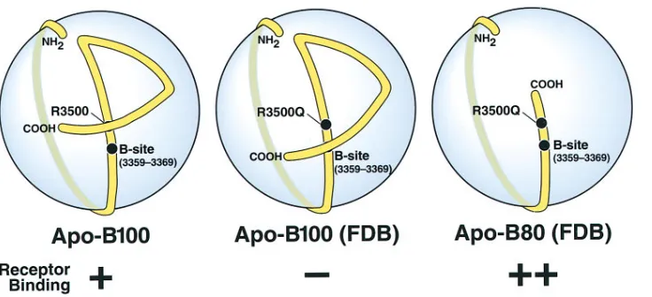
The results from this study together with the elegant map-ping of apo-B100 on the LDL particle (29) now allow a broad understanding of the receptor-binding site of apo-B100 and explain how sequences and mutations distal to the receptor-binding region can modulate the affinity of apo-B100 for the LDL receptor. Based on this understanding, we developed the model illustrated in Fig. 6. Chatterton et al. (29) showed that the first 89% of apo-B100 enwraps the LDL particle like a belt and that the carboxyl-terminal 11% constitutes a bow that crosses over the belt and brings the carboxyl tail of apo-B100 close to amino acid residue 3,500. We propose that a region of apo-B100 carboxyl-terminal to Site B and encompassing resi-due 3,500 serves as a modulator that can alter the affinity of the receptor-binding domain of apo-B100. Moreover, our re-sults suggest that arginine-3,500 interacts with the carboxyl tail of apo-B100 and that this interaction is important for receptor binding of LDL. In our model, this modulator element, or more specifically arginine-3,500, interacts with the carboxyl tail that crosses over at or very close to residue 3,500. The sub-stitution of glutamine for arginine at residue 3,500 (i.e., site R3500Q in Fig. 6) abolishes this interaction, resulting in a con-formational change, disrupted receptor binding, and the clini-cal disorder FDB. Truncation mutations in which there is in-sufficient carboxyl tail to cross over near Site B do not need this interaction and therefore bind normally without arginine-3,500. This explains why LDL containing apo-B80 with the R3500Q mutation have normal or enhanced LDL receptor– binding activity (Fig. 6). Evidence that the R3500Q mutation significantly alters the microenvironment in the region around residue 3,500 of apo-B100 comes from both the MB47 studies showing enhanced binding to FDB LDL (20) and from 13C
nuclear magnetic resonance analysis of FDB LDL (30). Our receptor-binding studies with different types of
muta-Figure 6. Schematic of the
[image:9.612.59.427.571.739.2]tions at residue 3,500 demonstrate that it is arginine and not a positive charge at this position that is critical for receptor-bind-ing activity. The importance of arginine at this position is also indicated by the finding that arginine at residue 3,500 is con-served in all species that have been sequenced, including the frog X. laevis. The case of the frog is especially interesting be-cause synonymous codons are used for the frog and human apo-B genes to code for arginine-3,500. The finding that lysine cannot substitute for arginine at residue 3,500 suggests that arginine-3,500 does not form a salt bridge to an acidic residue but the nature of this interaction and the sequence(s) arginine-3,500 interacts with is an area we are investigating currently.
Our model also proposes that the carboxyl-terminal se-quence of apo-B100 functions as a negative modulator of ceptor binding and inhibits binding of VLDL to the LDL re-ceptors. We propose that during lipolysis of large VLDL to smaller LDL, the carboxyl terminus changes its conformation to allow interaction with the LDL receptor. Removal of the tail increases not only the receptor binding of VLDL more than sixfold but also, to a lesser extent, the binding of LDL to the LDL receptor. Interestingly, Milne and his colleagues have found that the only dramatic difference in the binding of mono-clonal antibodies to apo-B100 in VLDL and LDL occurs with antibodies binding to the carboxyl terminus of the protein (Wang, X., and R. Milne, personal communication). Further-more, Parhofer et al. (9) demonstrated enhanced clearance of both LDL and VLDL in a subject with apo-B89, indicating that our in vitro observations are most likely physiologically relevant. Our model further proposes, albeit more specula-tively, that the hydrolysis of VLDL permits arginine-3,500 to interact with the carboxyl tail, opening up Site B for receptor binding.
In addition to revealing the molecular basis for the genetic disorder FDB, our model explains the increased clearance of certain truncated forms of apo-B found in hypobetalipopro-teinemic patients. Our results show that truncation of the apo-B molecule enhanced its affinity for the LDL receptor and that apo-B80 VLDL had an affinity for the LDL receptor similar to that of apo-B100 LDL. The latter finding suggests that VLDL with truncated forms of apo-B containing Site B may bind to the LDL receptor without metabolic processing to LDL and explains the increased clearance of apo-B89 VLDL described by Parhofer et al. (9). Furthermore, the increased clearance of VLDL via the LDL receptor leads to a decreased formation of LDL. This mechanism explains why the plasma lipoprotein distribution in some hypobetalipoproteinemic patients is char-acterized by an almost complete lack of LDL.
The results from this study now allow a broad understand-ing of the receptor-bindunderstand-ing site of apo-B100 and explain how sequences and mutations distal to the receptor-binding region can modulate the affinity of apo-B100 for the LDL receptor. We suspect that the strategy applied in these studies will be useful for elucidating a wide range of apo-B structure and function issues.
Acknowledgments
The authors thank Dr. John Kane, Dr. Ross Milne, Dr. Robert W. Mahley, Dr. Martin Phillips, Dr. Stanley C. Rall, Dr. Verne Schu-maker, and Dr. Karl Weisgraber for stimulating discussions and com-ments on the manuscript, Dr. Stephen G. Young for generously pro-viding 125I-labeled C1.4 antibodies and for apo-B100 only mice, Dr.
Li-Ming Dong for generous help in expressing and purifying mouse apo-E, S. Ordway and G. Howard for editorial assistance, J. Carrol and B. Clark for help in preparation of the figures, and A. Corder and S. Gonzales for photographic work.
J. Borén is supported by the Howard Hughes Medical Institute with a Howard Hughes Postdoctoral Fellowship for Physicians. This work also was supported by National Heart, Lung, and Blood Insti-tute grant HL-47660.
References
1. Goldstein, J.L., M.S. Brown, R.G.W. Anderson, D.W. Russell, and W.J. Schneider. 1985. Receptor-mediated endocytosis: concepts emerging from the LDL receptor system. Annu. Rev. Cell Biol. 1:1–39.
2. Innerarity, T.L., R.W. Mahley, K.H. Weisgraber, T.P. Bersot, R.M. Krauss, G.L. Vega, S.M. Grundy, W. Friedl, J. Davignon, and B.J. McCarthy. 1990. Familial defective apolipoprotein B100: a mutation of apolipoprotein B that causes hypercholesterolemia. J. Lipid Res. 31:1337–1349.
3. Hobbs, H.H., M.S. Brown, and J.L. Goldstein. 1992. Molecular genetics of the LDL receptor gene in familial hypercholesterolemia. Hum. Mutat. 1: 445– 466.
4. Innerarity, T.L., K.H. Weisgraber, K.S. Arnold, R.W. Mahley, R.M. Krauss, G.L. Vega, and S.M. Grundy. 1987. Familial defective apolipoprotein B-100: low density lipoproteins with abnormal receptor binding. Proc. Natl. Acad. Sci. USA. 84:6919–6923.
5. Gaffney, D., J.M. Reid, I.M. Cameron, K. Vass, M.J. Caslake, J. Shep-herd, and C.J. Packard. 1995. Independent mutations at codon 3500 of the apo-lipoprotein B gene are associated with hyperlipidemia. Arterioscler. Thromb. Vasc. Biol. 15:1025–1029.
6. Pullinger, C.R., L.K. Hennessy, J.E. Chatterton, W. Liu, J.A. Love, C.M. Mendel, P.H. Frost, M.J. Malloy, V.N. Schumaker, and J.P. Kane. 1995. Famil-ial ligand-defective apolipoprotein B. Identification of a new mutation that de-creases LDL receptor binding affinity. J. Clin. Invest. 95:1225–1234.
7. Milne, R., R. Théolis, Jr., R. Maurice, R.J. Pease, P.K. Weech, E. Rassart, J.-C. Fruchart, J. Scott, and Y.L. Marcel. 1989. The use of monoclonal antibod-ies to localize the low density lipoprotein receptor-binding domain of apolipo-protein B. J. Biol. Chem. 264:19754–19760.
8. Welty, F.K., L. Seman, and F.T. Yen. 1995. Purification of the apolipo-protein B-67–containing low density lipoapolipo-protein particle and its affinity for the low density lipoprotein receptor. J. Lipid Res. 36:2622–2629.
9. Parhofer, K.G., A. Daugherty, M. Kinoshita, and G. Schonfeld. 1990. En-hanced clearance from plasma of low density lipoproteins containing a trun-cated apolipoprotein, apoB-89. J. Lipid Res. 31:2001–2007.
10. Krul, E.S., K.G. Parhofer, P.H.R. Barrett, R.D. Wagner, and G. Schon-feld. 1992. ApoB-75, a truncation of apolipoprotein B associated with familial hypobetalipoproteinemia: genetic and kinetic studies. J. Lipid Res. 33:1037– 1050.
11. Mahley, R.W., T.L. Innerarity, R.E. Pitas, K.H. Weisgraber, J.H. Brown, and E. Gross. 1977. Inhibition of lipoprotein binding to cell surface re-ceptors of fibroblasts following selective modification of arginyl residues in arginine-rich and B apoproteins. J. Biol. Chem. 252:7279–7287.
12. Weisgraber, K.H., T.L. Innerarity, and R.W. Mahley. 1978. Role of the lysine residues of plasma lipoproteins in high affinity binding to cell surface re-ceptors on human fibroblasts. J. Biol. Chem. 253:9053–9062.
13. Knott, T.J., S.C. Rall, Jr., T.L. Innerarity, S.F. Jacobson, M.S. Urdea, B. Levy-Wilson, L.M. Powell, R.J. Pease, R. Eddy, H. Nakai, et al. 1985. Human apolipoprotein B: structure of carboxyl-terminal domains, sites of gene expres-sion, and chromosomal localization. Science. 230:37–43.
14. Mahley, R.W. 1988. Apolipoprotein E: cholesterol transport protein with expanding role in cell biology. Science. 240:622–630.
15. Linton, M.F., R.V. Farese, Jr., G. Chiesa, D.S. Grass, P. Chin, R.E. Hammer, H.H. Hobbs, and S.G. Young. 1993. Transgenic mice expressing high plasma concentrations of human apolipoprotein B100 and lipoprotein(a). J. Clin. Invest. 92:3029–3037.
16. Borén, J., I. Lee, M.J. Callow, E.M. Rubin, and T.L. Innerarity. 1996. A simple and efficient method for making site-directed mutants, deletions, and fu-sions of large DNA such as P1 and BAC clones. Genome Res. 6:1123–1130.
17. Yao, Z., B.D. Blackhart, D.F. Johnson, S.M. Taylor, K.W. Haubold, and B.J. McCarthy. 1992. Elimination of apolipoprotein B48 formation in rat hepatoma cell lines transfected with mutant human apolipoprotein B cDNA constructs. J. Biol. Chem. 267:1175–1182.
18. McCormick, S.P.A., M.F. Linton, and S.G. Young. 1994. Expression of P1 DNA in mammalian cells and transgenic mice. Genet. Anal. Tech. Appl. 11: 158–164.
19. Arnold, K.S., T.L. Innerarity, R.E. Pitas, and R.W. Mahley. 1992. Lipo-protein–receptor interactions. In Lipoprotein Analysis. A Practical Approach. C.A. Converse, and E.R. Skinner, editors. Oxford University Press, Oxford. 145–168.
Famil-ial defective apolipoprotein B-100: enhanced binding of monoclonal antibody MB47 to abnormal low density lipoproteins. Proc. Natl. Acad. Sci. USA. 85: 9758–9762.
21. Forte, T.M., and R.W. Nordhausen. 1986. Electron microscopy of nega-tively stained lipoproteins. Methods Enzymol. 128:442–457.
22. Milne, R.W., R. Theolis, Jr., R.B. Verdery, and Y.L. Marcel. 1983. Char-acterization of monoclonal antibodies against human low density lipoprotein. Arteriosclerosis. 3:23–30.
23. Young, S.G., R.K. Koduri, R.K. Austin, D.J. Bonnet, R.S. Smith, and L.K. Curtiss. 1994. Definition of a nonlinear conformational epitope for the apolipoprotein B-100–specific monoclonal antibody, MB47. J. Lipid Res. 35: 399–407.
24. Farese, R.V., Jr., M.M. Véniant, C.M. Cham, L.M. Flynn, V. Pierotti, J.F. Loring, M. Traber, S. Ruland, R.S. Stokowski, D. Huszar, and S.G. Young. 1996. Phenotypic analysis of mice expressing exclusively apolipoprotein B48 or apolipoprotein B100. Proc. Natl. Acad. Sci. USA. 93:6393–6398.
25. Borchardt, R.A., and R.A. Davis. 1987. Intrahepatic assembly of very low density lipoproteins. Rate of transport out of the endoplasmic reticulum determines rate of secretion. J. Biol. Chem. 262:16394–16402.
26. Chiesa, G., D.F. Johnson, Z. Yao, T.L. Innerarity, R.W. Mahley, S.G. Young, R.H. Hammer, and H.H. Hobbs. 1993. Expression of human apolipo-protein B100 in transgenic mice. Editing of human apolipoapolipo-protein B100 mRNA. J. Biol. Chem. 268:23747–23750.
27. Law, A., and J. Scott. 1990. A cross-species comparison of the
apolipo-protein B domain that binds to the LDL receptor. J. Lipid Res. 31:1109–1120. 28. Weisgraber, K.H., and S.C. Rall, Jr. 1987. Human apolipoprotein B-100 heparin-binding sites. J. Biol. Chem. 262:11097–11103.
29. Chatterton, J.E., M.L. Phillips, L.K. Curtiss, R. Milne, J.-C. Fruchart, and V.N. Schumaker. 1995. Immunoelectron microscopy of low density lipo-proteins yields a ribbon and bow model for the conformation of apolipoprotein B on the lipoprotein surface. J. Lipid Res. 36:2027–2037.
30. Lund-Katz, S., T.L. Innerarity, K.S. Arnold, L.K. Curtiss, and M.C. Phil-lips. 1991. 13C NMR evidence that substitution of glutamine for arginine 3500 in
familial defective apolipoprotein B-100 disrupts the conformation of the recep-tor-binding domain. J. Biol. Chem. 266:2701–2704.
31. McCormick, S.P.A., J.K. Ng, M. Véniant, J. Borén, V. Pierotti, L.M. Flynn, D.S. Grass, A. Connolly, and S.G. Young. 1996. Transgenic mice that overexpress mouse apolipoprotein B. Evidence that the DNA sequences con-trolling intestinal expression of the apolipoprotein B gene are distant from the structural gene. J. Biol. Chem. 271:11963–11970.
32. Kleinman, Y., G. Schonfeld, D. Gavish, Y. Oschry, and S. Eisenberg. 1987. Hypolipidemic therapy modulates expression of apolipoprotein B epitopes on low density lipoproteins. Studies in mild to moderate hypertriglyc-eridemic patients. J. Lipid Res. 28:540–548.