Copyright © 1998, American Society for Microbiology. All Rights Reserved.
Characterization of Glycopeptide-Resistant Enterococci
from a Swiss Hospital
NADIA LIASSINE,1* RENO FREI,2ISABELLE JAN,1
ANDRAYMOND AUCKENTHALER1
Central Laboratory of Bacteriology, University Hospital, Geneva,1and
Bacteriology Laboratory, University Clinics, Basel,2Switzerland
Received 30 October 1997/Returned for modification 30 December 1997/Accepted 17 March 1998
Between August 1994 and September 1996, 28 glycopeptide-resistant enterococci (GRE) were isolated from 8 infected patients and 11 intestinal carriers hospitalized at the University Hospital of Geneva. Identification to the species was made by both phenotypic (API 20 STREP and Rapid ID 32 STREP systems, and Vitek Gram Positive Identification Card) and genotypic methods using a multiplex PCR assay developed also for the deter-mination of the genotype of glycopeptide resistance (vanA, vanB, vanC1, and vanC2-C3 genes). Fifteen isolates were identified as Enterococcus faecium, 8 as E. gallinarum, 4 as E. faecalis, and 1 as E. hirae. All of the pheno-typic identification methods failed to differentiate some isolates of E. gallinarum from E. faecium, or vice versa. Both vanA (n518) and vanB (n54) glycopeptide resistance genotypes were found. For the first time, the vanB determinant was found in two isolates of E. gallinarum. Two patients were colonized by two different species containing the vanA gene and one by two different species containing the vanB gene. All vanA isolates were high-ly resistant to both vancomycin and teicoplanin except for three isolates which were susceptible to teicoplanin. Molecular typing by pulsed-field gel electrophoresis showed identical or similar patterns among E. faecium isolates with the vanA gene in five patients for whom the epidemiological link could not be always elucidated. This study emphasizes the necessity of utilizing both phenotypic and genotypic methods to characterize GRE.
Enterococci now represent the second leading cause of nos-ocomial urinary tract infections and the third leading cause of nosocomial bacteremia (4, 22). Enterococci can survive for prolonged periods on environmental surfaces and on the hands of health care workers. In humans, the major reservoir of en-terococci is the gastrointestinal and genitourinary tracts (4, 22). The genus Enterococcus includes 20 species, but most human enterococcal infections are caused by E. faecalis (12). E.
fae-cium is the second most commonly identified species (12). A
few cases of infections caused by E. durans, E. gallinarum, and
E. casseliflavus have been reported (18, 26). Identification to
the species level of enterococci is not routinely performed except for E. faecalis and E. faecium (23), which may explain a possible underestimation of the frequency of occurrence of the other species.
The emergence of enterococci during the last decade has resulted mostly from their antimicrobial resistance and less from their virulence factors (17). The antimicrobial resistance spans different antimicrobial groups, includingb-lactam anti-biotics, macrolides, aminoglycosides, and glycopeptides. Glyco-peptide-resistant enterococci (GRE), first described in 1988 (19), have been reported in North America and several Euro-pean countries (2, 5, 14–16). In certain U.S. hospitals the in-cidence reached 14% in 1995 (24). Three types of acquired glycopeptide resistance are known: VanA and VanB are the most predominant (21, 34), whereas vanD has been reported only in one strain of E. faecium (29). VanC glycopeptide resistance is intrinsic in E. gallinarum, E. casseliflavus, and E. flavescens (20, 27). The resistance is due to the synthesis of modified pepti-doglycan precursors with reduced affinity to glycopeptides (21, 34).
The difficulty of treating infections due to GRE (10, 21), which might be resistant to all antimicrobial agents used for treatment of systemic infections (16, 28), emphasizes the need for detection of acquired GRE rapidly and accurately (31, 34). This also help to limit the intrahospital dissemination of GRE.
From August 1994 to September 1996, 28 GRE were iso-lated in our laboratory. The aim of the present study was to determine the correlation between phenotypic and genotypic identification methods, to characterize the phenotypes and ge-notypes of the glycopeptide-resistant isolates, and to explore the genetic relationship between the isolates.
(This study was presented in part at the 8th European Con-gress of Clinical Microbiology and Infectious Diseases, Lau-sanne, Switzerland, 25 to 28 May 1997.)
MATERIALS AND METHODS
Patients and bacterial isolates.The University Hospital of Geneva is a 1,300-bed health care center providing primary and tertiary care for the city and the surrounding area. GRE were isolated from infected patients by classical micro-biological techniques (12). Whenever possible, patients infected by GRE were subsequently screened for intestinal carriage. From stools, GRE were isolated with selective bile-esculin-azide agar (Difco, Detroit, Mich.) supplemented with 8 mg of vancomycin (Eli Lily) per liter (1). A second source of GRE were stools from immunocompromised patients hospitalized in the hematology-oncology and bone marrow transplant units which were controlled by surveillance cultures. If a predominant organism was found, it was further analyzed for identification. In total, 28 isolates of GRE were collected from nineteen hospitalized patients between August 1994 and September 1996. The origins of the isolates are sum-marized in Table 1.
Phenotypic identification.Identification of the isolates to the genus level was performed by Gram staining, catalase reaction, growth and blackening of bile-esculin agar, and growth in the presence of 6.5% NaCl. Identification to species level was performed by using API 20 STREP and rapid ID 32 STREP (bio-Me´rieux, Marcy l’Etoile, France) and the Vitek Gram Positive Identification Card (GPI) (bioMe´rieux Vitek Inc., Hazelwood, Mich.), according to the rec-ommendations of the manufacturers. Three categories of scores for species level identification were used: (i) excellent or very good, (ii) good, and (iii) uncertain. Antimicrobial susceptibility testing.Susceptibility tests were performed and interpreted according to guidelines from the National Committee for Clinical Laboratory Standards (NCCLS) (25). GRE were detected by their growth on brain heart infusion agar (BBL Microbiology Systems, Cockeysville, Md.)
con-* Corresponding author. Mailing address: Central Laboratory of Bacteriology, University Hospital, 25 rue Micheli-du-Crest, 1211 Ge-neva 4, Switzerland. Phone: (41) (22) 372 73 09. Fax: (41) (22) 372 73 04. E-mail: [email protected].
1853
on May 15, 2020 by guest
http://jcm.asm.org/
taining 6 mg of vancomycin per liter, as recommended by NCCLS (25). MICs were determined by the E-test method (AB Biodisk, Solna, Sweden) on Mueller-Hinton agar plates (Oxoid Ltd., Basingstoke, England). The following antibiotics were tested: penicillin, ampicillin, erythromycin, vancomycin, teicoplanin, gen-tamicin, and streptomycin. High-level resistance to aminoglycosides was deter-mined by use of the E-test with high drug concentrations (range, 0.064 to 1,024 mg/liter). The presence ofb-lactamase was determined with cefinase disks (BBL Microbiology Systems). E. faecalis ATCC 29212 and Staphylococcus aureus ATCC 29213 were used as quality control strains.
Genotypic identification and determination of glycopeptide resistance geno-type.The genes encodingD-alanine–D-alanine ligases specific for E. faecium
(ddlE. faecium) and for E. faecalis (ddlE. faecalis) and the glycopeptide resistance determinants vanA, vanB, vanC1, and vanC2-C3 were detected by a multiplex PCR assay, as described by Dutka-Malen et al. (9). The following well-charac-terized GRE strains belonging to genotypes vanA, vanB, and vanC were used as quality control strains: E. faecium BM4147 (vanA), E. faecalis V583 (vanB),
E. gallinarum BM4174 (vanC1), and E. casseliflavus ATCC 25788 (vanC2).
Genotyping.Molecular typing was performed by pulsed-field gel electrophore-sis (PFGE) with a temperature-controlled CHEF DR III System (Bio-Rad Lab-oratories, Hercules, Calif.). Genomic DNA was digested by the restriction en-donuclease SmaI (New England Biolabs, Beverly, Mass.) and additionally by
EagI for E. gallinarum. The molecular size standard (S. aureus NCTC 8325 DNA
digested with SmaI) was run in three lanes per gel. Following staining with ethidium bromide, restriction fragments were visualized by a UV transillumina-tor and documented by use of a video gel documentation system (MWG-BIO-TECH, Ebersberg, Germany). PFGE pattern analysis was performed with GelCompar 4.0 software (Applied Maths, Kortrijk, Belgium). The dendrograms were calculated by the unweighted pair group method using arithmetic averages. The restriction patterns were interpreted according to the method of Tenover et al. (32). PFGE types were designated by letters; subtypes (two to six band differences) were designated by numerals.
RESULTS
GRE isolates.From August 1994 to September 1996, a total of 28 GRE were collected from 19 patients hospitalized in various units, including 4 isolates from the orthopedic unit, 11
isolates from the surgical intensive care unit, 4 isolates from the hematology-oncology unit, and nine isolates from various other units of the medical or surgery departments (Table 1). Three patients were infected by GRE (patients 7, 8, and 16), 5 were simultaneously infected and colonized (patients 2, 3, 17, 18, and 19), and 11 were colonized (patients 1, 4 through 6, and 9 through 15). Three infected patients were colonized by more than one species of GRE (patients 17, 18, and 19).
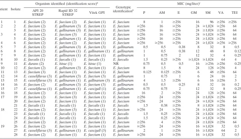
Identification.Molecular identification showed that 15 iso-lates were E. faecium, 4 were E. faecalis, and 8 were E.
galli-narum (see Table 2). One isolate (isolate 11) which could not
be identified by the PCR multiplex assay was identified as
E. durans by API 20 STREP and as E. hirae by rapid ID 32
STREP and Vitek GPI. The strain was considered to be E.
hi-rae because of its capacity to metabolize sucrose and raffinose
(13). The four isolates identified as E. faecalis by PCR were confirmed by API 20 STREP, rapid ID 32 STREP, and Vitek GPI. Discrepancies in the species identification of E. faecium and E. gallinarum isolates were observed between the geno-typic and phenogeno-typic methods. Moreover, differences in iden-tification were observed between API 20 STREP, rapid ID 32 STREP, and Vitek GPI. Four of the eight isolates identified as
E. gallinarum by PCR assay did not ferment raffinose. They
were identified as E. faecium by API 20 STREP with a very good identification score, and the other four isolates were identified as E. casseliflavus (isolates 14, 15, 17, and 27). In contrast, all eight of these isolates were identified as E.
galli-narum by the rapid ID 32 STREP method, five with an
excel-lent score (99.9%; T 5 0.85), one with a very good score (77.3%; T 5 0.56), one with a doubtful score (99.9%; T 5
[image:2.612.51.551.80.369.2]0.69), and one with good identification to the genus (57.2%; TABLE 1. Characteristics of 28 GRE isolated at the University Hospital of Geneva
Patient Isolate Site (mo/yr)Date Warda Genotypic identificationb
MIC (mg/liter)c
Glycopeptide
genotype patternPFGE
VA TEI
1 1 Stool 8/94 Medecine E. faecium $256 $256 vanA A1
2 2 Prosthesis 2/95 Orthopedics E. faecium $256 64 vanA B1
3 Stool 3/95 Orthopedics E. faecium $256 64 vanA B2
3 4 Ulcer 3/95 Orthopedics E. faecium $256 64 vanA B1
5 Stool 3/95 Orthopedics E. faecium $256 64 vanA B1
4 6 Stool 3/95 Hemat/onco E. faecium $256 $256 vanA B3
5 7 Stool 6/95 Medecine E. gallinarum 8 0.5 vanC1 C1
6 8 Stool 6/95 Surg/ICU E. gallinarum 8 0.12 vanC1 D
7 9 Ascites 6/95 Surg/ICU E. gallinarum 4 0.25 vanC1 E
8 10 Wound 9/95 Med/ICU E. faecalis 64 4 vanA F
9 11 Stool 9/95 Medecine NR $256 0.25 vanA Not done
10 12 Stool 10/95 Long-term H E. faecium $256 4 vanA A2
11 13 Stool 12/95 Surgery E. faecium $256 64 vanA G
12 14 Stool 12/95 Medecine E. gallinarum 16 2 vanC1 H
13 15 Stool 1/96 Medecine E. gallinarum $256 3 vanC1, vanB I
14 16 Stool 1/96 Hemat/onco E. gallinarum 16 0.2 vanC1 C1
15 17 Stool 2/96 Surg/ICU E. gallinarum 8 0.25 vanC1 C2
16 18 Peritoneum 4/96 Surg/ICU E. faecium $256 64 vanA K
17 19 Peritoneum 5/96 Surg/ICU E. faecium $256 64 vanA B4
20 Stool 7/96 Surg/ICU E. faecium $256 64 vanA B5
21 Stool 7/96 Surg/ICU E. faecalis $256 64 vanA L
18 22 Urethra 5/96 Surg/ICU E. faecium $256 64 vanA B4
23 Stool 6/96 Surg/ICU E. faecalis $256 64 vanA L
24 Wound 7/96 Surg/ICU E. faecalis $256 64 vanA L
25 Stool 7/96 Surg/ICU E. faecium $256 64 vanA B6
19 26 Stool 7/96 Hemat/onco E. faecium 32 0.5 vanB M1
27 Stool 7/96 Hemat/onco E. gallinarum 64 2 vanC1, vanB N
28 Gallbladder 9/96 Med/ICU E. faecium 32 0.5 vanB M2
aHemat/onco, hematology-oncology; Med/ICU, medical intensive care unit; Surg/ICU, surgical intensive care unit; long-term H, long-term hospitalization unit. bNR, no result obtained.
cVA, vancomycin; TEI, teicoplanin.
on May 15, 2020 by guest
http://jcm.asm.org/
T5 0.49). The Vitek GPI method identified two isolates as
E. gallinarum (one with a good identification score and one
with a presumptive identification score), two as E. casseliflavus/
gallinarum, and four as E. faecium, one of which had a good
identification score. Of the 15 isolates identified as E. faecium by PCR, all were identified as such by API 20 STREP and Vitek GPI, but four isolates were identified as E. gallinarum by the Rapid ID 32 STREP assay (isolates 2, 3, 5, and 12).
Antimicrobial susceptibility testing.Table 2 presents MICs of vancomycin for the 28 GRE isolates. Six had intermediate resistance (MIC, .4 to,32 mg/liter), and 22 were resistant (MIC $ 32 mg/liter). Thirteen of these were susceptible to
teicoplanin (MIC#8 mg/liter). All E. faecalis and E.
gallina-rum isolates were susceptible to penicillin (MIC#8 mg/liter) and ampicillin (MIC#8 mg/liter). In contrast, 11 of 15 E.
[image:3.612.56.548.81.364.2]fae-cium isolates were highly resistant to penicillin (MIC$ 256 mg/liter) and 1 of them was resistant to ampicillin (MIC$256 mg/liter). None of the strains showed b-lactamase activity. Only 1 isolate was highly resistant to gentamicin, whereas 16 of 28 isolates were highly resistant to streptomycin. Erythromycin resistance was present in 20 of 28 isolates of GRE. One isolate of E. faecium recovered from a stool sample (isolate 6) was highly resistant to all antimicrobial agents tested except gen-tamicin.
[image:3.612.138.459.567.705.2]FIG. 1. PFGE restriction fragment patterns of SmaI-digested genomic DNA obtained from glycopeptide-resistant E. faecium isolates and a dendrogram showing similarities. Numbering of isolates corresponds to the numbering in Tables 1 and 2.
TABLE 2. Comparative identification and susceptibility to antimicrobial agents of 28 GRE isolated at the University Hospital of Geneva
Patient Isolate
Organism identified (identification score)a
Genotypic identificationb
MIC (mg/liter)c
API 20
STREP Rapid ID 32STREP Vitek GPI P AM E GM SM VA TEI
1 1 E. faecium (2) E. faecium (2) E. faecium (1) E. faecium 8 1 $256 16 96 $256 $256 2 2 E. faecium (2) E. gallinarum (3) E. faecium (1) E. faecium $256 16 $256 24 $1,024 $256 64 3 E. faecium (2) E. gallinarum (3) E. faecium (1) E. faecium $256 16 $256 24 $1,024 $256 64 3 4 E. faecium (2) E. faecium (3) E. faecium (1) E. faecium $256 16 $256 24 $1,024 $256 64 5 E. faecium (2) E. gallinarum (1) E. faecium (1) E. faecium $256 16 $256 24 $1,024 $256 64 4 6 E. faecium (2) E. faecium (2) E. faecium (1) E. faecium $256 $256 $256 24 $1,024 $256 $256 5 7 E. faecium (2) E. gallinarum (3) E. faecium (3) E. gallinarum 0.5 0.5 0.38 8 32 8 0.5 6 8 E. faecium (2) E. gallinarum (1) E. gallinarum (1) E. gallinarum 1 0.5 0.38 12 48 8 0.12 7 9 E. faecium (2) E. gallinarum (1) E. faecium (1) E. gallinarum 1 0.75 2 12 32 4 0.25 8 10 E. faecalis (1) E. faecalis (1) E. faecalis (1) E. faecalis 1.5 0.25 $256 $1,024 $1,024 64 4 9 11 E. durans (2) E. hirae (1) E. hirae (1) NR 0.75 0.5 0.5 16 $256 $256 0.25 10 12 E. faecium (2) E. gallinarum (3) E. faecium (1) E. faecium 8 1.5 8 24 128 $256 4 11 13 E. faecium (3) E. faecium (1) E. faecium (1) E. faecium 0.125 0.125 $256 6 48 $256 64 12 14 E. casseliflavus (3) E. gallinarum (3) E. faecium (3) E. gallinarum 1 0.75 1 6 24 16 2 13 15 E. casseliflavus (3) E. gallinarum (1) E. gallinarum (3) E. gallinarum 4 1.5 $256 6 16 $256 3 14 16 E. faecium (2) E. gallinarum (3) E. faecium (3) E. gallinarum 0.5 0.5 0.25 8 32 16 0.2 15 17 E. casseliflavus (1) E. gallinarum (1) E. cass/gall (1) E. gallinarum 0.75 0.75 2 12 32 8 0.25 16 18 E. faecium (2) E. faecium (1) E. faecium (1) E. faecium 16 2 $256 24 128 $256 64 17 19 E. faecium (2) E. faecium (3) E. faecium (1) E. faecium $256 24 $256 24 $1,024 $256 64 20 E. faecium (2) E. faecium (1) E. faecium (1) E. faecium $256 24 $256 24 $1,024 $256 64 21 E. faecalis (1) E. faecalis (1) E. faecalis (1) E. faecalis 1.5 0.38 $256 8 $1,024 $256 64 18 22 E. faecium (2) E. faecium (3) E. faecium (1) E. faecium $256 24 $256 24 $1,024 $256 64 23 E. faecalis (1) E. faecalis (1) E. faecalis (1) E. faecalis 1.5 0.25 $256 24 $1,024 $256 64 24 E. faecalis (1) E. faecalis (1) E. faecalis (1) E. faecalis 1.5 0.25 $256 24 $1,024 $256 64 25 E. faecium (2) E. faecium (1) E. faecium (1) E. faecium $256 4 $256 24 $1,024 $256 64 19 26 E. faecium (2) E. faecium (1) E. faecium (1) E. faecium $256 24 $256 16 $1,024 32 0.5
27 E. casseliflavus (3) E. gallinarum (1) E. cass/gall (3) E. gallinarum 2 1 $256 16 $1,024 64 2 28 E. faecium (2) E. faecium (1) E. faecium (1) E. faecium $256 24 $256 16 $1,024 32 0.5
aIdentification scores of phenotypic identifications: 1, excellent or very good identification; 2, good identification; 3, uncertain identification to the species level.
E. cass/gall, E. casseliflavus/gallinarum.
bNR, no result obtained.
cP, penicillin; AM, ampicillin; E, erythromycin; GM, gentamicin; SM, streptomycin; VA, vancomycin; TEI, teicoplanin.
on May 15, 2020 by guest
http://jcm.asm.org/
Genotype of glycopeptide resistance. The vanA gene was detected in 18 of 28 isolates (Table 1) including 14 E. faecium, 3 E. faecalis, and 1 E. hirae. These 18 vanA isolates were all highly resistant to vancomycin (MIC$64 mg/liter). Fifteen of these were highly resistant to teicoplanin (MIC$64 mg/liter), whereas the other three (isolates 10, 11, and 12) were suscep-tible to this compound (MIC#4 mg/liter). The vanB gene was detected in 4 of 28 isolates, including two E. faecium isolates (isolates 26 and 28) and two E. gallinarum isolates (isolates 15 and 27). MICs of vancomycin for vanB strains ranged from 32 to 256 mg/liter, whereas those of teicoplanin remained in the susceptibility range (MIC#4 mg/liter). All eight E. gallinarum isolates contained the vanC1 gene. The six isolates of E.
galli-narum containing the vanC1 gene without the additional
gly-copeptide resistance gene (isolates 7 through 9, 14, 16, and 17) showed intermediate resistance to vancomycin (MIC58 to 16 mg/liter) and susceptibility to teicoplanin (MIC#2 mg/liter). When patients were colonized by different species of entero-cocci, similar genotypes were observed for the different species of a single patient (vanA for patients 17 and 18 and vanB for patient 19).
Genotyping.The results of genotyping are presented in Ta-ble 1 and Fig. 1, 2, and 3. Analysis of PFGE patterns obtained with the 15 E. faecium isolates showed a cluster of 9 isolates of
E. faecium vanA (isolates 2 through 6, 19, 20, 22, and 25) with
identical or similar banding patterns (PFGE pattern B, zero to six fragment differences) (Fig. 1). These isolates came from five patients (patients 2 through 4, 17, and 18) who were hos-pitalized in three different wards. In addition, the E. faecium stool isolates (isolates 1 and 12) from two other patients (pa-tients 1 and 10) differed by only three fragments and may be considered probably genetically related (PFGE pattern A) (Fig. 1). The two isolates of E. faecium vanB (isolates 26 and 27) from the same patient (patient 19) present differences in six bands and were interpreted as possibly related (PFGE pattern M) (Fig. 1). The other E. faecium isolates (isolates 13 and 18), from patients 11 and 16, were not related. The E.
fae-calis isolates (isolates 21, 23, and 24) from patients 17 and 18,
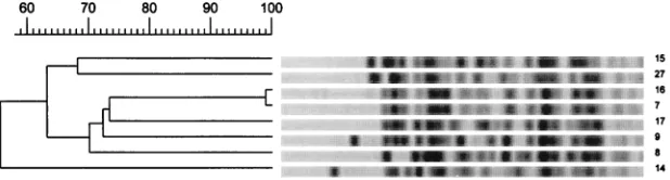
who were hospitalized in the surgical intensive care unit in the same time period, appeared to be identical (PFGE pattern L) (Fig. 2). Among the E. gallinarum isolates, two isolates (iso-lates 7 and 16) from two different patients (5 and 14) showed identical banding patterns (PFGE pattern C1) (Fig. 3). The six
other isolates (isolates 8, 9, 14, 15, 17, and 27), from patients 6, 7, 12, 13, 15, and 19, including the two E. gallinarum isolates with the vanB gene, were not related (Fig. 3).
DISCUSSION
The present study emphasizes the difficulties of phenotypical characterization of clinical isolates of GRE. Characterizing GRE allows the distinction between acquired GRE and intrin-sic GRE to be made. This is important for clinicians and for implementation of infection control measures (21, 31, 34). Acquired GRE are more difficult to treat than intrinsic GRE due to their broad-spectrum antimicrobial resistance. More-over, only acquired resistance is transferable to other entero-cocci and is associated with nosocomial epidemics (21, 34). In the present study, three limitations of the phenotypic methods used for both the identification of enterococci and the deter-mination of the type of glycopeptide resistance are highlighted. First, enterococci are not correctly identified to the species level by phenotypic methods. Second, because intrinsic GRE can acquire additional genes of vancomycin resistance, the identification of enterococci to the species level does not pre-dict the glycopeptide resistance type. Third, discrepancies be-tween the VanA and VanB phenotypes and their correspon-dent vanA and vanB genotypes may exist.
Our results showed that three commercially available kits frequently used in microbiology laboratories for identification of enterococci (API 20 STREP, Rapid ID 32 STREP, and Vitek GPI), failed to differentiate some E. gallinarum isolates from E. faecium, or vice versa: E. gallinarum isolates that did not ferment raffinose were misidentified as E. faecium by API 20 STREP and Vitek GPI. Rapid ID 32 STREP, which per-formed well for the identification of E. gallinarum, failed to identify some E. faecium isolates, which it misidentified as
E. gallinarum. These findings, previously reported for API 20
STREP (15) and Rapid ID 32 STREP (33), emphasize that based on biochemical reactions, it is not possible to differen-tiate between E. faecium and E. gallinarum, which belong to the same biochemical group, group II (12). With regard to these results, the characterization of GRE based on phenotypic iden-tification (31) of enterococci cannot be recommended.
[image:4.612.134.466.71.128.2]For the first time, as far as we are aware, two E. gallinarum isolates were found harboring the vanB gene. The two strains
FIG. 2. PFGE restriction fragment patterns of SmaI-digested genomic DNA obtained from glycopeptide-resistant E. faecalis isolates and a dendrogram showing similarities. Numbering of isolates corresponds to the numbering in Tables 1 and 2.
FIG. 3. PFGE restriction fragment patterns of EagI-digested genomic DNA obtained from glycopeptide-resistant E. gallinarum isolates and a dendrogram showing similarities. Numbering of isolates corresponds to the numbering in Tables 1 and 2.
on May 15, 2020 by guest
http://jcm.asm.org/
[image:4.612.144.453.624.707.2]were isolated from two different patients 6 months apart and were not related by PFGE. Dutka-Malen et al. have previously reported the transfer of the vanA gene to E. gallinarum and
E. casseliflavus (8). The acquisition of additional glycopeptide
resistance genes by intrinsic GRE emphasizes that the charac-terization of intrinsic GRE should be based on genotypic anal-ysis of the van type. Using phenotypic methods, VanB resis-tance is difficult to detect because MICs of vancomycin may be only moderately increased, comparable to those of the VanC type (11, 21).
Of the 18 GRE with the vanA genotype analyzed in the present study, three isolates were susceptible to teicoplanin, corresponding to a VanB phenotype. This finding was previ-ously reported by others (34). Conversely, it has been reported that some strains of the vanB genotype can have a VanA phenotype, with coresistance to both vancomycin and teicopla-nin (33). Thus, for unusual isolates the phenotype cannot be inferred from the genotype, and vice versa.
The multiplex PCR assay proposed by Dutka-Malen et al. (9) and used in the present study presents a clear advantage over phenotypic methods with regard to specificity and rapid-ity. Identification of the most frequently occurring species of enterococci and determination of vanA and vanB genotypes were performed in a single reaction. In conjunction with the determination of MICs of vancomycin and teicoplanin, this approach is useful for microbiology laboratories facing the need to characterize GRE. In the present study only one iso-late, a strain of E. hirae, could not be identified by the multi-plex PCR assay, because the primers of theD-alanine–D
-ala-nine ligase gene specific to this species were not included in the assay.
No epidemiological conclusion can be drawn from this study concerning extent of colonization, because GRE were not looked for prospectively in the stools. However, of the five infected patients analyzed for stool carriage of GRE, all were found to be positive with a similar or related strain, confirming the role of the gastrointestinal tract as a reservoir of GRE. In addition, three patients were colonized by several GRE be-longing to different species, two with E. faecium and E. faecalis containing the vanA gene and one with E. faecium and E.
galli-narum containing the vanB gene. This suggests an in vivo
transfer of glycopeptide resistance in the intestinal tract, as previously reported for the vanA determinant (8). It also em-phasizes that, with regard to the recent report of a vanB trans-ferable determinant in Streptococcus bovis in France (30), the dissemination of vanB resistance to enterococcal species other than E. faecium and E. faecalis or to other genera should be closely monitored.
The results of the present study showed a predominance of
E. faecium with the vanA gene among acquired GRE with both
sporadic cases and clusters of cases. The major cluster of five patients included four of the five patients infected with E.
fae-cium vanA. The dissemination of the same strain of E. faefae-cium vanA within a single unit may be easily explained by
cross-contamination. However, we have no explanation for the re-covery of a similar strain of E. faecium vanA after an interval of 1 year in two different units. It emphasizes that the mech-anisms by which resistance is disseminated within hospitals have not been fully elucidated (21) and are more complex than initially thought. In Europe, the food chain has been suspected to be a source of GRE (3, 21) in relation to the use of avopar-cin as a food additive for animals (21). The isolation in the present study of an E. hirae strain supports this hypothesis, as this species is predominant in the digestive tracts of poultry and cattle (6, 7).
In conclusion, this study shows the importance of
character-izing GRE by both phenotypic and genotypic methods. With the rapid increase in the occurrence of such isolates, the use of both types of methods will provide useful information for cli-nicians and will implement infection control measures.
ACKNOWLEDGMENTS
We thank P. Courvalin for providing reference strains and P. Maj-cherczyk and P. Moreillon for kindly reviewing the manuscript.
REFERENCES
1. Barton, A. L., and G. V. Doern. 1995. Selective media for detecting gastro-intestinal carriage of vancomycin-resistant enterococci. Diagn. Microbiol. Infect. Dis. 23:119–122.
2. Boyce, J. M., S. M. Opal, J. W. Chow, M. J. Zervos, G. Potter-Bynoe, C. B. Sherman, R. L. C. Romulo, S. Fortna, and A. A. Medeiros.1994. Outbreak of multidrug-resistant Enterococcus faecium with transferable vanB class vancomycin resistance. J. Clin. Microbiol. 32:1148–1153.
3. Chadwick, P. R., N. Woodford, E. B. Kaczmarski, S. Gray, R. A. Barrell, and B. A. Oppenheim.1996. Glycopeptide-resistant enterococci isolated from uncooked meat. J. Antimicrob. Chemother. 38:908–909.
4. Chenoweth, C., and D. Shaberg. 1990. The epidemiology of Enterococci. Eur. J. Clin. Microbiol. Infect. Dis. 9:80–89.
5. Clark, N. C., R. C. Cooksey, B. C. Hill, J. M. Swenson, and F. C. Tenover. 1993. Characterization of glycopeptide-resistant enterococci from U.S. hos-pitals. Antimicrob. Agents Chemother. 37:2311–2317.
6. Devriese, L. A., J. Hommez, R. Wijfels, and F. Haesebrouck. 1991. Compo-sition of the enterococcal and streptococcal intestinal flora of poultry. J. Appl. Bacteriol. 71:46–50.
7. Devriese, L. A., L. Laurier, P. De Herdt, and F. Haesebrouck. 1992. Entero-coccal and streptoEntero-coccal species isolated from faeces of calves, young cattle and diary cows. J. Appl. Bacteriol. 72:29–31.
8. Dutka-Malen, S., B. Blaimont, G. Wauters, and P. Courvalin. 1994. Emer-gence of high-level resistance to glycopeptides in Enterococcus gallinarum and Enterococcus casseliflavus. Antimicrob. Agents Chemother. 38:1675– 1677.
9. Dutka-Malen, S., S. Evers, and P. Courvalin. 1995. Detection of glycopep-tide resistance genotypes and identification to the species level of clinically relevant enterococci by PCR. J. Clin. Microbiol. 33:24–27.
10. Eliopoulos, G. M. 1993. Increasing problems in the therapy of enterococcal infections. Eur. J. Clin. Microbiol. Infect. Dis. 12:409–412.
11. Endtz, H. P., N. Van Den Braak, A. Van Belkum, W. H. Goessens, D. Kreft, A. B. Stroebel, and H. A. Verbrugh.1998. Comparison of eight methods to detect vancomycin resistance in enterococci. J. Clin. Microbiol. 36:592–594. 12. Facklam, R. R., and D. F. Sahm. 1995. Enterococcus, p. 308–314. In P. R. Murray, E. J. Barron, M. A. Pfaller, F. C. Tenover, and R. H. Yolken (ed.), Manual of clinical microbiology, 6th ed. American Society for Microbiology, Washington, D.C.
13. Farrow, J. A. E., and M. D. Collins. 1985. Enterococcus hirae, a new species that includes amino acid assay strain NCDO 1258 and strains causing growth depression in young chickens. Int. J. Syst. Bacteriol. 35:73–75.
14. Frieden, T. R., S. S. Munsiff, and D. E. Low. 1993. Emergence of vancomy-cin-resistant enterococci in New York City. Lancet 342:76–79.
15. Gordts, B., H. Van Landuyt, M. Ieven, P. Vandame, and H. Goossens. 1995. Vancomycin-resistant enterococci colonizing the intestinal tracts of hospi-talized patients. J. Clin. Microbiol. 33:2842–2846.
16. Handwerger, S., B. Raucher, D. Altarac, J. Monka, S. Marchione, K. V. Singh, B. E. Murray, J. Wolff, and B. Walters.1993. Nosocomial outbreak due to Enterococcus faecium highly resistant to vancomycin, penicillin, and gentamicin. Clin. Infect. Dis. 16:750–755.
17. Jett, B. D., M. M. Huycke, and M. S. Gilmore. 1994. Virulence of entero-cocci. Clin. Microbiol. Rev. 7:462–478.
18. Kaplan, A. H., P. H. Gilligan, and R. R. Facklam. 1988. Recovery of resistant enterococci during vancomycin prophylaxis. J. Clin. Microbiol. 26:1216– 1218.
19. Leclercq, R., E. Derlot, J. Duval, and P. Courvalin. 1988. Plasmid-mediated resistance to vancomycin and teicoplanin in Enterococcus faecium. N. Engl. J. Med. 319:157–161.
20. Leclercq, R., S. Dutka-Malen, J. Duval, and P. Courvalin. 1992. Vancomycin resistance gene vanC is specific to Enterococcus gallinarum. Antimicrob. Agents Chemother. 36:2005–2008.
21. Leclercq, R., and P. Courvalin. 1997. Resistance to glycopeptides in entero-cocci. Clin. Infect. Dis. 24:545–556.
22. Moellering, R. C. 1992. Emergence of Enterococcus as a significant patho-gen. Clin. Infect. Dis. 14:1173–1178.
23. Murray, B. E. 1990. The life and times of the enterococcus. Clin. Microbiol. Rev. 3:46–65.
24. Murray, B. E. 1995. What can we do about vancomycin-resistant entero-cocci? Clin. Infect. Dis. 20:1134–1136.
25. National Committee for Clinical Laboratory Standards. 1997. Performance
on May 15, 2020 by guest
http://jcm.asm.org/
standards for antimicrobial disk susceptibility tests. Approved standard M100-S7. National Committee for Clinical Laboratory Standards, Wayne, Pa.
26. Nauschvetz, W. F., S. B. Trevino, L. S. Harrison, R. N. Longfield, L. Fletcher, and W. G. Wartham.1993. Enterococcus casseliflavus as an agent of noso-comial bloodstream infections. Med. Microbiol. Lett. 2:102–108. 27. Navarro, F., and P. Courvalin. 1994. Analysis of genes encodingD-alanine–
D-alanine ligase-related enzymes in Enterococcus casseliflavus and Entero-coccus flavescens. Antimicrob. Agents Chemother. 38:1788–1793.
28. Pegues, D. A., C. F. Pegues, P. L. Hibberd, D. S. Ford, and D. C. Hooper. 1997. Emergence and dissemination of a highly vancomycin-resistant vanA strain of Enterococcus faecium at a large teaching hospital. J. Clin. Microbiol. 35:1565–1570.
29. Perichon, B., P. Reynolds, and P. Courvalin. 1997. VanD-type glycopeptide-resistant Enterococcus faecium BM4339. Antimicrob. Agents Chemother. 41: 2016–2018.
30. Poyart, C., C. Pierre, G. Quesne, B. Pron, P. Berche, and P. Trieu-Cuot.
1997. Emergence of vancomycin resistance in the genus Streptococcus: char-acterization of a vanB transferable determinant in Streptococcus bovis. An-timicrob. Agents Chemother. 41:24–29.
31. Sahm, D. F., L. Free, C. Smith, M. Eveland, and L. M. Mundy. 1997. Rapid characterization schemes for surveillance isolates of vancomycin-resistant enterococci. J. Clin. Microbiol. 35:2026–2030.
32. Tenover, F. C., R. D. Arbeit, R. V. Goering, P. A. Mickelsen, B. E. Murray, D. H. Persing, and B. Swaminathan.1995. Interpreting chromosomal DNA restriction patterns produced by pulsed-field gel electrophoresis: criteria for bacterial strain typing. J. Clin. Microbiol. 33:2233–2239.
33. Vandamme, P., E. Vercauteren, C. Lammens, N. Pensart, M. Ieven, B. Pot, R. Leclercq, and H. Goossens.1996. Survey of enterococcal susceptibility patterns in Belgium. J. Clin. Microbiol. 34:2572–2576.
34. Woodford, N., A. P. Johnson, D. Morrison, and D. C. E. Speller. 1995. Current perspectives on glycopeptide resistance. Clin. Microbiol. Rev. 8: 585–615.