epitope processing
Ulrike Seifert, … , Peter-M. Kloetzel, Barbara Rehermann
J Clin Invest. 2004;
114(2)
:250-259.
https://doi.org/10.1172/JCI20985
.
The high incidence of hepatitis C virus (HCV) persistence raises the question of how HCV
interferes with host immune responses. Studying a single-source HCV outbreak, we
identified an HCV mutation that impaired correct carboxyterminal cleavage of an
immunodominant HLA-A2–restricted CD8 cell epitope that is frequently recognized by
recovered patients. The mutation, a conservative HCV nonstructural protein 3 (NS3)
tyrosine to phenylalanine substitution, was absent in 54 clones of the infectious source, but
present in 15/21 (71%) HLA-A2–positive and in 11/24 (46%) HLA-A2–negative patients with
chronic hepatitis C. In order to analyze whether the mutation affected the processing of the
HLA-A2–restricted CD8 cell epitope, mutant and wild-type NS3 polypeptides were digested
in vitro with 20S constitutive proteasomes and with immunoproteasomes. The presence of
the mutation resulted in impaired carboxyterminal cleavage of the epitope. In order to
analyze whether impaired epitope processing affected T cell priming in vivo, HLA-A2–
transgenic mice were infected with vaccinia viruses encoding either wild-type or mutant
HCV NS3. The mutant induced fewer epitope-specific, IFN-
g
;–producing and fewer
tetramer
+cells than the wild type. These data demonstrate how a conservative mutation in
the flanking region of an HCV epitope impairs the induction of epitope-specific CD8
+T cells
and reveal a mechanism that may contribute to viral sequence evolution in infected patients.
Article
Infectious disease
Find the latest version:
Research article
250 The Journal of Clinical Investigation http://www.jci.org Volume 114 Number 2 July 2004
Hepatitis C virus mutation affects
proteasomal epitope processing
Ulrike Seifert,1 Heike Liermann,2 Vito Racanelli,3 Anne Halenius,4 Manfred Wiese,5
Heiner Wedemeyer,3 Thomas Ruppert,1 Kay Rispeter,6 Peter Henklein,1 Alice Sijts,1
Hartmut Hengel,4 Peter-M. Kloetzel,1 and Barbara Rehermann2,3
1Institute of Biochemistry, Charité, Humboldt University, Berlin, Germany. 2Department of Gastroenterology, Hepatology and Endocrinology, Medizinische Hochschule Hannover (MHH), Hannover, Germany. 3Liver Diseases Section, Digestive Diseases Branch, National Institute for Diabetes and Digestive and Kidney Diseases (NIDDK), NIH, Department of Health and Human Services (DHHS), Bethesda, Maryland, USA. 4Robert Koch-Institute, Berlin,
Germany. 5II. Klinik für Innere Medizin, Städtisches Klinikum St. Georg, Leipzig, Germany. 6Institute for Virology, Universitätsklinikum, Essen, Germany.
The high incidence of hepatitis C virus (HCV) persistence raises the question of how HCV interferes with host
immune responses. Studying a single-source HCV outbreak, we identified an HCV mutation that impaired
correct carboxyterminal cleavage of an immunodominant HLA-A2–restricted CD8 cell epitope that is
fre-quently recognized by recovered patients. The mutation, a conservative HCV nonstructural protein 3 (NS3)
tyrosine to phenylalanine substitution, was absent in 54 clones of the infectious source, but present in 15/21
(71%) HLA-A2–positive and in 11/24 (46%) HLA-A2–negative patients with chronic hepatitis C. In order to
analyze whether the mutation affected the processing of the HLA-A2–restricted CD8 cell epitope, mutant and
wild-type NS3 polypeptides were digested in vitro with 20S constitutive proteasomes and with
immunoprote-asomes. The presence of the mutation resulted in impaired carboxyterminal cleavage of the epitope. In order
to analyze whether impaired epitope processing affected T cell priming in vivo, HLA-A2–transgenic mice
were infected with vaccinia viruses encoding either wild-type or mutant HCV NS3. The mutant induced fewer
epitope-specific, IFN-
γ
–producing and fewer tetramer
+cells than the wild type. These data demonstrate how
a conservative mutation in the flanking region of an HCV epitope impairs the induction of epitope-specific
CD8
+T cells and reveal a mechanism that may contribute to viral sequence evolution in infected patients.
Introduction
Hepatitis C virus (HCV) is a 9.6 kb positive-stranded RNA virus of the flavivirus family and the leading cause of chronic hepati-tis worldwide. Whereas recovery from acute HCV infection has been associated with multispecific T cell responses that protect upon reexposure to the virus (1, 2), these responses are either not induced or not maintained in the large number of patients who develop chronic infection (3–5).
We have therefore asked whether HCV interferes with the induc-tion of antigen-specific T cells. Inducinduc-tion of CD8+ T cells depends
on the generation of MHC class I ligands by the proteasome, the major cytosolic proteinase. The proteasome cleaves short peptides from longer polypeptide precursors that are then translocated into the endoplasmic reticulum and bind to newly synthesized MHC class I molecules (6). 26S proteasomes contain a 20S catalytic core, arranged as 2 heptameric outer rings with 7 α-subunits each and 2 heptameric inner rings with 7 β-subunits each. Proteasome activity is closely regulated by cytokines that are produced in viral infec-tions (7–12). In response to IFN-γ, for example, the constitutive catalytic subunits β1, β2, and β5 are replaced by low molecular weight protein 2 (LMP2) (iβ1), LMP7 (iβ5), and multicatalytic endopeptidase complex-like–1 (MECL-1) (iβ2) to form immuno-proteasomes with altered cleavage properties (13).
HCV circulates in an abundant number of quasispecies because of its high replication rate (14) and its lack of polymerase
proof-reading capacity. Individual HCV sequences have been described as abolishing recognition by T cell receptors (TCRs) and antibodies, thus interfering with the effector arms of the cellular (2, 15, 16) and humoral immune responses (17). In contrast, the possibility that HCV mutations affect the induction of T cell responses has not been investigated. Indirect evidence for this hypothesis stems from descriptive reports that HCV isolates from persistently infected patients often encode less immunogenic sequences than prototype peptides used for in vitro analysis (18, 19). Whether the decreased immunogenicity results from viral mutations or from infection with less immunogenic strains has not been analyzed because the sequence of the infecting virus is not known in most human stud-ies and also because the route of infection and inoculum size differ among the studied individuals.
Having studied a cohort of patients accidentally infected by a single-source HCV with known sequence, we here demonstrate that an HCV mutation located in the flanking region of a fre-quently recognized HCV epitope (4, 20–25) impairs the induction of HCV-specific CD8+ T cells by affecting the sophisticated
protea-somal antigen-processing machinery.
Results
The tyrosine/phenylalanine mutation at residue HCV nonstructural pro-tein 31082 is common in patients who develop persistent infection after a
single-source outbreak of HCV. In order to study viral mutations dur-ing the natural course of HCV infection in humans, we analyzed a cohort of patients that had accidentally been infected with HCV in 1978/1979 during a single-source outbreak due to a contaminated anti–D immunoglobulin (26). Because the precise time of infection as well as the genotype and the sequence of the original infectious virus were known and identical for all patients, this cohort was
Nonstandard abbreviations used: hepatitis C virus (HCV); nonstructural protein 3 (NS3); phenylalanine (F); tyrosine (Y).
Conflict of interest: The authors have declared that no conflict of interest exists.
suitable for studying candidate mechanisms of viral persistence. As previously described for this and other cohorts, the strength of the cellular immune response correlated with the outcome of infection, and HCV nonstructural protein 3 (NS3) peptides were among the most frequently recognized (3, 5, 20, 27, 28).
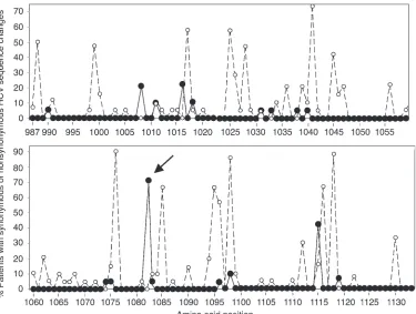
When we compared the HCV NS3987–1133 sequence of the
[image:3.585.50.425.81.364.2]infec-tious source with sequences that we isolated from the sera of persis-tently infected, HLA-A2–positive patients 18 years after the single-source outbreak, we found the highest ratio of nonsynonymous to synonymous HCV mutations at amino acid position NS31082
(Figure 1). Position NS31082 was located directly adjacent to the
carboxyterminus of an HLA-A2–restricted CD8+ T cell epitope
recognized by recovered patients of this (20) and other cohorts (4, 20–25). Whereas 54 molecular clones from 3 independent PCRs of the infectious source encoded a tyrosine (Y) in position NS31082,
serum isolates from 15 of 21 (71%) HLA-A2–positive, persistently infected patients encoded a phenylalanine (F) at position NS31082
(Table 1). Because the Y/F mutant was also observed in a substan-tial number of HLA-A2–negative, persistently infected patients (11 of 24 [46%]; P = 0.07; data not shown), the epidemiological data alone did not indicate whether HLA-A2–restricted T cell selection pressure could have contributed to the viral sequence evolution in this patient cohort. We therefore decided to analyze the molecular and immunological effects of the Y/F mutation in vitro by study-ing proteasomal processstudy-ing of antigen and in vivo by studystudy-ing its effect on T cell priming in an HLA-transgenic mouse model.
The Y/F mutation at residue N31082 impairs carboxyterminal processing of
the NS31073–1081 epitope. Most MHC class I–restricted peptides are
lib-erated from antigenic precursor sequences by the 20S core particle of the proteasome. To analyze whether the NS31082 Y/F substitution
affected proteasomal processing of the NS31073–1081 epitope, we built
on our previous demonstration that proteasome-dependent in vitro
processing of epitope-harboring polypeptides reflects the in vivo sit-uation with high fidelity (10, 12, 29, 30) and compared proteasome-dependent in vitro processing of the NS3 wild-type polypeptide, designated NS3(Wt)1062–1095, with processing of the NS3 mutant
polypeptide, designated NS3(Mut)1062–1095. For this purpose,
immu-noproteasomes were purified from IFN-γ–stimulated HepG2 human hepatoma cells and from murine mouse embryonal cells–217 (MEC-217) transfected with the immunoproteasome subunits LMP2, LMP7 and MECL-1 (10). Constitutive proteasomes were purified from unstimulated HepG2 cells and from murine MEC-18 cells.
Synthetic NS3(Wt)1062–1095 and NS3(Mut)1062–1095
polypep-tides (Figure 2a) were then incubated with 20S immunoprotea-somes. The digestion products were separated and analyzed by reverse phase–HPLC and mass spectrometry. NS3(Wt)1062–1095
and NS3(Mut)1062–1095 polypeptide substrates were digested with
the same kinetics with nearly 40–50% turn-over of each substrate within 4–8 hours (Figure 2B). Because longer digestion times resulted in secondary cleavage of processing intermediates, 4-hour and 8-hour digestion times were considered optimal for further biochemical analyses. Processing with constitutive proteasomes yielded the same qualitative results but lower amounts of digest product than processing with immunoproteasomes (not shown).
In contrast to the substrates, the relative abundance of the NS31073–1081 epitope could not be precisely determined because
the epitope’s 2 cysteine residues formed aggregates via disulfide bonds. These aggregates interfered with reliable identification of the NS31073–1081 epitope and its immediate precursors by mass
spectrometry. As an indirect measure of the generation of the NS31073–1081 epitope, we therefore determined the relative
abun-dance of those cleavage products that flanked its amino- and car-boxyterminus. Cleavage product NS31062–1070, for example, flanked
the amino-terminus of the NS31073–1081 epitope. Cleavage product
Figure 1
Prevalence of synonymous (open circles) and non-synonymous (filled circles) HCV mutations within the HCV NS3987–1133 sequence
research article
252 The Journal of Clinical Investigation http://www.jci.org Volume 114 Number 2 July 2004
NS31062–1070 was generated from both the NS3(Wt)1062–1095 and
NS3(Mut)1062–1095 polypeptide substrates with comparable
effi-ciency (Figure 2C). Generation of cleavage product NS31062–1070
was associated with the generation of the complementary cleav-age product NS31071–1095 (Figure 2D), consistent with a
protea-somal cut between amino acid positions 1070 and 1071 in both the NS3(Wt)1062–1095 and NS3(Mut)1062–1095 polypeptide substrates
(see dotted line in Figure 2A). This proteasomal cut resulted in
an amino-terminal elongation of the NS31073–1081 epitope by 2
amino acids.
A second cleavage product that flanked the amino-terminus of the NS31073–1081 epitope was
peptide NS31062–1072. As
indicat-ed in Figure 2E, cleavage product NS31062–1072 was also liberated
from both the NS3(Wt)1062–1095
and NS3(Mut)1062–1095
polypep-tide substrates in comparable amounts (Figure 2E), indicat-ing that the proteasome cut both the NS3(Wt)1062–1095 and
NS3(Mut)1062–1095 polypeptide
between amino acid positions 1072 and 1073 as indicated by the dotted line in Figure 2A. This proteasomal cut resulted in the generation of the correct amino-terminus of the NS31073–1081
epitope, irrespective of the pres-ence or abspres-ence of the NS31082
mutation. The demonstration that the NS31073–1081 epitope and
its amino-terminally elongated forms were generated at the same time is consistent with previous findings for other epitopes (31, 32). In cases of amino-terminally elongated peptides, the precise amino-terminus has been shown to be further defined by postpro-teasomal trimming by aminoexo-peptidases (31, 32).
In contrast to the amino-termi-nus, the correct carboxyterminus of the NS31073–1081 epitope was
generated only from the wild-type polypeptide. Figure 2 (F, G, and H) shows the relative abundance of those cleavage products whose generation defined the epitope’s carboxyterminus at amino acid position NS31082. The generation
of cleavage product NS31082–1095
indicated the correct processing of the epitope’s carboxyterminus with a proteasomal cut between amino acid positions 1081 and 1082 (Figure 2F). Impor-tantly, cleavage product NS31082–1095 was only liberated from the
NS3(Wt)1062–1095 and not from theNS3(Mut)1062–1095 polypeptide
substrate (Figure 2F). In contrast, cleavage product NS31083–1095 was
liberated from the NS3(Mut)1062–1095 more efficiently than from the
NS3(Wt)1062–1095 polypeptide substrate (Figure 2G). This result
indi-cated that, in the presence of the NS31082 mutation, the proteasome
[image:4.585.50.465.114.649.2]cut between amino acid positions 1082 and 1083 rather than between amino acid positions 1081 and 1082 and thus generated
Table 1
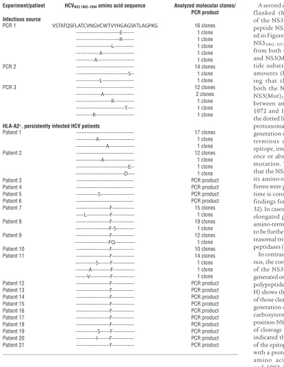
HCV NS31062–1095 sequence of the infectious source and HLA-A2+ HCV-infected patients
Experiment/patient HCVNS3 1062–1094 amino acid sequence Analyzed molecular clones/
PCR product Infectious source
PCR 1 VSTATQSFLATCVNGVCWTVYHGAGSKTLAGPKG 16 clones
---E--- 1 clone ---R--- 1 clone ---L--- 1 clone ---A--- 1 clone ---A--- 1 clone
PCR 2 --- 14 clones
---S-- 1 clone ---L--- 1 clone
PCR 3 --- 12 clones
---A--- 2 clones ---R--- 1 clone ---T---- 1 clone ---R--- 1 clone
HLA-A2+, persistently infected HCV patients
Patient 1 --- 17 clones
---A--- 1 clone ---A--- 1 clone
Patient 2 --- 12 clones
---A--- 1 clone ---E-- 1 clone ---D---- 1 clone Patient 3 --- PCR product Patient 4 --- PCR product Patient 5 ---S--- PCR product Patient 6 --- PCR product
Patient 7 ---F--- 15 clones
---L---F--- 1 clone
Patient 8 ---F--- 19 clones
---F-S--- 1 clone
Patient 9 ---F--- 12 clones
a carboxyterminally elongated NS3epitope. Finally, cleavage prod-uct NS31084–1095 was liberated from both the NS3(Wt)1062–1095 and
NS3(Mut)1062–1095 polypeptide substrates in comparable amounts
(Figure 2H), indicating that the proteasomal cut between amino acid 1083 and 1084 was not affected by the NS31082 mutation.
In summary, an incorrect carboxyterminus of the NS31073–1081
epitope appeared to be generated from the NS3(Mut)1062–1095 but
not from the NS3(Wt)1062–1095 polypeptide substrate.
NS31073–1081-specific CD8+ T cells recognize aminoterminally, but not
carboxyterminally elongated forms of the minimal optimal epitope. To study recognition of the processing products by CD8+ T cells, we
generated NS31073–1081-specificCD8+ T cell lines from the blood of
HCV-recovered patients. These T cell lines recognized target cells pulsed with the minimal optimal NS31073–1081-epitope in a standard
cytotoxicity assay (Figure 3). Epitope-specific T cell lines also recog-nized the corresponding aminoterminally elongated peptides, but not the carboxyterminally elongated mutant and wild-type peptides (Figure 3), thereby confirming that correct carboxyterminal cleav-age by the proteasome was indispensable (33, 34).
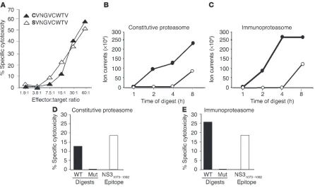
Direct biochemical and immunological detection of the NS31073–1081 epitope
inproteasomal digests. To directly assess and quantitate the in vitro generation of the NS31073–1081 epitope, it was necessary to prevent
the formation of disulfide bonds between the cysteine residues. We therefore synthesized a serine variant of the epitope with a cysteine to serine exchange at position NS31073. This NS31073–1081 serine
vari-ant was equally well recognized by cytotoxic T cells as the original NS31073–1081 epitope (Figure 4A).
When the corresponding NS3(Wt)1062–1095 and NS3(Mut)1062–1095
polypeptides with a serine in position NS31073 were subjected to
diges-tion by constitutive proteasomes, the producdiges-tion of the NS31073– 1081 serine variant could be directly assessed by mass spectrometry.
As demonstrated in Figure 4B, a significantly larger amount of the NS31073–1081SVNGVCWTV epitope was generated from the
NS3(Wt)1062–1095 polypeptide than from the NS3(Mut)1062–1095
[image:5.585.67.531.85.412.2]polypeptide. For immunological analysis, the complete 8-hour proteasomal digests were then loaded onto transporter associated with antigen processing–deficient (TAP-deficient), T2-target cells and tested for recognition by NS31073–1081-specific, cytotoxic T cell
Figure 2
Digestion of NS3(Wt)1062–1095 polypeptide and NS3(Mut)1062–1095 polypeptide with purified 20S immunoproteasomes. (A) The amino acid sequences
of the 34-mer NS3(Wt)1062–1095 andNS3(Mut)1062–1095 polypeptides are shown in single-letter code. The HLA-A2 restricted CD8+ T cell epitope
NS31073–1081 is framed, and amino acid position NS31082 containing the wild-type tyrosine or the mutant phenylalanine is circled. The dashed
lines indicate the location of proteasomal cuts as deduced from the data shown in panels C–H. (B) NS3(Wt)1062–1095 polypeptide (black bars) and
NS3(Mut)1062–1095 polypeptide (white bars) were digested for 1, 2, 4, and 8 hours with 20S immunoproteasomes. Kinetic analysis of polypeptide
substrate turnover is indicated. (C–H) Kinetic analysis of cleavage product generation. Relative abundances of cleavage products derived from immunoproteasomal digestion of NS3(Wt)1062–1095 polypeptide (filled circles) and NS3(Mut)1062–1095 polypeptide (open circles) are plotted. The relative
research article
254 The Journal of Clinical Investigation http://www.jci.org Volume 114 Number 2 July 2004
lines. Only the wild-type and not the mutant polypeptide digests were recognized by NS31073–1081-specific cytotoxic T cells (Figure
4D), thus confirming the biochemical data in Figure 2.
The same qualitative results were obtained when NS3(Wt)1062–1095
and NS3(Mut)1062–1095 polypeptides were digested with
immuno-proteasome instead of constitutive immuno-proteasome. Again, a signifi-cantly larger amount of the NS31073–1081SVNGVCWTV epitope
was generated from NS3(Wt)1062–1095 than from NS3(Mut)1062–1095
polypeptide (Figure 4, C and E). Overall, the immunoproteasome appeared to digest the wild-type polypeptide more rapidly than the constitutive proteasome did, as indicated by a plateau-phase
of epitope liberation from the wild-type polypeptide in the bio-chemical analysis (Figure 4C) and by the higher cytotoxicity in the immunological analysis (Figure 4E).
The HCV NS31082 Y/F mutant reduces the induction of HCV NS31073–1081
-specific CD8+ T cells in HLA-A2 transgenic mice. To analyze whether the
altered proteasomal cleavage of the NS31082 Y/F mutant affected
the generation of the NS31073–1081 epitope in vivo, we employed a
humanized mouse model. Specifically, we used transgenic mice that expressed the α1 and α2 chains of the human HLA-A2 mol-ecule and the α3 chain of the murine Kd molecule (35). Upon
immunization, these mice generate T cells against the same
HLA-Figure 3
NS31073–1081 epitope–specific CD8+ T cells recognize
aminoterminally extended, but not carboxyterminally extended, peptides of the minimal optimal NS31073–1081
epitope. NS31073–1081–specific CD8+ T cells were
expand-ed from PBMCs of an HCV-recoverexpand-ed patient by several weeks of peptide stimulation and tested against amino- and carboxyterminally extended peptides in a standard
51Cr-release assay. The mutant sequence at AA position
[image:6.585.46.322.81.243.2]1082 is underlined.
Figure 4
Biochemical and immunological analysis of proteasomal digests. (A) NS31073–1081–specific CD8+ T cells lyse T2 cells loaded with either the
NS31073–1081 serine variant SVNGVCWTV or the NS31073–1081 epitope CVNGVCWTV. (B–C) Generation of NS31073–1081 serine variant S
VNGVC-WTV from NS3(Wt)1062–1095 (filled circles) and NS3(Mut)1062–1095 (open circles) serine variant polypeptides NS31062–1095 by digestion with
constitu-tive 20S proteasome (B) and by digestion with 20S immunoproteasome (C). (D–E) 8-hour–proteasome (D) and immunoproteasome (E) digests of NS3(Wt)1062–1095 and NS3(Mut)1062–1095 serine variant polypeptides were loaded onto TAP-deficient T2-target cells and incubated with NS31073– 1081 epitope–specific, cytotoxic T cells at an effector to target ratio of 13 to 1. For comparison, 10-5 M NS31073–1081 serine variant was loaded onto
[image:6.585.73.519.398.664.2]A2–restricted epitopes as HLA-A2–positive humans (36). In con-trast to what occurs in human studies, however, this mouse model allows the induction of T cells in the context of a single, defined HLA molecule and a single, defined viral sequence. Therefore, the HLA-A2–transgenic mouse model is not subject to the influences of additional factors such as selection pressure in the context of other HLA molecules and epitopes and/or preservation of viral rep-lication fitness as may be operating in vivo in infected humans.
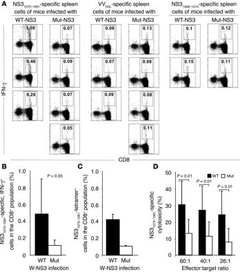
To take advantage of this model, we immunized HLA-A2–trans-genic mice with vaccinia viruses that encoded full-length HCV NS3 sequences with either the Y wild type or the F mutant at amino acid position NS31082. Two weeks after immunization, spleen cells
were isolated, and the frequency of NS31073–1081-specific T cells was
assessed by ex vivo intracellular IFN-γ–staining and by tetramer analysis. As shown in Figure 5A for individual mice and in Fig-ure 5, B and C, for all mice, the frequency of NS31073–1081-specific,
IFN-γ–producing and tetramer+ cells was significantly higher in
mice infected with wild-type than in those infected with mutant NS3 encoding virus. In contrast, the frequency of CD8+ cells that
recognized either a vaccinia virus epitope or an unrelated HCV NS3 epitope did not differ between both groups of mice, thus con-firming the specificity of the observation.
In separate experiments, additional immunizations with recom-binant DNA-expression vectors were performed to increase the number of NS31073–1081-specific T cells and to establish T cell lines
suitable for cytotoxicity analyses. At all effector/target ratios, cytotoxic T cell responses of mice that had been immunized with wild-type NS3 sequences were significantly stronger than cytotoxic T cell responses of mice immunized with mutant NS3 sequences (Figure 5D). Collectively, these results demonstrate that the NS31082 Y/F substitution reduced the generation of the
NS31073–1081 epitope in vitro when polypeptides were digested with
purified 20S proteasomes. In addition, the NS31082 substitution
impaired the induction of epitope-specific T cells in vivo when full-length NS3 protein was endogenously expressed, ubiqui-tinylated, and processed by the 26S proteasome in the presence of additional cytosolic proteases.
Discussion
[image:7.585.45.393.83.477.2]Most MHC class I ligands are liberated from strings of polypeptides and ubiquitinylated proteins by the proteasome, the main cytosolic protease (13). Whereas the amino-terminus of each epitope can be further defined by postproteasomal aminoexopeptidases (31, 32, 37), the carboxyterminus needs to be defined precisely with the first
Figure 5
Immunization of HLA-A2 transgenic mice with the wild-type HCV NS3 sequence induces more IFN-γ–secreting and cyto-toxic HCV NS31073–1081–specific CD8 T
cells than immunization with the mutant HCV NS3 sequence. (A and B) Ex vivo analysis of NS31073–1081–specific, IFN-γ
producing CD8 T cells demonstrated a significantly greater response in mice immunized with wild-type than of mice immunized with mutant NS3 encoding vaccinia virus. In contrast, the response against the vaccinia virusH3L (VVH3L)
epitope and the HCV NS31406–1414 epitope
did not differ between both groups of mice. (A) Dot plots from individual mice tested in the same experiment. (B) Mean and standard deviation of the results of all 8 mice per group. (C) Frequency of NS31073–1081-tetramer–specific T cells is
higher in mice immunized with wild-type than in mice immunized with mutant HCV NS3 sequences. Mean and stan-dard deviation of the results of 8 mice per group are shown. (D) NS31073–1081-
Author index
256 The Journal of Clinical Investigation http://www.jci.org Volume 113 Number 12 June 2004
cut (33, 34). Studying a single source outbreak of HCV, we identified an HCV mutation that interfered with the correct carboxyterminal cleavage of an immunodominant, HLA-A2 restricted HCV epitope from its mutated polypeptide precursor.
The emergence of viral mutations in immunogenic sequences has long been discussed as a potential immune escape mecha-nism. As regards HCV, sequences that do not bind to the MHC and/or the T cell receptor and thus are not recognized by HCV-specific CD8+ T cells have been observed in chimpanzees and in
humans (15, 16, 18). In addition, mutations that generate partial agonists or antagonists to the T cell receptor and downregulate wild-type–specific T cell responses have been described in HCV (18) as well as in hepatitis B virus (HBV) (38) and HIV infections (39). Perhaps even more efficient mechanisms of viral escape are mutations in epitope-flanking residues that interfere with antigen processing and presentation of MHC class I–restricted epitopes because, in these cases, the induction phase rather than the effec-tor phase of HCV-specific T cell responses can be impaired. Indeed, the literature provides several examples showing that amino acid residues in the flanking regions of T cell epitopes impair protea-somal processing of those epitopes (40–43). On the other hand, there are also examples showing that extensive sequence changes in the flanking regions of other immunodominant CD8 T cell epitopes do not influence antigen processing (44). In our study, we observed impairment of antigen processing by an exchange of two very similar amino acids. Although the Y/F substitution is a conservative one, it impaired correct carboxyterminal cleavage of the NS31073–1081 epitope not only by constitutive proteasomes, but
also by immunoproteasomes. Importantly, the same effects were observed in vitro when polypeptides were digested with purified 20S proteasomes and in vivo when full length HCV NS3 protein was endogenously expressed in an animal model, ubiquitinylated, and processed by the 26S proteasome in the presence of additional cytosolic proteases. Thus, the data demonstrate a mechanism by which a conservative HCV mutation can interfere with the induc-tion of epitope-specific CD8+ T cells.
The observation that this specific HCV mutation was also found in a substantial number of HLA-A2–negative patients is consistent with a recent report on an HIV mutation in an HLA-B51–restricted epitope, which was also less frequent in HLA-B*51-negative than in HLA-B*51-positive persons, but not completely absent in negative persons. Overall, 29% of HLA-B*51-negative persons carried the HIV mutation as compared to 98% of HLA-B*51-positive persons (43). Notably, with more than 400 patients enrolled, the HIV study was much larger than our study, and differences in the prevalence of the HIV mutation between patient subgroups were remarkable and statistically significant (43). In the HIV study as well as in our study, however, the pres-ence of the mutation in a subpopulation of patients without the relevant HLA haplotype is consistent with the influence of mul-tiple selection forces that drive the evolution of viral sequences in humans. These selection forces include pressure on additional, overlapping, or adjacent T cell epitopes that are presented in the context of other MHC class I and II alleles as well as selection pressure to preserve viral replication fitness (45). Because the HCV NS31073–1082 sequence is located directly downstream of the HCV
protease domain, it is, for example, possible that the Y/F mutation affects HCV replication and was therefore also found in HLA-A2– negative patients. For these reasons, the HLA-A2–transgenic mouse model provides a valuable tool for analyzing the effect of
a single mutation on the induction of epitope-specific CD8+ cells
in the context of a single, defined HLA molecule.
Although this study demonstrated a mechanism of altered anti-gen processing and impaired induction of HCV-specific T cells, significantly larger, population-based studies will be required to analyze the contribution of this mechanism to the overall selec-tion pressure that drives HCV sequence evoluselec-tion in infected patients. As a starting point for these studies, mutations in the flanking region of this particular HCV NS31073–1081 epitope are
intriguing for several reasons. In addition to being the single most vigorously recognized CD8+ T cell epitope in all published studies
(4, 20–25), the NS31073–1081 epitope is one of only a few described
epitopes that are recognized by circulating T cells as well as by nonspecifically expanded intrahepatic T cells (46). Moreover, it is also one of only two epitopes for which a TCR antagonist based on an intraepitope mutation has been described in persistently infected patients (18). The Y/F mutation in the flanking region of the epitope described here should, however, not be regarded as a main cause of HCV persistence. Rather, it should be regarded as an example of a viral escape mechanism that might also occur in the flanking regions of other CD4+ and CD8+ T cell epitopes. If
many of these mutations occur throughout the HCV polyprotein, they may collectively contribute to the evolution of HCV quasi-species in persistently infected patients and to the characteristic weakness of the HCV-specific immune response.
Methods
Clinical samples. Sera of patients with persistent HCV infection were analyzed 18 years after an accidental single-source outbreak (genotype 1b, AJ32996) due to a contaminated anti–D immuno-globulin (20, 26). The patients gave informed consent to this analysis. Patient samples were analyzed at Medizinische Hoch-schule Hannover (MHH) under a protocol approved by the MHH Ethics Committee. At the time of analysis, none of the patients had developed liver cirrhosis. HCV persistence was defined by detection of serum HCV RNA by RT-PCR, and by detection of HCV antibodies by enzyme immunoassay (HCV Version 3.0; Abbott Diagnostika GmbH, Wiesbaden, Germany). HLA typing was performed with Terasaki HLA-typing trays (One Lambda Inc., Canoga Park, California, USA).
Peptides. The wild-type NS3(Wt)1 0 6 2 – 1 0 9 5
VSTATQS-FLATCVNGVCWTVYHGAGSKTLAGPKG polypeptide, the mutant NS3(Mut)1062–1095 VSTATQSFLATCVNGVCWTVF
H-GAGSKTLAGPKG polypeptide, the NS31062–1095 serine
vari-ants VSTATQSFLATSVNGVCWTVYHGAGSKTLAGPKG and VSTATQSFLATSVNGVCWTVFHGAGSKTLAGPKG, the NS31073–1081 epitope CVNGVCWTV and the corresponding
car-boxyterminally and/or amino-terminally elongated peptides, the NS31073–1081 serine variant SVNGVCWTV, and the control
peptides vaccinia virusH3L SLSAYIIRV (47) and HCV NS31406–1415
KLVALGINAV were synthesized using standard Fmoc method-ology on an Applied Biosystems 433A automated synthesizer at >90% purity (Applied Biosystems, Darmstadt, Germany).
prim-ers HCV43S (CCCTGTGAGGAACT[AT]CTGTCTTCACGC) and HCV318AS (GGTGCACGGTCTACGAGACCT) and nested primers HCV78S (TCTAGCCATGGCGTTAGTCAG[CT]GA) and HCV288AS (CACTCGCAAGCACCCTATCAGGCAGT) (BioteZ Berlin-Buch GmbH, Berlin, Germany) in 1 cycle with 5 minutes at 93°C, 2 minutes at 52°C, 3 minutes at 72°C; in 35 cycles with 1 minute at 93°C, 1 minute at 52°C, 2 minutes at 72°C; and in 1 cycle with 4 minutes at 72°C. PCR products were visualized on an ethidium bromide–stained 1.5% agarose gel.
For sequencing analysis, reverse transcription of RNA isolated from the inoculum was performed with the HCV-specific primer NS3as (CAGCATGCCTCGTGACCA); reverse transcription of RNA isolated from the serum of persistently infected patients was performed with Superscript II Reverse Transcriptase (Gibco BRL) and random hexamers as previously described (48, 49). The NS3987–1133 region containing the NS31073–1081 epitope was
amplified with primers NS3s (GAGGCCACTATGTCCAAATG) and NS3as (CAGCATGCCTCGTGACCA) and nested primers NS3s-nested (GTAGAGCCCGTCGTCTTCTC) and NS3as-nested (GCCTCGTGACCAAGTAAA-GG). PCR conditions for the outer primer pair were 1 cycle with 2 minutes at 94°C; 30 cycles with 30 seconds at 94°C, 90 seconds at 50°C, and 2 minutes at 72°C; and 1 cycle with 4 minutes at 72°C. PCR conditions for the inner primer pair were 1 cycle with 2 minutes at 94°C; 30 cycles with 30 seconds at 94°C, 90 seconds at 59°C, and 2 minutes at 72°C; and 1 cycle with 4 minutes at 72°C. PCR products were either sequenced directly using the ABI PRISM BigDye Terminator Cycle Ready Reaction Kit and the ABI PRISM 310 sequencer (PerkinEl-mer, Rodgau-Jügesheim, Germany) or cloned into pCRII Vector (Topo TA Cloning Kit; Invitrogen, Carlsbad, California, USA) for sequencing of molecular clones. Sequence analysis was performed with software Factura and Sequence Navigator (PerkinElmer).
Generation of recombinant vaccinia viruses encoding wild-type and mutant HCV NS3. The HCV clone HCV-AD78P1 (Genbank number AJ132997) (50) was used as a template to amplify the full-length NS3 sequence by PCR using primers NS3FL-s (CCGCTAGC-CACCATGGCGCCCATCACGGCCTATTCC; nt 3081–3101) and NS3FL-as (CCGCGGCCGCTTAGGTGACGACCTCCAGGTCAGC; nt 4974–4954) under the following PCR conditions: 1 cycle with 2 minutes at 95°C; 5 cycles with 30 seconds at 94°C, 1 minute at 52°C, and 4 minutes at 72°C; followed by 25 cycles with 30 seconds at 94°C, 1 minute at 60°C, and 4 minutes at 72°C; and 1 cycle with 4 minutes at 72°C. The primer pair included a KpnI (5′) and NotI (3′) restriction site, respectively, for cloning into the pEF1/myc-His vector (Invitrogen). Site-directed mutagenesis was performed with the QuickChange Site-Directed Mutagenesis Kit (Stratagene, La Jolla, California, USA) and primers GCTGGAGTGTCTACCATG-GCGCTGGC and GCCAGCGCCATGGTAGACAGTCCAGC to substitute the tyrosine for phenylalanine at position NS31082.
NS3-sequences were then subcloned into plasmid p7.5K131 (51) using EcoRI restriction sites to generate the recombinant vaccinia viruses VVNS3-Wt and VVNS3-Mut by homologous recombination with the
Copenhagen strain and selection on 143TK– cells. Synthesis of
HCV-NS3–specific mRNA was confirmed by Northern blot analysis of CV-1 cells infected with vaccinia viruses (MOI 10) using radioactive-ly labeled HCV NS3-cDNA and standard techniques (52). HCV-NS3 expression of wild-type and mutant NS3-encoding vaccinia virus was comparable as determined by flow cytometry (not shown).
Proteasome purification and peptide digestion. Constitutive 20S proteasomes were isolated from HepG2 human hepatoma cells
and murine MEC-18 cells. Immunoproteasomes were isolated from HepG2 that had been cultured with 200 U/l human IFN-γ
(Roche Diagnostics GmbH, Mannheim, Germany) for 72 hours and from MEC-18 cells that had been transfected with LMP2, LMP7, and MECL-1 under a tetracycline-regulated promoter (10). The purity of isolated proteasomes was greater than 90% (11). Twenty micrograms of wild-type and mutant NS31062–1095
peptides were incubated with 2 µg purified proteasomes in 150
µl assay buffer (20 mM Hepes/KOH, pH 7.8, 2 mM MgAc2, 1 mM dithiothreitol) at 37°C. The reaction was terminated by the addi-tion of 0.1% trifluoroacetic acid (TFA). Forty microliters of the digests were separated by reversed-phase chromatography on a
µRPC C2/C18 2.1/10 column (Pharmacia Biotech, Freiburg, Ger-many) and analyzed online with an ion trap mass spectrometer (LCQ; Electron Corp., Dreieich, Germany) with an electrospray ion source. Peptides were identified by tandem mass spectrometry (MS/MS) experiments and the amount of generated NS31073–1081
epitope was calculated by comparison to a defined amount of synthetic NS31073–1081 peptide.
In vivo induction of HCV-specific CD8+ T cells in HLA-A2–transgenic
mice. AAD mice, which express the α1 and α2 domains of the HLA-A2.1 molecule and the α3 domain of the murine H-2Dd molecule
(35), were used to test the in vivo effect of the HCV mutation on the induction of NS31073–1081-specific T cells. The animal protocol was
approved by the National Institute for Diabetes, Digestive and Kid-ney Diseases (NIDDK) Animal Care and Use Committee.
For ex vivo analysis of NS31073–1081-specific T cells, 6- to
8-week-old AAD mice were intraperitoneally injected with 107 PFU
recombinant vaccinia virus (VVNS3-Wt and VV NS3-Mut respectively)
in 200 µl PBS. Spleens were isolated two weeks after immuniza-tion, injected with 400 µg/ml of Liberase CI (Roche Diagnostics, Indianapolis, Indiana, USA), incubated at 37°C for 30 min and forced through a cell strainer (Falcon; BD Biosciences, Franklin Lakes, New Jersey). Single-cell suspensions were subsequently incubated with purified anti-mouse CD16/CD32 (FcgIII/IIR; BD Pharmingen, San Diego, California, USA) for 15 minutes at 4°C, then washed with PBS/2% FBS and stained with the HLA-A2/ HCV NS31073 tetramer (NIAID Tetramer Facility, Atlanta,
Geor-gia, USA) for 30 minutes at room temperature. After two washes, cells were stained with FITC-conjugated anti-CD8 for an addi-tional 30 minutes at 4°C, washed again, and resuspended in 500
�l PBS/0.5% paraformaldehyde (PFA) for flow cytometry.
For analysis of cytokine production, CD8+ T cells were isolated
from spleen cells using MACS CD8+ T Cell Isolation Kit and
Col-umns (Miltenyi Biotec, Auburn, California, USA), according to the manufacturer’s instructions. C1R-AAD cells (106) (35) that had
been pulsed overnight with 10 �g/ml of either HCV NS31073–1081,
VV H3L35–43, or HCV NS31406–1415 peptide were used to stimulate
2 × 10⁶ purified CD8+ T cells for 12 hours in 1 ml RPMI1640
con-taining 10% fetal bovine serum (BioWhittaker, Walkersville, Mary-land, USA), 2 mM L-glutamine, 100 U/ml penicillin, 100 µg/ml
research article
258 The Journal of Clinical Investigation http://www.jci.org Volume 114 Number 2 July 2004
PE-conjugated IgG1 isotype control. After one additional wash in 1× Perm/Wash buffer, cells were resuspended in PBS and analyzed using a Becton-Dickinson FACSCalibur with CellQuest (BD, San Jose, California, USA) and FlowJo software (Tree Star, San Carlos, California, USA). At least 10,000 events were acquired in a forward and side-scatter gate set to exclude cell debris.
For the generation of T cell lines and for in vitro analysis of cytotoxicity, mice were immunized twice with 107 PFU
recom-binant vaccinia virus (VVNS3-Wt or VV NS3-Mut) in 200 µl PBS at
4-week intervals. An additional intramuscular immunization with NS3-Wt and NS3-Mut encoding pEF1/myc-His plasmids (Invitrogen) was performed prior to the vaccinia virus immu-nization in some experiments, but did not further enhance the NS31073–1081-specific T cell response. Spleen cells were harvested
7 days after the last immunization and stimulated in T-25 flasks (3 × 10⁷ cells/flask) in standard medium containing 50 µg/ml
synthetic NS31073–1081 peptide. On day 2 of culture, 10%
Rat-T-Stim (Collaborative Biomedical Products, Bedford, Massachu-setts, USA) was added. On day 7, a standard 51Cr release assay was performed using NS31073–1081-peptide–pulsed, 51Cr-labeled
CIR-AAD cells as target cells, and a 40-fold excess of unlabeled CIR-AAD as previously described (36). Percent specific lysis was calculated as (experimental release – spontaneous release)
×100/(maximum release – spontaneous release), in which sponta-neous and maximum release reflect target cell lysis in the absence of effector cells and in the presence of 10% Triton X-100 (Sigma-Aldrich, St. Louis, Missouri, USA), respectively. Nonspecific lysis in the absence of peptide was less than 10% in all assays.
Cytotoxicity assay using human T cell lines. HCV NS31073–1081
–spe-cific T cell lines were used to detect the NS31073–1081 epitope in
the proteasome digests. For this purpose, HCV NS31073–1081
–spe-cific, cytotoxic CD8+ T cell lines were established by repetitive
NS31073–1081 peptide stimulation (20) from PBMCs of
HCV-recovered patients. HCV-HCV-recovered patients were followed in the Liver Diseases Section, NIDDK, NIH, and gave informed consent according to a protocol approved by the NIDDK Insti-tutional Review Board. HCV-specific T cell lines were tested in a serum-free 51Cr-release assay (20) against TAP-deficient T2
target cells that had either been loaded with 1 �M peptide over-night or incubated with 8-hour–proteasome digests for 12 hours in serum-free RPMI 1640 medium prior to labeling with 50 �Ci
51Cr. Only NS3
1073–1081-specific T cell lines with a sensitivity level
of at least 0.1 �M peptide were tested against target cells loaded with the proteasomal digests.
Statistical analysis. Student’s t test (two-tailed) was used to com-pare the frequency of NS31073–1081-specific cells with their CTL
activity in mice immunized with recombinant vaccinia virus expressing wild-type or mutant NS3, respectively. The Fisher exact probability test (two-tailed) was used to compare the frequency of the Y/F mutation in HLA-A2–positive and –negative patients.
Acknowledgments
The authors thank L. Müller and S. Bigl (Landesuntersuchun-gsanstalt, Chemnitz, Germany) for patient samples; M. Roggendorf (Universitätsklinikum Essen, Germany) for PCR products of the infectious source; and A. Elstner for expert technical assistance. The HLA-A2 tetramer was produced at the National Institute of Allergy and Infectious Diseases Tetramer Facility of the NIH AIDS Research and Reference Reagent Program. This study was supported by grant Se885/1-1 of the Deutsche Forschungsgemeinschaft, Bonn, Germany, to U. Seifert; grant 1297 (01KI 9659/5 Pathogenesis of HCV infection) of the Bundesministerium für Bildung, Wissenschaft, Forschung und Technologie (BMBF) to B. Rehermann; Sonderforschungsbereich 421 to P. Kloetzel and H. Hengel; and by the NIDDK, NIH, Department of Health and Human Services (DHHS) intramural research program. Received for publication January 5, 2004, and accepted in revised form May 18, 2004.
Address correspondence to: Barbara Rehermann, Liver Diseases Section, NIDDK, NIH, DHHS, 10 Center Drive, Room 9B16, Bethesda, Maryland 20892, USA. Phone: (301) 402-7144; Fax: (301) 402-0491; E-mail: [email protected].
Heiner Wedemeyer’s present address is: Department of Gastro-enterology, Hepatology and Endocrinology, Medizinische Hoch-schule Hannover (MHH), Hannover, Germany.
Thomas Ruppert’s present address is: Zentrum für Molekulare Biol-ogie Heidelberg, University of Heidelberg, Heidelberg, Germany. Alice Sijts’s present address is: David H. Smith Center for Vaccine Biology and Immunology, Aab Institute for Biomedical Sciences, University of Rochester Medical Center, Rochester, New York, USA.
1. Shoukry, N., et al. 2003. Memory CD8+ T cells are required for protection from persistent hepatitis C virus infection. J. Exp. Med.197:1645–1655. 2. Grakoui, A., et al. 2003. HCV persistence and
immune evasion in the absence of memory T cell help. Science.302:659–662.
3. Diepolder, H.M., et al. 1995. Possible mechanism involving T lymphocyte response to non-structur-al protein 3 in virnon-structur-al clearance in acute hepatitis C virus infection. Lancet.346:1006–1007.
4. Lechner, F., et al. 2000. CD8+ T lymphocyte responses are induced during acute hepatitis C virus infection but are not sustained. Eur. J. Immunol.30:2479–2487.
5. Thimme, R., et al. 2001. Determinants of viral clearance and persistence during acute hepatitis C virus infection. J. Exp. Med.194:1395–1406. 6. Pamer, E., and Cresswell, P. 1998. Mechanisms of
MHC class I-restricted antigen processing. Annu. Rev. Immunol.16:323–358.
7. Khan, S., et al. 2001. Immunoproteasomes largely replace constitutive proteasomes during an
antivi-ral and antibacterial immune response in the liver.
J. Immunol.167:6859–6868.
8. Schwarz, K., et al. 2000. Overexpression of the proteasome subunits LMP2, LMP7, and MECL-1, but not PA28 alpha/beta, enhances the pre-sentation of an immunodominant lymphocytic choriomeningitis virus T cell epitope. J. Immunol.
165:768–778.
9. Groettrup, M., Khan, S., Schwarz, K., and Schmidtke, G. 2001. Interferon-gamma inducible exchanges of 20S proteasome active site subunits: why? Biochimie.83:367–372.
10. Sijts, A.J., et al. 2000. Efficient generation of a hepatitis B virus cytotoxic T lymphocyte epitope requires the structural features of immunoprotea-somes. J. Exp. Med.191:503–514.
11. Groettrup, M., et al. 1995. The interferon-gamma-inducible 11 S regulator (PA28) and the LMP2/ LMP7 subunits govern the peptide production by the 20 S proteasome in vitro. J. Biol. Chem.
270:23808–23815.
12. Sijts, A.J., et al. 2000. MHC class I antigen
process-ing of an adenovirus CTL epitope is linked to the levels of immunoproteasomes in infected cells.
J. Immunol.164:4500–4506.
13. Kloetzel, P.M. 2001. Antigen processing by the proteasome. Nat. Rev. Mol. Cell Biol.2:179–187. 14. Neumann, A.U., et al. 1998. Hepatitis C viral
dynamics in vivo and the antiviral efficacy of inter-feron-alpha therapy. Science.282:103–107. 15. Weiner, A., et al. 1995. Persistent hepatitis C virus
infection in a chimpanzee is associated with emer-gence of a cytotoxic T lymphocyte escape variant.
Proc. Natl. Acad. Sci. U. S. A.92:2755–2759. 16. Erickson, A.L., et al. 2001. The outcome of hepatitis
C virus infection is predicted by escape mutations in epitopes targeted by cytotoxic T lymphocytes.
Immunity.15:883–895.
17. Farci, P., et al. 2000. The outcome of acute hepatitis C predicted by the evolution of the viral quasispe-cies. Science.288:339–344.
virus. J. Clin. Invest.100:2376–2385.
19. Tsai, S.L., et al. 1998. Hepatitis C virus variants circumventing cytotoxic T lymphocyte activ-ity as a mechanism of chronicactiv-ity. Gastroenterology.
115:954–965.
20. Takaki, A., et al. 2000. Cellular immune responses persist, humoral responses decrease two decades after recovery from a single source outbreak of hepatitis C. Nat. Med.6:578–582.
21. Lechner, F., et al. 2000. Analysis of successful immune responses in persons infected with hepa-titis C virus. J. Exp. Med.191:1499–1512. 22. Gruener, N.H., et al. 2000. Association of hepatitis
C virus-specific CD8+ T cells with viral clearance in acute hepatitis C. J. Infect. Dis.181:1528–1536. 23. Cerny, A., et al. 1995. Cytotoxic T lymphocyte
response to hepatitis C virus - derived peptides containing the HLA A2.1 binding motif. J. Clin. Invest.95:521–530.
24. Cucchiarini, M., et al. 2000. Vigorous peripheral blood cytotoxic T cell response during the acute phase of hepatitis C virus infection. Cell Immunol.
203:111–123.
25. Prezzi, C., et al. 2001. Virus-specific CD8(+) T cells with type 1 or type 2 cytokine profile are related to different disease activity in chronic hepatitis C virus infection. Eur. J. Immunol.31:894–906. 26. Wiese, M., Berr, F., Lafrenz, M., Porst, H., and
Oesen, U. 2000. Low frequency of cirrhosis in a hepatitis C (genotype 1b) single-source outbreak in Germany: a 20-year multicenter study. Hepatology.
32:91–96.
27. Wertheimer, A.M., et al. 2003. Novel CD4+ and CD8+ T-cell determinants within the NS3 protein in subjects with spontaneously resolved HCV infec-tion. Hepatology.37:577–589.
28. Wedemeyer, H., et al. 2002. Impaired effector func-tion of hepatitis C virus-specific CD8+ T cells in chronic hepatitis C virus infection. J. Immunol.
169:3447–3458.
29. Seifert, U., et al. 2003. An essential role for tripeptidyl peptidase in the generation of an MHC class I epitope. Nat. Immunol.4:375–379. 30. Kuckelkorn, U., et al. 2002. Link between
organ-specific antigen processing by 20S proteasomes and CD8(+) T cell-mediated autoimmunity. J. Exp.
Med.195:983–990.
31. Mo, X.Y., Cascio, P., Lemerise, K., Goldberg, A.L., and Rock, K. 1999. Distinct proteolytic processes generate the C and N termini of MHC class I-bind-ing peptides. J. Immunol.163:5851–5859. 32. Beninga, J., Rock, K.L., and Goldberg, A.L. 1998.
Interferon-gamma can stimulate post-protea-somal trimming of the N terminus of an antigenic peptide by inducing leucine aminopeptidase. J. Biol. Chem.273:18734–18742.
33. Niedermann, G., et al. 1995. Contribution of proteasome-mediated proteolysis to the hierarchy of epitopes presented by major histocompatibility complex class I molecules. Immunity.2:289–299. 34. Craiu, A., Akopian, T., Goldberg, A., and Rock, K.L.
1997. Two distinct proteolytic processes in the gen-eration of a major histocompatibility complex class I-presented peptide. Proc. Natl. Acad. Sci. U. S. A.
94:10850–10855.
35. Newberg, M.H., et al. 1996. Importance of MHC class 1 alpha2 and alpha3 domains in the rec-ognition of self and non-self MHC molecules.
J. Immunol.156:2473–2480.
36. Wedemeyer, H., et al. 2001. Oral immunization with HCV-NS3-transformed salmonella: induction of HCV-specific CTL in a transgenic mouse model.
Gastroenterology.121:1158–1166.
37. Stoltze, L., et al. 2000. Two new proteases in the MHC class I processing pathway. Nat. Immunol.
1:413–418.
38. Bertoletti, A., et al. 1994. Natural variants of cyto-toxic epitopes are T-cell receptor antagonists for antiviral cytotoxic T cells. Nature.369:407–410. 39. Klenerman, P., et al. 1994. Cytotoxic T-cell
activ-ity antagonized by naturally occurring HIV-1 Gag variants. Nature.369:403–407.
40. Del Val, M., Schlicht, H.J., Ruppert, T., Reddehase, M.J., and Koszinowski, U.H. 1991. Efficient pro-cessing of an antigenic sequence for presentation by MHC class I molecules depends on its neighbor-ing residues in the protein. Cell.66:1145–1153. 41. Theobald, M., et al. 1998. The sequence alteration
associated with a mutational hotspot in p53 pro-tects cells from lysis by cytotoxic T lymphocytes specific for a flanking peptide epitope. J. Exp. Med.
188:1017–1028.
42. Beekman, N.J., et al. 2000. Abrogation of CTL epitope processing by single amino acid substitu-tion flanking the C-terminal proteasome cleavage site. J. Immunol.164:1898–1905.
43. Moore, C.B., et al. 2002. Evidence of HIV-1 adap-tation to HLA-restricted immune responses at a population level. Science.296:1439–1443. 44. Brander, C., et al. 1999. Efficient processing of
the immunodominant, HLA-A*0201-restricted human immunodeficiency virus type 1 cytotoxic T-lymphocyte epitope despite multiple varia-tions in the epitope flanking sequences. J. Virol.
73:10191–10198.
45. Friedrich, T.C., et al. 2004. Reversion of CTL escape-variant immunodeficiency viruses in vivo.
Nat. Med.10:275–281.
46. Koziel, M.J., et al. 1995. HLA class I-restricted cyto-toxic T lymphocytes specific for hepatitis C virus. Identification of multiple epitopes and character-ization of patterns of cytokine release. J. Clin. Invest.
96:2311–2321.
47. Drexler, I., et al. 2003. Identification of vaccinia virus epitope-specific HLA-A*0201-restricted T cells and comparative analysis of smallpox vac-cines. Proc. Natl. Acad. Sci. U. S. A.100:217–222. 48. Rehermann, B., et al. 1996. Quantitative analysis
of the peripheral blood cytotoxic T lymphocyte response, disease activity and viral load in patients with chronic hepatitis C virus infection. J. Clin. Invest.98:1432–1440.
49. Rehermann, B., et al. 1996. Differential cytotoxic T lymphocyte responsiveness to the hepatitis B and C viruses in chronically infected patients. J. Virol.
70:7092–7102.
50. Rispeter, K., Lu, M., Lechner, S., Zibert, A., and Roggendorf, M. 1997. Cloning and characteriza-tion of a complete open reading frame of the hepa-titis C virus genome in only two cDNA fragments.
J. Gen. Virol.78:2751–2759.
51. Schlicht, H.J., and Schaller, H. 1989. The secretory core protein of human hepatitis B virus is expressed on the cell surface. J. Virol.63:5399–5404. 52. Sambrook, J., Fritsch, E.F., and Maniatis, T. 1989.