0095-1137/96/$04.0010
Copyrightq1996, American Society for Microbiology
Revised Approach for Identification and Detection of Ampicillin
and Vancomycin Resistance in Enterococcus Species
by Using MicroScan Panels
PETER C. IWEN,* DIANNE M. KELLY, JAMES LINDER,
ANDSTEVEN H. HINRICHS
Department of Pathology and Microbiology, University of Nebraska Medical Center, Omaha, Nebraska
Received 31 October 1995/Returned for modification 6 December 1995/Accepted 1 April 1996
The frequency of antimicrobial agent-resistant enterococci is increasing, making accurate identification and screening for susceptibility essential. We evaluated the ability of MicroScan Positive Breakpoint Combo Type 6 panels (Dade MicroScan Inc., West Sacramento, Calif.) to identify Enterococcus species and to detect ampicillin and vancomycin resistance. A total of 398 well-characterizedEnterococcusisolates from two insti-tutions were inoculated into MicroScan panels, into conventional biochemical assays, and into ampicillin and vancomycin agar dilution media. Resistance was verified by the broth macrodilution method. MicroScan panels accurately detected resistance to ampicillin in 132 of 132 enterococcal isolates, while three isolates for which the MICs were <16mg/ml were classified incorrectly by MicroScan panels as resistant. No beta-lactamase-producing enterococci were detected. All 64 isolates showing resistance to vancomycin (MICs>32mg/ml) were correctly classified by MicroScan panels. Seven isolates for which the vancomycin MICs were 8 and 16mg/ml were incorrectly classified as susceptible by MicroScan panels, while eight isolates for which the MICs were 4
mg/ml were incorrectly labeled as intermediate. Fourteen of these 15 isolates were subsequently identified as motile enterococci. Overall, there were three major errors in susceptibility testing for ampicillin and 15 minor errors for vancomycin. Conventional testing confirmed the identity of 181Enterococcus faecalisisolates, 157E.
faeciumisolates, and 60 isolates of other species; however, 56 of these 60 isolates were misidentified by the
MicroScan panels. After recognition of this problem, a reviased approach which included tests for pigment, motility, and sucrose fermentation was devised. In combination with these additional assays, the conventional MicroScan panels accurately identified the 56 originally misidentified isolates. In summary, the ability of MicroScan panels to detect vancomycin and ampicillin resistance in enterococci was confirmed. Our study found that the inability of MicroScan panels to identify enterococci other thanE. faecalisandE. faeciumcan be compensated for by the addition of standard assays.
Currently, 14 species of enterococci recovered from humans have been identified (6, 7, 9, 17, 18). Enterococcus faecalis accounts for 80 to 90% of enterococcal infections, with E.
faecium being responsible for 10 to 15% (18). The number of
other species is generally reported at less than 5%, although this number may be higher since methods to identify entero-cocci other than E. faecalis and E. faecium are not widely utilized. Additionally, enterococci are significant nosocomial pathogens and have the capacity to develop and transfer anti-microbial resistance (5, 12, 14, 19). Enterococci resistant to high levels of aminoglycosides and the beta-lactam antimicro-bial agents—and, more recently, the glycopeptides, including vancomycin and teicoplanin—have emerged (1, 2, 4, 5, 14, 19, 21). Currently, there are no known effective agents to treat infections caused by vancomycin-resistant enterococci; preven-tion and early detecpreven-tion are the best approaches to control (5, 10, 19).
Automated systems, including the Vitek and MicroScan sys-tems, have been developed to identify and to determine the antimicrobial susceptibility of enterococci (1, 2, 18, 22, 23, 25–28). Previous studies have shown conventional MicroScan panels to be reliable in the identification of Enterococcus
spe-cies, even though the data bank includes only E. faecalis, E.
faecium, E. durans, and E. avium (24). There are, however,
conflicting reports on the reliability of the MicroScan system to detect ampicillin- and vancomycin-resistant strains of
Entero-coccus species (13, 22, 23, 27).
Recently, the Food and Drug Administration expressed con-cerns with the ability of both of the conventional MicroScan Dried Positive panels to detect ampicillin and vancomycin re-sistance in strains of enterococci. MicroScan personnel subse-quently requested that their panels not be used to detect am-picillin and vancomycin resistance in enterococci until their performance could be verified. MicroScan reported acceptable performance results with enterococcal challenge strains from the Centers for Disease Control and Prevention, and clearance was granted from the Food and Drug Administration to re-move the ampicillin and vancomycin limitation for these panels (results on file at MicroScan).
The purpose of this study was to evaluate the ability of the conventional MicroScan Positive Breakpoint Combo Type 6 panel to identify Enterococcus species and to detect vancomy-cin and ampicillin resistance. After recognition of a problem in identifying enterococci, we devised a modified approach whereby the MicroScan panels, in combination with supple-mental testing, were successful in identifying Enterococcus species not included in the present data management sys-tem.
(This research was presented at the 95th General Meeting of
* Corresponding author. Mailing address: Department of Pathology and Microbiology, University of Nebraska Medical Center, 600 South 42nd St., Omaha, NE 68198-6495. Phone: (402) 559-7774. Fax: (402) 559-4077.
1779
on May 15, 2020 by guest
http://jcm.asm.org/
the American Society for Microbiology, Washington, D.C., May 1995.)
MATERIALS AND METHODS
Organisms.Three hundred ninety-eight enterococcal isolates were evaluated, including 370 randomly selected clinical isolates of enterococci identified in the Clinical Microbiology Laboratory at the University of Nebraska Medical Center (195 from blood, 94 from urine, and 81 from tissue and other body fluids). An additional 28 well-characterized Enterococcus isolates (4 E. faecium vanB iso-lates, 4 E. faecium vanA isoiso-lates, 4 E. faecalis vanB, isoiso-lates, 1 E. faecalis vanA isolate, 9 E. gallinarum vanC1 isolates, and 6 E. casseliflavus isolates) were kindly supplied by Daniel F. Sahm (Jewish Hospital of St. Louis, St. Louis, Mo.). Only one isolate per patient was tested. The isolates included were pyrrolidonyl aryl-amidase-positive, catalase-negative, and Gram stain-positive specimens which had been stored frozen for up to 2 years at2708C. A bacitracin-susceptible, beta-hemolytic, pyrrolidonyl-arylamidase-positive isolate was identified as a group A streptococcus and excluded from evaluation. Prior to testing, the iso-lates were passed twice on sheep blood agar to ensure a pure culture.
MicroScan panels. Conventional MicroScan panels (Positive Breakpoint Combo Type 6; Dade MicroScan Inc., West Sacramento, Calif.) were inoculated with fresh isolates by the turbidity standard technique. The panels were incu-bated for a full 24 h at 358C in ambient air and read with the MicroScan autoSCAN-4 reader. All procedures were performed according to the manufac-turer’s directions.
Conventional biochemicals.The abbreviated conventional biochemical iden-tification scheme of Facklam and Collins was used as a basis for species identi-fication (9). Briefly, tests for the following were performed on all enterococcal isolates: tolerance to and hydrolysis of bile-esculin; growth in brain heart infusion broth with 6.5% NaCl; deamination of arginine (1%) in Moeller decarboxylase base; fermentation of 1% lactose, 1% mannitol, 1% sorbose, 1% sorbitol, 1% glycerol, 1% sucrose, 1% raffinose, 1% ribose, and 1% arabinose in heart infu-sion broth; and motility at 308C and pigmentation (detected by swabbing sheep blood agar following a 48-h incubation). For selected isolates, the utilization of pyruvate was tested in 1% pyruvate broth and the ability to grow and reduce tellurite (indicated by blackening of the medium) in Todd-Hewitt broth supple-mented with 0.05% potassium tellurite and to ferment 1% inulin was tested. Unless specified, the inoculated media were incubated at 358C in ambient air for up to 7 days. Modifications to Facklam and Collins’ identification scheme in-cluded the following differentiations: lactose-negative strains of E. faecalis (tel-lurite reduction and ribose fermentation positive) from E. solitarius (negative for both), yellow-pigmented and motile E. casseliflavus (ribose fermentation posi-tive) from yellow-pigmented and motile E. flavescens (ribose fermentation neg-ative), and E. hirae (pyruvate utilization negative) from E. dispar (pyruvate utilization positive) (1, 2, 6, 9, 17, 18, 25). When identification discrepancies between MicroScan panels and conventional biochemicals occurred, the follow-ing MicroScan panel biochemical results were determined: lactose, ribose, ar-abinose, and raffinose fermentations and pyruvate utilization.
Agar dilution method.The agar dilution procedure of the National Committed for Clinical Laboratory Standards (NCCLS) using the direct colony suspension method for preparing the inoculum was used with the following modifications: Mueller-Hinton agar (Difco Laboratories, Detroit, Mich.) was supplemented with 4mg of either vancomycin (Sigma Chemical Co., St. Louis, Mo.) or
ampi-cillin (Sigma) per ml, and a 0.001-ml calibrated loop was used to place an aliquot of the organism on the surface of the media for an inoculum of approximately 105
CFU per spot (15). The plates were incubated at 358C in ambient air and read at 24 and 48 h, and any growth or haze was considered to indicate resistance to the antibiotic used. All resistant isolates on the agar screen plate and any isolates with discrepant agar dilution method and MicroScan panel results were subse-quently retested by the broth macrodilution method to verify resistance.
Broth macrodilution method.All enterococcal isolates resistant (intermedi-ate) to ampicillin and/or vancomycin as determined by the agar dilution method and/or MicroScan panels were subsequently retested by the broth macrodilution method according to NCCLS recommendations (15). A standardized inoculum was prepared by making a saline suspension of colonies from an 18- to 24-h growth on sheep blood agar adjusted to match the 0.5 McFarland turbidity standard and then diluting the suspension 1:200 in Mueller-Hinton broth. The final inoculum in the growth control well contained approximately 53105
CFU/ml (range of 33105to 73105CFU/ml). E. faecalis ATCC 29212 was used
as a control organism; the ampicillin MIC for this organism is 0.5 to 2.0mg/ml, and the vancomycin MIC for this organisms is 1.0 to 4.0mg/ml. Isolates were considered resistant to ampicillin if the MIC was$16mg/ml and resistant to vancomycin if the MIC was$32mg/ml (all isolates for which the vancomycin MIC was 8 to 16mg/ml were classified as intermediate as recommended by the NCCLS). A minor error in testing was defined as an MIC of,8mg/ml for vancomycin classified by the MicroScan panel as intermediate or an MIC of 8 to 16mg/ml for vancomycin classified by the MicroScan panel as susceptible. A major error was considered to be an MIC for ampicillin of#8mg/ml classified by the MicroScan panel as resistant. The broth macrodilution results were also utilized to resolve discrepancies between MicroScan panels and agar dilution testing.
Beta-lactamase detection. Beta-lactamase production was detected by the chromogenic cephalosporin (nitrocefin) disc method (BBL Microbiology, Cock-eysville, Md.).
RESULTS
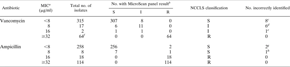
[image:2.612.58.555.93.209.2]All 132 isolates resistant to ampicillin by the broth macrodi-lution method were correctly identified by MicroScan panels, while 3 isolates (MIC,16mg/ml) were reported incorrectly by MicroScan panels as resistant to ampicillin (Table 1). No beta-lactamase-producing enterococci were identified. Additionally, all isolates (64 of 64) resistant to vancomycin were correctly classified by MicroScan panels. Seven isolates for which the vancomycin MICs were 8 and 16 mg/ml (intermediate) were incorrectly identified as susceptible by MicroScan panels. Eight motile Enterococcus isolates were classified incorrectly by Mi-croScan panel as intermediate; the vancomycin MICs for these isolates were 4 mg/ml. Thus, a total of 15 discrepancies in vancomycin susceptibility results between MicroScan panels and the broth macrodilution test were identified. Of 315 iso-lates susceptible to vancomycin, 32 grew on the agar screen
TABLE 1. Performance of conventional MicroScan Positive Breakpoint Combo Type 6 panels for detection of ampicillin and vancomycin resistance in Enterococcus species
Antibiotic MIC a
(mg/ml)
Total no. of isolates
No. with MicroScan panel resultb
NCCLS classification No. incorrectly identified
S I R
Vancomycin ,8 315 307 8 0 S 8c
8 17 6 11 0 I 6d
16 2 1 1 0 I 1e
$32 64f 0 0 64 R 0
Ampicillin ,8 258 256 2 S 2g
8 8 7 1 S 1h
16 18 0 18 R 0
$32 114 0 114 R 0
aDetermined by the broth macrodilution method by using NCCLS procedures. bAbbreviations: S, susceptible; R, resistant; I, intermediate.
cSeven isolates were E. gallinarum and one was E. casseliflavus; the MICs for all were 4mg/ml. dFour isolates were E. gallinarum, one was E. faecalis, and one was E. casseliflavus.
eIsolate was E. gallinarum. f
Sixty isolates identified as E. faecium (52 vanA and 8 vanB) and 4 identified as E. faecalis (3 vanB and 1 vanA). g
One isolate each of E. faecium and E. faecalis; the MICs for both were 4mg/ml. h
Isolate was E. raffinosus.
on May 15, 2020 by guest
http://jcm.asm.org/
plate containing 4mg of vancomycin per ml. As determined by the broth microdilution method, the MICs for all of these isolates were 2 to 4mg/ml, and all but three E. faecalis isolates were identified as motile enterococci.
Three hundred ninety-eight Enterococcus isolates were iden-tified by conventional biochemicals as 181 E. faecalis isolates (21 failed to ferment lactose), 157 E. faecium isolates, 27 E.
gallinarum isolates, 19 E. casseliflavus isolates, 6 E. raffinosus
isolates, 4 E. hirae isolates, 3 E. durans isolates, and 1 E. avium isolate (Table 2). Fifty-six enterococcal isolates from species not included in the data management system were misidenti-fied by MicroScan panels. All six E. raffinosus isolates were misidentified as E. avium by MicroScan panels, and all four E.
hirae isolates were incorrectly identified as E. durans.
Forty-five of the motile enterococcal isolates detected (27 E.
gallina-rum isolates and 18 E. casseliflavus isolates) were identified by
MicroScan panels as E. faecium, with 1 E. casseliflavus isolate identified as a group D Enterococcus species. A new approach to identify the 14 clinical species of enterococci known to be isolated from human sources by utilizing conventional bio-chemical assays, as suggested by other investigators, is outlined in Table 3 (1, 2, 6, 9, 18, 25). Fifty-three of the misidentified isolates were identifiable through a combination of results from the MicroScan panels and tests for motility and pigment production. Additional testing for sucrose fermentation was required to identify the three E. hirae isolates which were raffinose fermentation negative as indicated by MicroScan pan-els.
DISCUSSION
The ability to accurately identify enterococci at the species level is important not only for epidemiological purposes but also to recognize species such as E. faecium and the motile enterococci, which tend to show resistance to antimicrobial agents commonly used for therapy. Automated systems, such as the Vitek system and both the conventional and rapid Mi-croScan systems, are reported to provide accurate identifica-tion for both E. faecalis and E. faecium; however, they are not considered reliable for the identification of the other
Entero-coccus species (1, 2, 18, 24, 26). In addition, using automated
systems to perform susceptibility testing of enterococci for antimicrobial agents, such as ampicillin, vancomycin, and the aminoglycosides, has had conflicting results (13, 21–23, 26, 27). This study evaluated the ability of the conventional MicroScan Positive breakpoint Combo Type 6 panels to identify entero-cocci and to detect resistance to both ampicillin and vancomy-cin.
[image:3.612.57.299.93.175.2]The results of the present study showed that 132 of 132
TABLE 2. Ability of conventional MicroScan Positive Breakpoint Combo Type 6 panels to identify Enterococcus speciesa
MicroScan panel identification
No. of isolates correct/no. tested
No. of errors (correct identification)
E. faecalisb 181/181 None
E. faecium 157/202 27 (E. gallinarumc)
18 (E. casseliflavusc)
E. avium 1/7 6 (E. raffinosusd)
E. durans 3/7 4 (E. hiraee)
Group D Enterococcus 0/1 1 (E. casseliflavusc,f)
aSpecies of Enterococcus known to be isolated from human sources which
were not detected in this study included E. solitarius, E. flavescens, E. mundtii, E.
malodoratus, E. pseudoavium, and E. dispar. Asaccharolytic variants of E. faecalis
also were not detected.
bTwenty-one isolates did not ferment lactose, but all fermented ribose and
reduced tellurite.
cAll isolates exhibited motility at 308C, with E. casseliflavus having a distinct yellow pigment.
dAll isolates were raffinose fermentation positive on the MicroScan panel. eOne isolate was raffinose fermentation positive by the MicroScan panel, and
all fermented both raffinose and sucrose in conventional biochemical tests. fNegative for arginine dihydrolase.
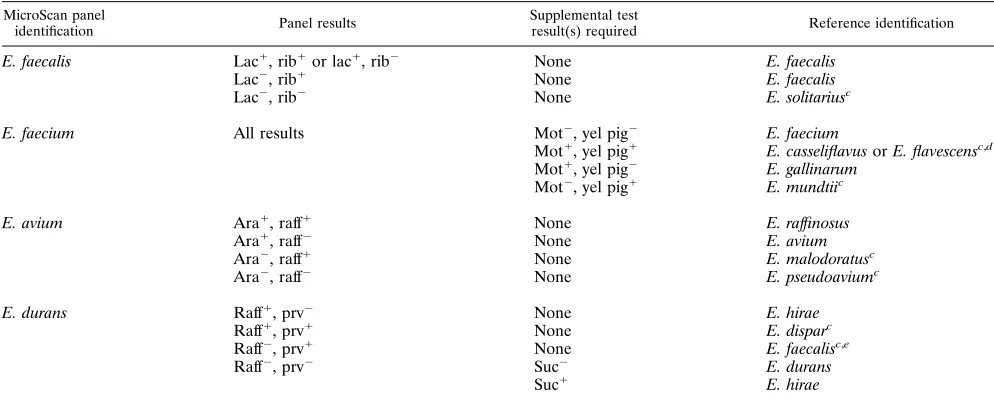
TABLE 3. Revised approach to identification of Enterococcus species using the MicroScan Positive Breakpoint Combo Type 6 panel results along with supplemental testinga,b
MicroScan panel
identification Panel results
Supplemental test
result(s) required Reference identification
E. faecalis Lac1, rib1or lac1, rib2 None E. faecalis
Lac2, rib1 None E. faecalis
Lac2, rib2 None E. solitariusc
E. faecium All results Mot2, yel pig2 E. faecium
Mot1, yel pig1 E. casseliflavus or E. flavescensc,d
Mot1, yel pig2 E. gallinarum Mot2, yel pig1 E. mundtiic
E. avium Ara1, raff1 None E. raffinosus
Ara1, raff2 None E. avium
Ara2, raff1 None E. malodoratusc
Ara2, raff2 None E. pseudoaviumc
E. durans Raff1, prv2 None E. hirae
Raff1, prv1 None E. disparc
Raff2, prv1 None E. faecalisc,e
Raff2, prv2 Suc2 E. durans
Suc1 E. hirae
a
Symbols:1, positive result;2, negative result. Abbreviations: lac, lactose fermentation; rib, ribose fermentation; mot, motility; yel pig, yellow pigment; ara, arabinose fermentation; raff, raffinose fermentation; prv, pyruvate utilization; suc, sucrose fermentation.
b
In cases of questionable identification, i.e., group D Enterococcus and Streptococcus species, additional conventional biochemical tests may be warranted. c
Tentative identification based on conventional biochemical testing from other investigators (1, 2, 6, 9, 18, 25). d
Evaluation of MicroScan result for ribose fermentation to differentiate E. casseliflavus (positive) from E. flavescens (negative). e
Asaccharolytic variants.
on May 15, 2020 by guest
http://jcm.asm.org/
[image:3.612.57.554.477.677.2]ampicillin-resistant enterococcal isolates were detected by con-ventional MicroScan panels. One isolate for which the ampi-cillin MIC was 8 mg/ml, which is at the high ends of the susceptible category as described by the NCCLS, and two iso-lates for which the MICs were 4 mg/ml were classified by MicroScan panels as resistant to ampicillin. The overall per-formance (sensitivity of 100% and specificity of 98.9%) was similar to those reported by Holmsen et al., who also showed that Enterococcus isolates for which the MICs were 8mg/ml were classified as resistant by the MicroScan Rapid Positive MIC panels (11). Additionally, Louie et al. showed that the MicroScan Positive MIC Type 6 panels demonstrated poor sensitivity (83.3%) for detection of ampicillin resistance when readings relied on the Walkaway System (13).
Since no beta-lactamase-positive enterococci were identified in this study, the reliability of conventional MicroScan panels for detecting strains which produce this enzyme is not known. Following multilaboratory testing of various Enterococcus iso-lates, Tenover et al. demonstrated that MicroScan panels using the autoSCAN-4 reader correctly detected susceptibility to am-picillin when the MIC was#4 or.256mg/ml but were unable to identify a beta-lactamase-producing strain in a majority of cases (23). The NCCLS recommends that all clinically signifi-cant ampicillin-susceptible enterococci be tested for beta-lac-tamase production by using a direct, nitrocefin-based test (15). Currently, the identification of these strains is considered rare, and the cost-effectiveness of this approach has been challenged (1, 18, 20).
Vancomycin resistance in enterococci has recently become a clinical problem due to a lack of alternative therapies (4, 5, 14, 19). Three major phenotypes of vancomycin-resistant entero-cocci have been described (12, 28). These are the vanA strains, which show high-level vancomycin resistance (MICs of .32
mg/ml) and resistance to teicoplanin; vanB strains, which have variable resistance to vancomycin (MICs of 4 to$128mg/ml) and susceptibility to teicoplanin; and vanC motile strains, which show intrinsic resistance to low levels of vancomycin (MICs of 2 to 32mg/ml) and susceptibility to teicoplanin. From studies of vancomycin resistance, Willey et al. showed the MicroScan Positive MIC Type 6 panels to have a sensitivity and specificity of 99 and 96%, respectively, when the visual inspection method of detection is used (27). In addition, other studies have shown these panels to be reliable except in cases in which low-level vancomycin resistance (MICs of 4 to 8mg/ ml) was present (22). Tenover et al. showed in multilaboratory testing, using MicroScan panels and the autoSCAN-4 reader and enterococci for which the vancomycin MICs were 16 to 64
mg/ml, that laboratories had only 36.8 to 57.9% agreement with the reference MIC result (23). In our study, 15 isolates had vancomycin results misclassified by the MicroScan system; 8 as intermediate, with MICs being 4mg/ml, and 7 as susceptible, with MICs being 8 and 16mg/ml, for an overall sensitivity and specificity of 91.6 and 97.5%, respectively. Of these, 14 were identified as motile enterococci and 1 as E. faecalis (vanB phenotype) with a vancomycin MIC of 8mg/ml. All isolates for which the vancomycin MICs were$32mg/ml were classified as resistant to vancomycin by the MicroScan panel. This group included 53 vanA enterococcal isolates (52 E. faecium isolates and 1 E. faecalis isolate) and 11 vanB enterococcal isolates (8
E. faecium isolates and 3 E. faecalis isolates).
Since low-level vancomycin resistance has been described as intrinsic to certain motile species of enterococci, proper iden-tification of these organisms may help in recognizing this low-level vancomycin resistance characteristic (12). However, iden-tification of the vanB enterococci for which the vancomycin MICs are in the range of 8 to 16mg/ml has proved to be a
challenge for the MicroScan system and other automated sys-tems. The present study showed that the conventional Mi-croScan panel misclassified 7 of the 19 isolates for which the MICs were 8 to 16 mg/ml. Similar results were found by Tenover et al. when they evaluated automated systems (22).
Currently, the NCCLS recommends testing all clinically sig-nificant enterococci with an agar screen plate containing 6mg of vancomycin per ml or by using the disc diffusion method (10, 15, 21). We evaluated the use of an agar dilution plate con-taining 4mg of vancomycin per ml as a supplemental test to the MicroScan panel in order to detect enterococci with low-level resistance to vancomycin. Enterococci which grew on this van-comycin-agar screen medium were retested by the disc diffu-sion method as described by the NCCLS to verify resistance. Additionally, all E. faecium bacteria which grew on the screen plate were tested for motility and pigment production. In this study, all motile enterococci grew on the agar screen medium. This medium appears to be useful to identify motile species for which the MICs are less than 4mg/ml, which was the case with six isolates of E. casseliflavus, for which the MICs were 2mg/ml. Three false-resistant E. faecalis isolates which grew on the vancomycin screen medium were verified as susceptible by the broth macrodilution method (MICs of 2mg/ml).
Of the 398 Enterococcus isolates tested, MicroScan panels correctly identified 342 of an error rate of 8.6%, which is similar to that reported by others (1, 24). The 56 misidentified species were not included in the present MicroScan data man-agement system. All of the misidentified species were identi-fiable by results of supplemental tests for motility, pigment production, and/or sucrose fermentation in combination with panel results. The six E. raffinosus isolates were distinguished from E. avium isolates by their ability to ferment raffinose on the MicroScan panel. The 27 E. gallinarum and 19 E.
casselifla-vus isolates were correctly identified through the use of a
motility test and determination of pigment production. One of four E. hirae isolates was distinguished from E. durans isolates by the ability to ferment raffinose on the MicroScan panel. It has previously been shown that 75 to 85% of E. hirae strains ferment both raffinose and sucrose whereas E. durans does not ferment either sugar (24). The addition of sucrose fermenta-tion testing helped distinguish three of four E. hirae isolates from the raffinose-negative E. durans isolates. Identification of other characteristics may be needed to identify some entero-cocci since nonmotile, nonpigmented E. casseliflavus bacteria and motile, pigmented E. flavescens bacteria have been de-scribed (3, 7, 8, 12, 25). DNA-based assays have been devel-oped to identify these species; however, Cartwright et al. have shown using DNA amplification testing that tests for pigmen-tation and motility were sufficiently reliable for identification of these species (3, 5, 11). Six of the 14 Enterococcus were not included in this study; additional studies are needed to evalu-ate our revised approach for identification for these other species.
The results of this study confirm that the conventional Mi-croScan Positive Breakpoint Combo Type 6 panels are not reliable in identifying species of enterococci other than E.
faecalis and E. faecium. However, supplemental testing in
com-bination with MicroScan panel biochemical results was useful to correctly identify these species. Recognition of resistance to both ampicillin and vancomycin was acceptable with these pan-els. This study demonstrated than an agar screen plate con-taining 4mg of vancomycin per ml was useful to identify
En-terococcus species with low-level vancomycin resistance. The
clinical significance of detecting these enterococcal isolates is not known, even though serious infections caused by the
on May 15, 2020 by guest
http://jcm.asm.org/
level vancomycin-resistant motile enterococci have been re-ported (16).
ACKNOWLEDGMENTS
We thank both Laura Baker and Marjorie Boyden for secretarial assistance, James Mockler, for providing technical support with this project, and Daniel F. Sahm for kindly providing isolates of entero-cocci for testing.
REFERENCES
1. Bryce, E. A., S. J. V. Zemcov, and A. M. Clarke. 1991. Species identification and antibiotic resistance patterns of the enterococci. Eur. J. Clin. Microbiol. Infect. Dis. 10:745–747.
2. Buschelman, B. J., M. J. Bale, and R. N. Jones. 1993. Species identification and determination of high-level aminoglycoside resistance among entero-cocci: comparison study of sterile body fluid isolates, 1985–1991. Diagn. Microbiol. Infect. Dis. 16:119–122.
3. Cartwright, C. P., F. Stock, G. A. Fahle, and V. J. Gill. 1995. Comparison of pigment production and motility tests with PCR for reliable identification of intrinsically vancomycin-resistant enterococci. J. Clin. Microbiol. 33:1931– 1399.
4. Centers for Disease Control and Prevention. 1993. Nosocomial enterococci resistant to vancomycin; United States, 1989–1993. Morbid. Mortal. Weekly Rep. 42:597–599.
5. Centers for Disease Control and Prevention. 1995. Recommendations for preventing the spread of vancomycin resistance; recommendations of the hospital control practices advisory committee. Morbid. Mortal. Weekly Rep.
44:(RR-12):1–3.
6. Collins, M. D., V. M. Rodriques, N. E. Pigott, and R. R. Facklam. 1991.
Enterococcus dispar sp. nov., a new Enterococcus species from human
sources. Lett. Appl. Microbiol. 12:95–98.
7. Donabedian, S., J. W. Chow, D. M. Shlaes, M. Green, and M. J. Zervos. 1995. DNA hybridization and contour-clamped homogeneous electric field elec-trophoresis for identification of enterococci to the species level. J. Clin. Microbiol. 33:141–145.
8. Dutka-Malen, S., S. Evers, and P. Courvalin. 1995. Detection of glycopep-tide resistance genotypes and identification to the species level of clinically relevant enterococci by PCR. J. Clin. Microbiol. 33:24–27.
9. Facklam, R. R., and M. D. Collins. 1989. Identification of Enterococcus species isolated from human infections by a conventional test scheme. J. Clin. Microbiol. 27:731–734.
10. Free, L., and D. F. Sahm. 1995. Investigation of the reformulated Remel Synergy Quad plate for detection of high-level aminoglycoside and vanco-mycin resistance among enterococci. J. Clin. Microbiol. 33:1643–1645. 11. Holmsen, K., S. Cullen, L. Nea, F. Julio, M. Mccray, D. Ballou, and B.
Vanderhoof.1995. Detection of ampicillin resistance in enterococci using MicroScanwRapid Pos MIC panels (RPM), abstr. C-267, p. 47. In Abstracts of the 95th General Meeting of the American Society for Microbiology 1995. American Society for Microbiology, Washington, D.C.
12. Leclercq, R., S. Dutka-Malen, J. Duval, and P. Courvalin. 1992. Vancomycin resistance gene vanC is specific to Enterococcus gallinarum. Antimicrob. Agents Chemother. 36:2005–2008.
13. Louie, M., A. E. Simor, S. Szeto, M. Patel, B. Kreiswirth, and D. E. Low. 1992. Susceptibility testing of clinical isolates of Enterococcus faecium and
Enterococcus faecalis. J. Clin. Microbiol. 30:41–45.
14. Montecalvo, M. A., H. Horowitz, C. Gedris, C. Carbonaro, F. C. Tenover, A.
Issha, P. Cook, and G. P. Wormser.1994. Outbreak of vancomycin-, ampi-cillin-, and aminoglycoside-resistant Enterococcus faecium bacteremia in an adult oncology unit. Antimicrob. Agents Chemother. 38:1363–1367. 15. National Committee for Clinical Laboratory Standards. 1993. Methods for
dilution antimicrobial susceptibility tests for bacteria that grow aerobically. Publication M7-A3, 3rd ed. National Committee for Clinical Laboratory Standards, Villanova, Pa.
16. Patterson, J. E., A. H. Sweeney, M. Simms, N. Carley, R. Mangi, J. Sabetta,
and R. W. Lyons.1995. An analysis of 110 serious enterococcal infections; epidemiology, antibiotic susceptibility, and outcome. Medicine (Baltimore)
74:191–199.
17. Pompei, R., F. Berlutti, M. C. Thaller, A. Ingianni, G. Cortis, and B.
Dainelli.1992. Enterococcus flavescens sp. nov., a new species of enterococci of clinical origin. Int. J. Syst. Bacteriol. 42:365–369.
18. Ruoff, K. L., L. de la Maza, M. J. Murtagh, J. D. Spargo, and M. J. Ferrano. 1990. Species identities of enterococci isolated from clinical specimens. J. Clin. Microbiol. 28:435–437.
19. Spera, R. V. Jr., and B. F. Farber. 1994. Multidrug-resistant Enterococcus
faecium: an untreatable nosocomial pathogen. Drugs 48:678–688
20. Steffee, C. H., R. M. Morrell, and B. L. Wasilauskas. 1995. Routine testing for beta-lactamase positive enterococci: it is really necessary? A paradigm shift in the clinical detection of rare organisms, abstr. C-119, p. 21. In Abstracts of the 95th General Meeting of the American Society for Micro-biology 1995. American Society for MicroMicro-biology, Washington, D.C. 21. Swenson, J. M., N. C. Clark, M. J. Ferraro, D. F. Sahm, G. Doern, M. A.
Pfaller, L. B. Reller, M. P. Weinstein, R. J. Zabransky, and F. C. Tenover.
1994. Development of a standardized screening method for detection of vancomycin-resistant enterococci. J. Clin. Microbiol. 32:1700–1704. 22. Tenover, F. C., J. M. Swenson, C. M. O’Hara, and S. A. Stocker. 1995. Ability
of commercial and reference antimicrobial susceptibility testing methods to detect vancomycin resistance in enterococci. J. Clin. Microbiol. 33:1524– 1527.
23. Tenover, F. C., J. Tokars, J. Swenson, S. Paul, K. Spitalny, and W. Jarvis. 1993. Ability of clinical laboratories to detect antimicrobial agent-resistant enterococci. J. Clin. Microbiol. 31:1695–1699.
24. Tritz, D. M., P. C. Iwen, and G. L. Woods. 1990. Evaluation of MicroScan for identification of Enterococcus species. J. Clin. Microbiol. 28:1477–1478. 25. Vincent, S., R. G. Knight, M. Green, D. F. Sahm, and D. M. Shales. 1991.
Vancomycin susceptibility and identification of motile enterococci. J. Clin. Microbiol. 29:2335–2337.
26. Willey, B. M., B. N. Kreiswirth, A. E. Simor, Y. Faur, M. Patel, G. Williams,
and D. E. Low.1993. Identification and characterization of multiple species of vancomycin-resistant enterococci, including an evaluation of Vitek soft-ware version 7.1. J. Clin. Microbiol. 31:2777–2779.
27. Willey, B. M., B. N. Kreiswirth, A. E. Simor, G. Williams, S. R. Scriver, A.
Phillips, and D. E. Low.1992. Detection of vancomycin resistance in
En-terococcus species. J. Clin. Microbiol. 30:1621–1624.
28. Zabransky, R. J., A. R. Dinuzzo, and G. L. Woods. 1995. Detection of vancomycin resistance in enterococci by the Alamar MIC system. J. Clin. Microbiol. 33:791–793.