Ventilation and oxygenation induce endothelial
nitric oxide synthase gene expression in the
lungs of fetal lambs.
S M Black, … , J Bristow, S J Soifer
J Clin Invest.
1997;
100(6)
:1448-1458.
https://doi.org/10.1172/JCI119665
.
At birth, ventilation and oxygenation immediately decrease pulmonary vascular resistance
(PVR) and increase pulmonary blood flow (PBF); more gradual changes occur over the next
several hours. Nitric oxide, produced by endothelial nitric oxide synthase (eNOS), mediates
these gradual changes. To determine how ventilation and oxygenation affect eNOS gene
expression, 12 fetal lambs were ventilated for 8 h without changing fetal descending aortic
blood gases or pH (rhythmic distension) or with 100% oxygen (O2 ventilation). Vascular
pressures and PBF were measured. Total RNA, protein, and tissue sections were prepared
from lung tissue for RNase protection assays, Western blotting, and in situ hybridization. O2
ventilation increased PBF and decreased PVR more than rhythmic distension (P < 0.05).
Rhythmic distension increased eNOS mRNA expression; O2 ventilation increased eNOS
mRNA expression more and increased eNOS protein expression (P < 0.05). To define the
mechanisms responsible for these changes, ovine fetal pulmonary arterial endothelial cells
were exposed to 1, 21, or 95% O2 or to shear stress. 95% O2 increased eNOS mRNA and
protein expression (P < 0.05). Shear stress increased eNOS mRNA and protein expression
(P < 0.05). Increased oxygenation but more importantly increased PBF with increased shear
stress induce eNOS gene expression and contribute to pulmonary vasodilation after birth.
Research Article
Find the latest version:
J. Clin. Invest.
© The American Society for Clinical Investigation, Inc. 0021-9738/97/09/1448/11 $2.00
Volume 100, Number 6, September 1997, 1448–1458 http://www.jci.org
Ventilation and Oxygenation Induce Endothelial Nitric Oxide Synthase Gene
Expression in the Lungs of Fetal Lambs
Stephen M. Black, Michael J. Johengen, Zhi-Dong Ma, James Bristow, and Scott J. Soifer
Department of Pediatrics and the Cardiovascular Research Institute, University of California, San Francisco, San Francisco, California 94143-0106
Abstract
At birth, ventilation and oxygenation immediately decrease
pulmonary vascular resistance (PVR) and increase
pulmo-nary blood flow (PBF); more gradual changes occur over the
next several hours. Nitric oxide, produced by endothelial
ni-tric oxide synthase (eNOS), mediates these gradual changes.
To determine how ventilation and oxygenation affect eNOS
gene expression, 12 fetal lambs were ventilated for 8 h
with-out changing fetal descending aortic blood gases or pH
(rhythmic distension) or with 100% oxygen (O
2ventilation).
Vascular pressures and PBF were measured. Total RNA,
protein, and tissue sections were prepared from lung tissue
for RNase protection assays, Western blotting, and in situ
hybridization. O
2ventilation increased PBF and decreased
PVR more than rhythmic distension (
P
,
0.05). Rhythmic
distension increased eNOS mRNA expression; O
2ventila-tion increased eNOS mRNA expression more and increased
eNOS protein expression (
P
,
0.05). To define the
mecha-nisms responsible for these changes, ovine fetal pulmonary
arterial endothelial cells were exposed to 1, 21, or 95% O
2or
to shear stress. 95% O
2increased eNOS mRNA and protein
expression (
P
,
0.05). Shear stress increased eNOS mRNA
and protein expression (
P
,
0.05). Increased oxygenation
but more importantly increased PBF with increased shear
stress induce eNOS gene expression and contribute to
pul-monary vasodilation after birth. (
J. Clin. Invest.
1997. 100:
1448–1458.) Key words: nitric oxide
•endothelial nitric oxide
synthase
•endothelial cells
•pulmonary circulation
•devel-opment
Introduction
After birth, with initiation of ventilation by the lungs, and the subsequent increase in pulmonary and systemic arterial blood oxygen tensions, pulmonary vascular resistance decreases and pulmonary blood flow increases by 8–10-fold to match
sys-temic blood flow (1–4). This process is regulated by a complex and incompletely understood interplay between mechanical and metabolic factors (5). For example, the replacement of fluid with gas in the alveoli changes alveolar surface tension, which unkinks the small pulmonary arteries, and causes an im-mediate decrease in pulmonary vascular resistance and in-crease in pulmonary blood flow (2, 5). There is also release of vasoactive substances, such as prostacyclin (PGI2), which
de-crease pulmonary vascular resistance and inde-crease pulmonary blood flow (6, 7). Both rhythmic distension of the fetal lamb lung and ventilation without oxygenation produce partial pul-monary vasodilatation (2).
Ventilation of the fetus with air or oxygen produces nearly complete pulmonary vasodilatation (5). The increase in alveo-lar or pulmonary and systemic arterial blood oxygen tensions decreases pulmonary vascular resistance either directly by di-lating the small pulmonary arteries, or indirectly by stimudi-lating the production of vasodilator substances such as nitric oxide (NO)1 (8–10). Endothelial cells synthesize NO and l-citrulline
from l-arginine by the action of the enzyme endothelial nitric oxide synthase (eNOS) (11, 12). NO diffuses from the endo-thelial cell into the smooth muscle cell, activates guanylate cy-clase, increases cyclic guanosine-39,59-monophosphate produc-tion, and initiates a cascade that results in smooth muscle relaxation and vasodilation (13–15). Inhibition of eNOS atten-uates the increase in pulmonary blood flow due to oxygenation of fetal lambs either by maternal hyperbaric oxygen exposure or by ventilation of the fetus with air or oxygen (8–10, 16, 17). There is also an increase in eNOS mRNA and protein expres-sion in fetal rat lungs as they near term, which decreases 1 wk after birth (18, 19). These studies suggests an important role for NO in mediating pulmonary vascular tone in the perinatal period. The factors responsible for the changes in eNOS gene expression are not well understood.
An additional mechanism by which pulmonary vasodilation occurs relates to the increase in shear stress on the pulmonary vascular endothelium induced by the increase in pulmonary blood flow. Initially, NOS activity and NO production are increased when endothelial cells are exposed to increased shear stress (12, 14, 15, 20–23). If the shear is maintained there are then subsequent increases in eNOS mRNA and protein expression (24–26). The immediate increase in pulmonary blood flow at birth may increase shear forces on the pulmo-nary vascular endothelium inducing eNOS mRNA and protein expression. This would result in increased NO production and a continued, more gradual decrease in pulmonary vascular re-sistance and increase in pulmonary blood flow in the next hours and even days after birth.
This study had several purposes. First, to determine
This research was presented in part at the annual meeting of the Pedi-atric Academic Societies in Washington, DC, 5–10 May 1996.
Address correspondence to Scott J. Soifer, M.D., M-680, Box 0106, University of California, San Francisco, CA 94143-0106. Phone: 415-476-5153; FAX: 415-502-4186; E-mail: scott_soifer@pedcardgateway. ucsf.edu
Received for publication 22 July 1996 and accepted in revised form 10 July 1997.
whether eNOS mRNA and protein expression are induced in the fetal lung with ventilation and oxygenation at birth, RNase protection assays and Western blot analyses were performed on lung tissue from near-term fetal lambs ventilated in utero with a gas mixture which did not change fetal descending aor-tic blood gases or pH, or with 100% oxygen. Second, to deter-mine the cell type responsible for the increase in eNOS mRNA expression, in situ hybridization was performed on lung tissue sections from near-term fetal lambs ventilated with 100% oxy-gen. Breathing at birth produces increased oxygenation, in-creased pulmonary blood flow, and likely inin-creased shear stress; therefore, whole animal studies cannot determine which of these effects are responsible for the increase in eNOS mRNA and protein expression. Therefore, pulmonary arterial endothelial cells were harvested from near-term fetal lambs and maintained in culture. These cells were exposed to differ-ent oxygen environmdiffer-ents, or to shear stress, and changes in eNOS mRNA and protein levels were measured.
Our results show that the increases in descending aortic PO2 and pulmonary blood flow are greater after ventilation of
the near-term fetal lamb with 100% oxygen. Increases in eNOS mRNA and protein levels parallel these physiologic changes. The increase in eNOS expression is confined to the endothe-lium of small and large blood vessels. In cultured ovine fetal pulmonary arterial endothelial cells, shear stress induces eNOS mRNA and protein expression more than increased ox-ygenation does. Increased oxox-ygenation but more importantly increased pulmonary blood flow with increased shear stress in-duce eNOS gene expression and contribute to pulmonary va-sodilation after birth and the successful transition to extrauter-ine life.
Methods
Physiologic studies
Surgical preparation. All procedures and protocols were approved by the Committee on Animal Research of the University of Califor-nia at San Francisco. 12 mixed-breed Western ewes (136–138 d of ges-tation, term 5 145 days) were operated on under sterile conditions using local anesthesia (2% lidocaine hydrochloride), intramuscular sedation (20 mg valium, 600 mg ketamine hydrochloride, and 2 mg at-ropine), and intravenous anesthesia (1,000–2,000 mg ketamine hydro-chloride). A venous catheter was placed in the maternal hind limb for the administration of intraoperative anesthesia and postoperative an-tibiotics.
A laparotomy was performed. Through a small uterine incision, a fetal hind limb was exposed. 1% lidocaine hydrochloride was used for local anesthesia. Polyvinyl catheters were inserted into the pedal ar-tery and vein and advanced to the descending aorta and inferior vena cava, respectively. The fetus was then anesthetized with an intrave-nous injection of ketamine hydrochloride (25 mg), and spontaneous movement was prevented with an intravenous injection of succinyl-choline chloride (5 mg). The fetal skin and uterine incisions were closed in layers.
Through a separate uterine incision, the fetal left hemithorax was exposed. A left lateral thoracotomy was performed in the fourth in-tercostal space to expose the heart and great vessels. Polyvinyl cathe-ters were inserted into the main pulmonary trunk, left pulmonary ar-tery, and left atrium. A 6-mm ultrasonic flow transducer (Transonic Systems, Inc., Ithaca, NY) was placed around the left pulmonary ar-tery. Through a midline neck incision, the trachea was isolated and li-gated proximally. Through an incision in the midtrachea, a 4.5-mm endotracheal tube was inserted. Polyvinyl catheters were inserted into the carotid artery and jugular vein, and advanced to the
ascend-ing aorta and right atrium, respectively. A polyvinyl catheter was in-serted into the amniotic cavity.
The fetal, uterine, and maternal skin incisions were closed in lay-ers. Amniotic fluid losses were replaced with warmed 0.9% saline. To ensure unobstructed drainage of the tracheal fluid into the amniotic cavity, the endotracheal tube was connected by tubing to the amniotic cavity catheter (3, 10). All catheters and the flow transducer were ex-teriorized to the left flank of the ewe and placed in a vinyl pouch. To maintain catheter patency, heparin sodium (1,000–2,000 U) was in-stilled into the catheters immediately after surgery, and then daily. Penicillin G potassium (1,000,000 IU) and gentamicin sulfate (100 mg) were injected intravenously into the ewe and into the amniotic fluid during surgery, and then daily. 3 d were allowed for recovery.
Measurements. Descending aortic, left and right atrial, and pul-monary arterial pressures, zero referenced to amniotic pressure, were measured using P23 Db pressure transducers (Statham Instruments, Oxnard, CA) and recorded continuously on a direct-writing poly-graph (Gould Instruments, Valley View, OH). Mean pressures were obtained by electrical integration. Heart rate was measured by a car-diotachometer triggered from the phasic descending aortic pressure tracing. Left pulmonary arterial blood flow was measured with an ul-trasonic flow transducer and flowmeter (Transonic Systems, Inc.). Left pulmonary vascular resistance was calculated as the difference between mean left pulmonary arterial pressure and mean left atrial pressure divided by left pulmonary blood flow per kilogram of body weight. Descending aortic blood gases and pH were measured on a blood gas analyzer (Corning Medical and Scientific, Medfield, MA). Hemoglobin concentration and oxygen saturation were measured by a hemoximeter (Radiometer, Copenhagen, Denmark).
Drug preparation. Exosurf™ (108 mg colfosceril palmitate, 12 mg cetyl alcohol, 8 mg tyloxapol, and 48 mg sodium chloride; Bur-roughs-Wellcome, Research Triangle Park, NC) was dissolved in 10 ml of normal saline.
Experimental protocol. During the study, the ewes stood quietly in a cage with free access to food and water. Baseline measurements of the hemodynamic variables (descending aortic, left and right atrial, and pulmonary arterial pressures, heart rate, and left pulmonary arte-rial blood flow), descending aortic blood gases, pH, hemoglobin, and oxygen saturation were made. The fetal lambs were then randomized (using a table of random numbers) to one of two groups: rhythmic distension or O2 ventilation. In the rhythmic distension group (n 5 6),
the fetal lambs were ventilated with an N2/O2/CO2 mixture which did
not change fetal descending aortic blood gases or pH. In the O2
venti-lation group (n 5 6), the fetal lambs were ventilated with 100% oxygen. To ventilate the fetus, first, the endotracheal tubing was discon-nected from the amniotic cavity catheter to drain the tracheal fluid (3, 10). Then, Exosurf™ (10 ml) was instilled into the trachea. After 10 min, in utero ventilation was begun using a standard animal ventilator (Harvard Apparatus, Inc., South Natick, MA); the ventilator settings were a rate of 20–25 breaths/min, an inspiratory time-to-expiratory time ratio of 0.45, and a tidal volume of 30–50 ml.
The hemodynamic variables were measured continuously, and descending aortic blood gases, pH, hemoglobin concentration, and oxygen saturation were measured every 30 min. After 8 h of in utero ventilation, the ewe and fetus were killed with an intravenous injec-tion of pentobarbital sodium (Euthanasia CII; Central City Medical, Union City, CA) followed by bilateral thoracotomy. Catheter posi-tion was confirmed at autopsy.
Statistical analysis. The means6standard error were calculated for the hemodynamic variables, left pulmonary vascular resistance, and descending aortic blood gases, pH, hemoglobin concentration, and oxygen saturation during all experimental conditions (27). The maximum effects of rhythmic distension and O2 ventilation on these
variables were compared with their respective baselines by the paired
t test or the Wilcoxon signed rank test. During rhythmic distension
and O2 ventilation, the maximum effects on these variables were
Cellular and molecular studies
Tissue preparation.The fetal heart and lungs were removed en bloc. To prepare total RNA, lung tissue was removed, snap-frozen in liquid nitrogen, and stored at 2708C until used. Total RNA was prepared from snap-frozen lung tissue (after pulverization) or from cultured ovine fetal pulmonary arterial endothelial cells (see below) by brief homog-enization in 4 M guanidinium isothiocyanate, followed by extraction with acid phenol, and precipitation in isopropanol (28). To prepare protein, lung tissue was rinsed in cold (48C) normal saline to remove blood, then minced, and finally homogenized using a Tissuemizer (2 3 15 s at 80% power) at 4 vol/wet weight of Triton lysis buffer (20 mM Tris-HCl, pH 7.6, 0.5% Triton X-100, and 20% glycerol) supple-mented with protease inhibitors; cultured ovine fetal pulmonary arte-rial endothelial cells were sonicated in 3 vol/wet weight. Extracts were then centrifuged at 15,000 g for 15 min. The supernatant was then re-moved for protein determination and Western blot analysis (29, 30).
For in situ hybridization, the pulmonary vascular tree was rinsed with cold PBS to remove blood and fixed by perfusion with cold 4% paraformaldehyde. The pulmonary artery was then clamped. The air-ways were fixed at 20 cm of H2O pressure by filling the trachea with
cold 4% paraformaldehyde. When the lungs were distended at this pressure, the trachea was clamped. The lungs were fixed for 24 h at
48C by immersion in 4% paraformaldehyde. Representative slices
from each lobe were removed, placed in 30% sucrose until they sank, placed in OCT, frozen on dry ice, and stored at 2708C until sec-tioned. 10–20-mm sections were cut using a cryostat, transferred to aminoalkylsilane-treated slides (Superfrost Plus; Fisher Scientific, Santa Clara, CA), and stored at 2708C (30–32).
Generation of ovine eNOS cDNA and antiserum. Sequence com-parisons between the three isoforms of NOS (endothelial, inducible, and neuronal) were performed using dot-matrix homology plots to determine a region of minimal homology between them (15, 30). This region corresponded to an area within the heme binding domain. Oli-gonucleotides were synthesized (using the bovine eNOS as a tem-plate) to allow amplification of this region within the ovine eNOS
sequence. The sequences of the oligonucleotides were 59
-CCTCCG-GAGGGGCCCAAGTTCCCTCGC-39 for oligonucleotide 1 and
59-CACGTCGAAGCGCCGTTTCCGGGGGT-39 for
oligonucleo-tide 2. The region amplified corresponds to amino acids 62–288 of the eNOS protein (24, 33–35). Total RNA, prepared from ovine fetal lung, was used in RT-PCR reactions (kit from Perkin-Elmer, Foster City, CA). The cDNA fragment generated (681 bp) was then cloned directly into the pCR II vector (Invitrogen, San Diego, CA), se-quenced (Sequenase kit; United States Biochemical Corp., Cleve-land, OH), and then subcloned into pBluescript KS1 (Stratagene, La
Jolla, CA). The sequence in this region for ovine eNOS is 96.6% identical to bovine eNOS.
The eNOS cDNA fragment was subcloned in frame into the pET23a expression vector to overexpress the corresponding eNOS protein fragment (30) (Novagen, Madison, WI). After confirming the reading frame at both the 59- and 39-ends, the pET23a clone was trans-formed into the bacterial strain BL21(DE3)plys S, which contains a lysogen of T7 bacteriophage and a plasmid encoding lysozyme to re-duce constitutive expression of the T7 RNA polymerase. Cultures (1 liter) were grown from a single colony under ampicillin selection until the OD600 reached z 0.6; isopropyl b-D-thiogalactoside was then added (final concentration 0.4 mM). After 3 h, the cells were pelleted, resuspended in one-tenth volume of imidazole buffer, and sonicated to disrupt the cell membranes and shear chromosomal DNA. The ly-sate was cleared by centrifugation, passed over the Ni21 column,
washed, and eluted with 1 M imidazole. The eluted fraction was con-centrated by passage through a concentrator with the addition of ster-ile distilled water to reduce the imidazole concentration. The resultant partially purified eNOS protein fragment was then injected into New Zealand White rabbits (Animal Pharm. Services, Healdsberg, CA) to produce a polyclonal eNOS antiserum. The specificity of the antise-rum was assessed by Western blot analysis on protein extracts pre-pared from a variety of tissues.
RNA probe synthesis, RNase protection assay, and in situ hybrid-ization.The plasmid containing the eNOS cDNA fragment was linearized with the appropriate restriction enzyme (GIBCO BRL, Gaithersburg, MD). Antisense and sense radiolabeled single-stranded RNA eNOS probes were synthesized by in vitro transcrip-tion using either T3 or T7 RNA polymerases (Boehringer-Mann-heim, Indianapolis, IN) in the presence of cold rCTP, rGTP, and
rATP. RNA probes labeled with 32P-UTP (New England Nuclear,
Bos-ton, MA) were used for RNase protection assays (36); RNA probes labeled with 35S-UTP were used for in situ hybridization (33–35).
RNase protection assays were performed as described previously (36). Antisense radiolabeled single-stranded RNA eNOS probes
were hybridized overnight at 428C with total RNA isolated from
ovine fetal lung (50 mg) or from ovine fetal pulmonary arterial endo-thelial cells (20 mg) in 80% formamide, 50 mM Pipes (pH 6.4), 0.4 M NaCl, and 1 mM EDTA. Single-stranded RNA was digested for 1 h at
378C with an RNase A/T1 mixture (Ambion, Austin, TX). After
phe-nol/CHCl3 extraction and ethanol precipitation, the protected
frag-ments were analyzed by electrophoresis on a 6% denaturing poly-acrylamide gel. Also included was a probe for 18S to serve as a control for the amount of input total RNA and the recovery of pro-tected probe fragments.
In situ hybridization was performed as previously described (33– 35). Studies were done on serial sections of ovine fetal lung, using the antisense radiolabeled cRNA eNOS probe and the corresponding nonhybridizing sense radiolabeled cRNA eNOS probe. Frozen tissue sections were allowed to come to room temperature. All sections were fixed in 4% paraformaldehyde in PBS, treated with proteinase K, and acetylated (0.25% acetic anhydride in 0.1 M triethanolamine-HCl, pH 7.5). After washing in 0.53 SSC, the sections were covered with hybridization solution (50% deionized formamide, 0.3 M NaCl,
20 mM Tris, pH 8, 5 mM EDTA, 13 Denhardt’s, 10% dextran
sul-fate, and 10 mM DTT), and prehybridized for 1–3 h at 558C. In the presence of tRNA, the radiolabeled RNA probe (600,000 cpm/slide) was applied to the hybridization solution, and the sections were then hybridized for 12–18 h at 558C. After hybridization, the sections were
washed for 20 min (23 SSC, 10 mM b-mercaptoethanol, and 1 mM
EDTA), treated with RNase A (20 mg/ml), and washed for 20 min
(23 SSC, 10 mM b-mercaptoethanol, and 1 mM EDTA). The
sec-tions were then washed in high stringency buffer (50% deionized form-amide, 23 SSC, and 0.1 M DTT) for 30 min at 658C. The final wash was in 0.53 SSC for 20 min. The sections were dehydrated, the slides dipped in photographic emulsion (Ilford, St. Louis, MO), stored at 48C, developed after 2–10 wk of exposure, and counterstained with hematoxylin and eosin. For each experiment, four antisense and four sense slides (containing one to two sections) were studied. For each experiment, new radiolabeled probes were synthesized.
Western blot analysis. Western blot analysis was performed as de-scribed previously (30). Protein extracts isolated from ovine fetal lung (100 mg) or from cultured ovine fetal pulmonary arterial endothelial cells (25 mg) were separated on a 6% denaturing polyacrylamide gel and electrophoretically transferred to Hybond-PVDF membranes (Amersham, Arlington Heights, IL). The membranes were blocked with 5% nonfat dry milk in TBS containing 0.1% Tween. After block-ing, the membranes were incubated at room temperature with 1:1,000 dilution of the eNOS antiserum, washed with TBS containing 0.1% Tween, and then incubated with a mouse anti–rabbit IgG horseradish peroxidase conjugate. After washing, chemiluminescence was used to detect the eNOS protein bands.
bal-anced with N2. After 5 d, islands of endothelial cells were cloned to
ensure purity. Basic fibroblast growth factor (10 ng/ml, a gift from Denis Gospodarowicz, Chiron Corporation, Emeryville, CA) was added to the media every other day. When confluent, the cells were passaged to maintain them in culture or frozen in liquid nitrogen. En-dothelial cell identity was confirmed by their typical cobblestone ap-pearance, contact inhibition, specific uptake of DiI-Ac-LDL (Molec-ular Probes, Eugene, OR), and positive staining for von Willebrand factor (Dako, Carpinteria, CA). Ovine fetal pulmonary arterial en-dothelial cells were studied between passages 3 and 10.
Oxygen studies. Because tissue culture plastic adsorbs oxygen, ovine fetal pulmonary arterial endothelial cells were grown on glass plates (37). These plates were washed and baked to remove organic residue before use. Confluent cultures were placed in an airtight Plexiglas chamber inside a standard CO2 incubator. The chamber was
gassed with 1% O2/5% CO2 balanced with N2 (PO2 of the culture
me-dium 30–39 torr [1% O2]), or with 95% O2/5% CO2 (PO2 of the
cul-ture medium 549–644 torr [95% O2]). Ovine fetal pulmonary arterial
endothelial cells tolerate these treatments without change in mor-phology or cell death. Other confluent cultures were placed in 21% O2/5% CO2 balanced with N2 (PO2 of the culture medium 125–132
torr [21% O2]). The cells were harvested in triplicate after 0, 8, or 24 h
of exposure.
Shear stress studies. We have built a cone-plate viscometer to ac-cept 15-cm tissue culture plates (38). This apparatus consists of a cone of shallow angle (a) rotating at angular velocity (v) on top of a tissue culture plate, placed inside a standard CO2 incubator (21% O2/5%
CO2 balanced with N2). The monolayer of confluent ovine fetal
pul-monary arterial endothelial cells was subjected to a radially constant fluid shear stress as calculated using the formula t5 mv/a, where m is the viscosity of the medium covering the ovine fetal pulmonary arte-rial endothelial cells (38, 39). A cone angle of 0.58 was used to achieve laminar flow rates representing levels of shear stress within the physi-ological range (5–25 dynes/cm2). When rotated at 175 rpm, 20 dynes/
cm2 of shear stress was applied for 0, 2, 4, or 8 h.
Data analysis. Quantitation of autoradiographic results were per-formed by scanning (SCA Jet IICX; Hewlett Packard Inc., Palo Alto, CA) the bands of interest into an image editing software program (Adobe Photoshop, Adobe Systems, Mt. View, CA). Band intensities from RNase protection assays and Western blot analysis were ana-lyzed densitometrically on a Macintosh computer (model 9500; Apple Computer, Inc., Cupertino, CA) using the public domain NIH Image program (developed at the National Institutes of Health and avail-able on the Internet at http://rsb.info.nih.gov/nih-image) (40). For RNAase protection assays, to control for the amount of input RNA and the recovery of protected probe fragments, the eNOS signal was normalized to the corresponding 18S signal for each lane. Results from unventilated fetal lamb lungs or from 0 h of exposure in cell cul-ture experiments were assigned the value of 1 (relative eNOS mRNA). For Western blot analysis, to insure equal protein loading, duplicate polyacrylamide gels were run. One was stained with Coomassie blue. Results from unventilated fetal lamb lungs or from 0 h of exposure in cell culture experiments were assigned the value of 1 (relative eNOS
protein). The means6standard error were calculated for relative
eNOS mRNA and relative eNOS protein during all experimental conditions and were compared by the Kruskal-Wallis Test or
ANOVA. Multiple comparison testing was performed. P , 0.05 was
considered statistically significant.
Results
Physiologic studies. After 8 h of rhythmic distension of the fe-tal lung in utero by ventilation with a hypoxic gas mixture, there were no changes in fetal descending aortic PO2 (before
ventilation, 15.561.8 mmHg; during ventilation, 15.661.4 mmHg), PCO2 (before ventilation, 51.361.0 mmHg; during
ventilation, 49.061.7 mmHg), or pH (before ventilation,
7.3560.01; during ventilation, 7.3560.02). Rhythmic disten-sion increased left pulmonary arterial blood flow (P , 0.05, Fig. 1, top) without changing mean left pulmonary arterial pressure (before ventilation, 61.363.9 mmHg; during ventila-tion, 64.063.0 mmHg) or mean descending aortic pressure (before ventilation, 61.363.9 mmHg; during ventilation, 60.063.0 mmHg). Left pulmonary vascular resistance de-creased (P , 0.05, Fig. 1, bottom).
After 8 h of in utero ventilation of the fetal lung with 100% oxygen, fetal descending aortic PO2 (before ventilation, 18.861.9
mmHg; during ventilation, 327.2670.4 mmHg, P , 0.05) and pH increased (before ventilation, 7.2760.03; during ventila-tion, 7.4260.02, P , 0.05); fetal descending aortic PCO2
de-creased (before ventilation, 52.064.0 mmHg; during ventila-tion, 35.863.0 mmHg, P , 0.05). O2 ventilation increased left
[image:5.612.314.510.59.362.2]pulmonary arterial blood flow (P , 0.05, Fig. 1, top) and de-creased left pulmonary vascular resistance (P , 0.05, Fig. 1, bottom) more than rhythmic distension. Mean left pulmon-ary arterial pressure decreased (before ventilation, 63.764.6 mmHg; during ventilation, 46.361.7 mmHg, P , 0.05), while mean descending aortic pressure was unchanged (before venti-lation, 65.069.5 mmHg; during ventilation, 63.665.0 mmHg). Figure 1. The effects of in utero ventilation on left pulmonary artery blood flow (top) and left pulmonary vascular resistance (bottom). 12 fetal lambs were ventilated in utero for 8 h with a gas mixture which did not change fetal descending aortic blood gases or pH (rhythm dis-tension) or with 100% oxygen (O2 ventilation). Rhythm distension
in-creased left pulmonary blood flow and dein-creased left pulmonary vas-cular resistance. The effects were greater with O2 ventilation. Values
are mean6standard error. n 5 6. *P , 0.05 before ventilation vs. dur-ing ventilation (within treatments). †P , 0.05 during ventilation vs.
Cellular and molecular studies. After 8 h of in utero venti-lation, eNOS mRNA expression was increased in vivo in the fetal lung (Fig. 2). O2 ventilation increased eNOS mRNA
ex-pression by 5-fold compared with the unventilated fetal lamb lung (P , 0.05); rhythmic distension increased eNOS mRNA expression by 2.3-fold. This increase in eNOS mRNA expres-sion is confined to the endothelial cells of small and large ar-teries and veins (Fig. 3). After 8 h of rhythmic distension, there was no change in eNOS protein expression; O2 ventilation
in-creased eNOS protein expression by 1.9-fold compared with the unventilated fetal lamb lung (P , 0.05) (Fig. 4). When ovine fetal pulmonary arterial endothelial cells were exposed to 95% O2 for 24 h, there was a 3.3-fold increase in eNOS mRNA
ex-pression and a 2.3-fold increase in eNOS protein exex-pression (P , 0.05) (Fig. 5). Exposure to 1% O2 did not change eNOS
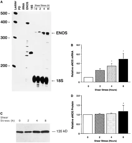
mRNA or protein expression. When ovine fetal pulmonary ar-terial endothelial cells were exposed to fluid shear stress to simulate the increase in pulmonary blood flow after birth, there were increases in eNOS mRNA expression of 2.5-, 3.4-, and 4.9-fold after 2, 4, and 8 h (P , 0.05), respectively (Fig. 6, A and B). eNOS protein expression was increased 1.2-fold af-ter 8 h (P , 0.05) (Fig. 6, C and D).
Discussion
The purpose of this study was to evaluate whether birth-related events induce changes in eNOS gene expression. This study shows that ventilation of the near-term fetal lamb with oxygen
to increase fetal descending aortic PO2 decreases pulmonary
vascular resistance and increases pulmonary blood flow more than rhythmic distension of the lung with a gas mixture which does not change fetal descending aortic blood gases or pH. As-sociated with these physiologic changes, eNOS mRNA and protein expression in the lung increases. This increase in eNOS mRNA expression is confined to the endothelium of small and large blood vessels. Both increased oxygenation and increased shear stress induce eNOS mRNA and protein expression in ovine fetal pulmonary arterial endothelial cells. The induction of eNOS mRNA by increased shear stress occurs more rapidly. Therefore, increased oxygenation, and more importantly, creased pulmonary blood flow with increased shear stress in-duce eNOS gene expression and contribute to pulmonary va-sodilation after birth.
There is increasing evidence that NO mediates pulmonary vascular tone in the perinatal period (8–10, 16, 17). First, in fetal lambs, acetylcholine decreases pulmonary vascular re-sistance and increases pulmonary blood flow (41); these hemo-dynamic effects are blocked by Nv
[image:6.612.56.406.82.399.2]-nitro-l-arginine or other l-arginine analogs which inhibit NO synthesis (41). Second, in-hibition of NO synthesis increases pulmonary vascular resis-tance in fetal lambs and attenuates the increase in pulmonary blood flow induced by maternal hyperbaric oxygen exposure, ventilation with air or oxygen (8–10, 16), or compression of the ductus arteriosus. Third, l-arginine, the precursor of NO, in-creases pulmonary blood flow in fetal and newborn lambs and augments endothelium-dependent pulmonary vasodilation (8,
Figure 2. RNase protection assay for eNOS mRNA in lung tissue from near-term fetal lambs: unventilated, and after 8 h of in utero with a gas mixture which did not change fetal descending aortic blood gases or pH (rhythm distension) or with 100% oxygen (O2 ventilation). (A) A
cRNA probe for ovine eNOS was hy-bridized overnight to 50 mg of total lung RNA prepared from three near-term fetal lambs (unventilated, rhythmic distention, and O2
ventila-tion). There is an increase in eNOS mRNA expression with rhythmic dis-tension and a greater increase with O2 ventilation. No protected
frag-ments were detected in the lanes where the probe was hybridized without RNA (PD) or in the pres-ence of tRNA. eNOS is undigested probe. A cRNA for ovine 18S was also hybridized to serve as a control for RNA loading. (B) The densito-metric values for relative eNOS mRNA (normalized to 18S mRNA and to unventilated) from five differ-ent experimdiffer-ents. Rhythmic disten-sion increased relative eNOS mRNA by 2.3-fold; O2 ventilation increased
42). Fourth, inhaled NO decreases pulmonary vascular resis-tance in fetal lambs and in newborn lambs with pulmonary hy-pertension (43, 44). Fifth, methylene blue, an inhibitor of solu-ble guanylate cyclase, increases pulmonary vascular resistance,
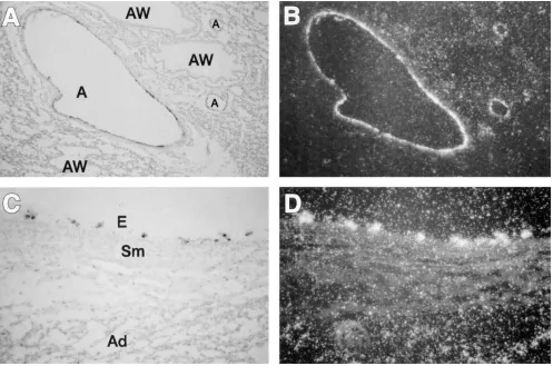
[image:7.612.60.557.59.388.2]while M&B 22948 and dipyridamole, inhibitors of cGMP phos-phodiesterase, decrease pulmonary vascular resistance and augment endothelium-dependent pulmonary vasodilation in fetal and newborn lambs (45–47). Finally, there are also Figure 3. eNOS mRNA expression in the lung in vivo from near-term fetal lambs after 8 h of ventilation in utero with 100% oxygen. A cRNA probe for ovine eNOS was hybridized to tissue sections prepared from the lung of fetal lambs ventilated in utero for 8 h with 100% oxygen. Ex-pression is seen as black grains in bright-field (A and C) and as silver grains in dark-field (B and D). A and B, 3200; C and D, 3400. Ovine eNOS mRNA is expressed only in the endothelium (E) of small and large pulmonary arteries (A) and veins but not in the smooth muscle (Sm) or ad-ventitia (Ad), or in the airways (AW). The result shown is representative of results from three experiments using different fetal lungs after 8 h of ventilation with 100% oxygen.
Figure 4. Western blot analysis for eNOS protein in lung tissue from near-term fetal lambs: unventi-lated, and after 8 h of in utero with a gas mixture which did not change fetal descending aortic blood gases or pH (rhythm distension) or with 100% oxygen (O2 ventilation). (A)
Protein extracts (100 mg), prepared from lung tissue from three near-term fetal lambs (unventilated, rhythmic distention, and O2
ventila-tion), were separated on a 6% dena-turing polyacrylamide gel, electro-phoretically transferred to Hybond membranes, and analyzed using a specific antiserum raised against eNOS. Human endothelial cell protein (Human EC, 20 mg) was used as a control. There was no change in eNOS protein (135 kD) with rhythmic distension; there was an increase with O2 ventilation. (B) The densitometric values for relative eNOS protein (normalized to unventilated) from
six different experiments. Rhythmic distension did not change eNOS protein; O2 ventilation increased eNOS protein by 1.9-fold (P , 0.05).
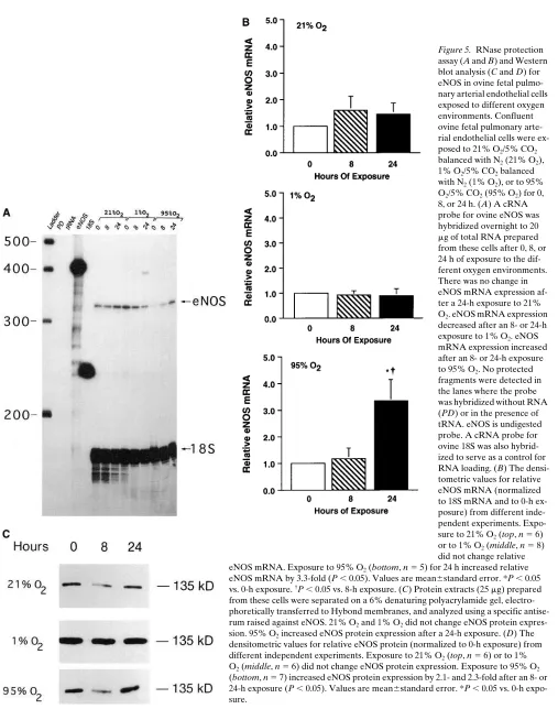
[image:7.612.59.459.533.691.2]Figure 5. RNase protection assay (A and B) and Western blot analysis (C and D) for eNOS in ovine fetal pulmo-nary arterial endothelial cells exposed to different oxygen environments. Confluent ovine fetal pulmonary arte-rial endothelial cells were ex-posed to 21% O2/5% CO2
balanced with N2 (21% O2),
1% O2/5% CO2 balanced
with N2 (1% O2), or to 95%
O2/5% CO2 (95% O2) for 0,
8, or 24 h. (A) A cRNA probe for ovine eNOS was hybridized overnight to 20 mg of total RNA prepared from these cells after 0, 8, or 24 h of exposure to the dif-ferent oxygen environments. There was no change in eNOS mRNA expression af-ter a 24-h exposure to 21%
O2. eNOS mRNA expression
decreased after an 8- or 24-h exposure to 1% O2. eNOS
mRNA expression increased after an 8- or 24-h exposure to 95% O2. No protected
fragments were detected in the lanes where the probe was hybridized without RNA (PD) or in the presence of tRNA. eNOS is undigested probe. A cRNA probe for ovine 18S was also hybrid-ized to serve as a control for RNA loading. (B) The densi-tometric values for relative eNOS mRNA (normalized to 18S mRNA and to 0-h ex-posure) from different inde-pendent experiments. Expo-sure to 21% O2 (top, n 5 6)
or to 1% O2 (middle, n 5 8)
did not change relative eNOS mRNA. Exposure to 95% O2 (bottom, n 5 5) for 24 h increased relative
eNOS mRNA by 3.3-fold (P , 0.05). Values are mean6standard error. *P , 0.05 vs. 0-h exposure. †P , 0.05 vs. 8-h exposure. (C) Protein extracts (25 mg) prepared
from these cells were separated on a 6% denaturing polyacrylamide gel, electro-phoretically transferred to Hybond membranes, and analyzed using a specific antise-rum raised against eNOS. 21% O2 and 1% O2 did not change eNOS protein
expres-sion. 95% O2 increased eNOS protein expression after a 24-h exposure. (D) The
densitometric values for relative eNOS protein (normalized to 0-h exposure) from different independent experiments. Exposure to 21% O2 (top, n 5 6) or to 1%
O2 (middle, n 5 6) did not change eNOS protein expression. Exposure to 95% O2
Figure 5 (Continued)
changes in NO production from isolated pulmonary arteries and endothelial cells with development. Basal and endothelial-dependent production of NO increase in newborn pulmonary arteries compared with near-term fetal pulmonary arteries, and in fetal pulmonary arterial endothelial cells exposed to hy-peroxia; production is decreased by hypoxia (48–50). There-fore, changes in NOS activity and NO production likely medi-ate, in part, pulmonary vascular tone in the perinatal period.
eNOS differs in structure and regulation from the two other isoforms of NOS (51). eNOS is present on the cell mem-brane of endothelial cells and its activity is calcium and cal-modulin dependent (11, 13–15, 23, 51). Human and bovine eNOS have been isolated and purified as a 135-kD protein whose clone identifies a 4.6–4.8-kb mRNA by Northern blot
analysis (24, 33–35). eNOS has been described as a constitutive enzyme; however, it is developmentally regulated. In rat and sheep lungs, eNOS mRNA expression increases near term and decreases after birth as do eNOS protein levels (18, 19, 52). This increased eNOS expression near term leads to increased NO production resulting in pulmonary vasodilation at birth. The results of this study show that in the ovine fetal lung, the increase in pulmonary blood flow and oxygenation induced by ventilation increases eNOS mRNA and protein expression. These increases in eNOS mRNA and protein expression are necessary for the more gradual and progressive pulmonary va-sodilation that occurs hours and days after birth. For example, mean pulmonary arterial pressure decreases to z 50% of
mean systemic arterial pressure by 24 h of age, and reaches adult values by 2–6 wk after birth (4).
eNOS is also regulated by changes in the oxygen environ-ment. eNOS activity and NO production are increased by hy-peroxia and decreased by hypoxia (10, 16, 48–50). In this study, eNOS mRNA and protein expression were increased in ovine fetal pulmonary arterial endothelial cells exposed to 95% oxygen for 24 h. In other studies, eNOS mRNA and pro-tein expression were also increased to a similar degree in bo-vine adult and obo-vine fetal pulmonary arterial endothelial cells exposed to a high oxygen environment for 24 to 48 h (53–55). These increases in gene expression are mediated through tran-scriptional and posttrantran-scriptional mechanisms. Since these oxygen-mediated increases in eNOS gene expression occur over 24 h, they likely will contribute to the gradual decrease in pulmonary vascular resistance after birth. In this study, eNOS mRNA and protein expression were unchanged in ovine fetal pulmonary arterial endothelial cells exposed to 1% oxygen for 24 h. However, in other studies, eNOS mRNA and protein ex-pression were decreased in bovine adult and human umbilical vein endothelial cells exposed to a low oxygen environment for 24 to 48 h (53–55). Since ovine fetal pulmonary arterial en-dothelial cells are exposed to a very low oxygen environment in vivo, a lower oxygen tension in the culture medium than achieved in this study (PO2, 40 torr) may be needed to
de-crease eNOS gene expression.
[image:9.612.58.226.59.548.2]shape secondary to a structural reorganization of the cytoskel-eton. There are also changes in gene expression. Shear stress induces PDGF-B mRNA and protein expression in endothe-lial cells (56–59). A 6-bp sequence (GAGACC) shear stress re-sponse element has been identified in the 59-flank of the
hu-man PDGF-B gene and in other shear stress–inducible endothelial cell genes. The 59-flank of the human NOS gene also contains the 6-bp shear stress response element (59). In this study, eNOS mRNA and protein expression were in-creased in ovine fetal pulmonary arterial endothelial cells ex-Figure 6. RNase protection assay (A and B) and Western blot analysis (C and D) for eNOS in ovine fetal pulmonary arterial endothelial cells ex-posed to fluid shear stress. Confluent ovine fetal pulmonary arterial endothelial cells were exex-posed for 0, 2, 4, and 8 h to radially constant fluid shear stress (20 dynes/cm2). (A) A cRNA probe for ovine eNOS was hybridized overnight to 20 mg of total RNA prepared from these cells. From
[image:10.612.56.481.58.518.2]posed to increased shear stress. In other studies, eNOS mRNA and protein expression were also increased to a similar degree and in a similar time course in bovine aortic and human umbil-ical vein endothelial cells exposed to increased shear stress. This shear stress–induced increase in eNOS gene expression in vitro was similar to the oxygen ventilation–induced increase in vivo, suggesting that the increase in pulmonary blood flow and the resulting increase in shear stress is more important than in-creased oxygenation in regulating eNOS gene expression.
There are multiple components contributing to the imme-diate decrease in pulmonary vascular resistance and increase in pulmonary blood flow with the initiation of ventilation and oxygenation at birth and the subsequent gradual (next hours) and progressive (days) transition to extrauterine life over the next hours to days. First, there is an immediate modest de-crease in pulmonary vascular resistance and inde-crease in pulmo-nary blood flow caused by physical expansion of the lung. This expansion produces partial pulmonary vasodilation indepen-dent of fetal oxygenation and NO production. This component of pulmonary vasodilation is not attenuated by inhibition of NOS activity. Next, there is further gradual pulmonary vasodi-lation associated with oxygenation, the initial increase in pul-monary blood flow, and the production of NO which results in a marked increase in pulmonary blood flow and decrease in pulmonary vascular resistance. This component of pulmonary vasodilation is attenuated by inhibition of NOS activity. Then, there is progressive pulmonary vasodilation induced by the in-crease in pulmonary blood flow and shear stress, and to a lesser degree, the increase in oxygenation. This is associated with increased eNOS mRNA and protein expression and fur-ther production of NO.
Acknowledgments
The authors thank Jennifer Seslar for editorial assistance.
This research was supported by the American Heart Association, California Affiliate (J. Bristow) and by grant HL-35518 from the Na-tional Heart, Lung and Blood Institute (S.J. Soifer).
References
1. Cassin, S., G.S. Dawes, J.C. Mott, B.B. Ross, and L.B. Strang. 1964. The vascular resistance of the foetal and newly ventilated lung of the lamb. J. Phys-iol. 171:61–79.
2. Dawes, G.S., J.C. Mott, J.G. Widdicombe, and D.G. Wyatt. 1953. Changes in the lungs of the newborn lamb. J. Physiol. 121:141–162.
3. Iwamoto, H.S., D. Teitel, and A.M. Rudolph. 1987. Effects of birth-related events on blood flow distribution. Pediatr. Res. 22:634–640.
4. Rudolph, A.M. 1979. Fetal and neonatal pulmonary circulation. Annu. Rev. Physiol. 41:383–395.
5. Fineman, J.R., S.J. Soifer, and M.A. Heymann. 1995. Regulation of pul-monary vascular tone in the perinatal period. Annu. Rev. Physiol. 57:115–134.
6. Leffler, C.W., J.R. Hessler, and R.S. Green. 1984. Mechanism of stimula-tion of pulmonary prostacyclin synthesis at birth. Prostaglandins. 28:877–887.
7. Leffler, C.W., J.R. Hessler, and R.S. Green. 1984. The onset of breathing at birth stimulates pulmonary vascular prostacyclin synthesis. Pediatr. Res. 18: 938–942.
8. Abman, S.H., B.A. Chatfield, S.L. Hall, and I.F. McMurtry. 1990. Role of endothelium-derived relaxing factor during transition of pulmonary circulation at birth. Am. J. Physiol. 259:H1921–H1927.
9. Cornfield, D.N., B.A. Chatfield, J.A. McQueston, I.F. McMurtry, and S.H. Abman. 1992. Effects of birth-related stimuli on l-arginine-dependent pul-monary vasodilation in ovine fetus. Am. J. Physiol. 262:H1474–H1481.
10. Moore, P., H. Velvis, J.R. Fineman, S.J. Soifer, and M.A. Heymann. 1992. EDRF inhibition attenuates the increase in pulmonary blood flow due to oxygen ventilation in fetal lambs. J. Appl. Physiol. 73:2151–2157.
11. Palmer, R.M.J., D.S. Ashton, and S. Moncada. 1988. Vascular endothe-lial cells synthesize nitric oxide from l-arginine. Nature (Lond.). 333:664–666.
12. Vane, J.R., E.E. Anggard, and R.M. Botting. 1990. Regulatory functions
of the vascular endothelium. N. Engl. J. Med. 323:27–36.
13. Ignarro, L.J. 1990. Biosynthesis and metabolism of endothelium-derived nitric oxide. Annu. Rev. Pharmacol. Toxicol. 30:535–560.
14. Moncada, S., R.M.J. Palmer, and E.A. Higgs. 1991. Nitric oxide: physi-ology, pathophysiology and pharmacology. Pharmacol. Rev. 43:109–142.
15. Moncada, S., and A. Higgs. 1993. The l-arginine–nitric oxide pathway. N. Engl. J. Med. 329:2002–2012.
16. Tikinsky, M.H., and F.C. Morin III. 1993. Increasing oxygen tension di-lates fetal pulmonary circulation via endothelium-derived relaxing factor. Am. J. Physiol. 265:H376–H380.
17. Fineman, J.R., J. Wong, F.C. Morin, L. Wright, and S.J. Soifer. 1994. Chronic nitric oxide inhibition in utero produces persistent pulmonary hyper-tension in newborn lambs. J. Clin. Invest. 93:2675–2683.
18. North, A.J., R.A. Star, T.S. Brannon, K. Ujiie, L.B. Wells, C.J. Lowen-stein, S.H. Snyder, and P.W. Shaul. 1994. Nitric oxide synthase type I and type III gene expression are developmentally regulated in rat lung. Am. J. Physiol. 266:L635–L641.
19. Kawai, N., D.B. Bloch, G. Filippov, D. Rabkina, H.C. Suen, P.D. Losty, S.P. Janssens, W.M. Zapol, S. de la Monte, and K.D. Bloch. 1995. Constitutive endothelial nitric oxide synthase gene expression is regulated during lung de-velopment. Am. J. Physiol. 268:L589–L595.
20. Noris, M., M. Morigi, R. Donadelli, S. Aiello, M. Foppolo, M. Todes-chini, S. Orisio, G. Remuzzi, and A. Remuzzi. 1995. Nitric oxide synthesis by cultured endothelial cells is modulated by flow conditions. Circ. Res. 76:536–543.
21. Korenaga, R., J. Ando, H. Tsuboi, W. Yang, I. Sakuma, T. Toyo-oka, and A. Kamiya. 1994. Laminar flow stimulates ATP- and shear stress-depen-dent nitric oxide production in cultured bovine endothelial cells. Biochem. Bio-phys. Res. Commun. 198:213–219.
22. Kuchan, M.J., H. Jo, and J.A. Frangos. 1994. Role of G proteins in shear stress-mediated nitric oxide production by endothelial cells. Am. J. Physiol. 267:C753–C758.
23. Kuchan, M.J., and J.A. Frangos. 1994. Role of calcium and calmodulin in flow-induced nitric oxide production in endothelial cells. Am. J. Physiol. 266: C628–C636.
24. Nishida, K., D.G. Harrison, J.P. Navas, A.A. Fisher, S.P. Dockery, M. Uematsu, R.M. Nerem, R.W. Alexander, and T.J. Murphy. 1992. Molecular cloning and characterization of the constitutive bovine aortic endothelial cell nitric oxide synthase. J. Clin. Invest. 90:2092–2096.
25. Ranjan, V., Z. Xiao, and S.L. Diamond. 1995. Constitutive NOS expres-sion in cultured endothelial cells is elevated by fluid shear stress. Am. J. Physiol. 269:H550–H555.
26. Uematsu, M., Y. Ohara, J.P. Navas, K. Nishida, T.J. Murphy, R.W. Al-exander, R.M. Nerem, and D.G. Harrison. 1995. Regulation of endothelial cell nitric oxide synthase mRNA expression by shear stress. Am. J. Physiol. 269: C1371–C1378.
27. Zar, J.H. 1974. Biostatistical Analysis. Prentice-Hall, Inc., Englewood Cliffs, NJ. 33–34, 101–129.
28. Chomczynski, P., and N. Sacchi. 1987. Single-step method of RNA isola-tion by acid guanidinium thiocyanate-phenol-chloroform extracisola-tion. Anal. Bio-chem. 162:156–159.
29. Bradford, M.M. 1976. A rapid and sensitive method for the quantitation of microgram quantities of protein utilizing the principle of protein-dye bind-ing. Anal. Biochem. 72:248–254.
30. Black, S.M., M.A. Bedolli, S. Martinez, J. Bristow, D.M. Ferriero, and S.J. Soifer. 1995. Expression of neuronal nitric oxide synthase corresponds to regions of selective vulnerability to hypoxia-ischemia in the developing rat cen-tral nervous system. Neurobiol. Dis. 2:145–155.
31. Nelkin, N.A., S.J. Soifer, J. O’Keefe, T.-K.H. Vu, I.F. Charo, and S.R. Coughlin. 1992. Thrombin receptor expression in normal and atherosclerotic human arteries. J. Clin. Invest. 90:1614–1621.
32. Soifer, S.J., K.G. Peters, J. O’Keefe, and S.R. Coughlin. 1994. Disparate temporal expression of the prothrombin and thrombin receptor genes during mouse development. Am. J. Pathol. 144:60–69.
33. Marsden, P.A., K.T. Schappert, H.S. Chen, M. Flowers, C.L. Sundell, J.N. Wilcox, S. Lamas, and T. Michel. 1992. Molecular cloning and characteriza-tion of human endothelial nitric oxide synthase. FEBS Lett. 307:287–293.
34. Lamas, S., P.A. Marsden, G.K. Li, P. Tempst, and T. Michel. 1992. En-dothelial nitric oxide synthase: molecular cloning and characterization of a dis-tinct constitutive enzyme isoform. Proc. Natl. Acad. Sci. USA. 89:6348–6352.
35. Sessa, W.C., J.K. Harrison, C.M. Barber, D. Zeng, M.E. Durieux, D.D. D’Angelo, K.R. Lynch, and M.J. Peach. 1992. Molecular cloning and expression of a cDNA encoding endothelial cell nitric oxide synthase. J. Biol. Chem. 267: 15274–15276.
36. Bristow, J., S.E. Gitelman, M.K. Tee, and W.L. Miller. 1993. Abundant adrenal-specific expression of the P450c21A “pseudogene.” J. Biol. Chem. 268: 12919–12924.
37. Li, H.T., C.S. Long, D.G. Rokosh, N.Y. Honbo, and J.S. Karliner. 1995. Chronic hypoxia differentially regulates alpha 1-adrenergic receptor subtype mRNAs and inhibits alpha 1-adrenergic receptor-stimulated cardiac hypertro-phy and signaling. Circulation. 92:918–925.
39. Resnick, N., and M.A. Gimbrone, Jr. 1995. Hemodynamic forces are complex regulators of endothelial gene expression. FASEB (Fed. Am. Soc. Exp. Biol.) J. 9:874–882.
40. Rasband, W. 1993. NIH Image Program, v1.49. National Institutes of Health, Bethesda, MD.
41. Titinsky, M.H., J.J. Cummings, and F.C. Morin. 1992. Acetylcholine in-creases pulmonary blood flow in intact fetuses via endothelium-dependent va-sodilation. Am. J. Physiol. 262:H406–H410.
42. Fineman, J.R., R. Chang, and S.J. Soifer. 1991. l-arginine, a precursor of EDRF in vitro, produces pulmonary vasodilation in lambs. Am. J. Physiol. 261: H1563–H1569.
43. Kinsella, J.P., J.A. McQueston, A.A. Rosenberg, and S.H. Abman. 1992. Hemodynamic effects of exogenous nitric oxide in ovine transitional pul-monary circulation. Am. J. Physiol. 263:H875–H880.
44. Zayek, M., D. Cleveland, and F.C. Morin III. 1993. Treatment of persis-tent pulmonary hypertension in the newborn lamb by nitric oxide. J. Pediatr. 122:743–750.
45. Fineman, J.R., M.R. Crowley, M.A. Heymann, and S.J. Soifer. 1991. In vivo attenuation of endothelium-dependent pulmonary vasodilation by methy-lene blue in the newborn lamb. J. Appl. Physiol. 71:735–741.
46. Braner, D.A., J.R. Fineman, R. Chang, and S.J. Soifer. 1993. M&B 22948, a cGMP phosphodiesterase inhibitor, is a pulmonary vasodilator in lambs. Am. J. Physiol. 264:H252–H258.
47. Steinhorn, R.H., R.C. Dukarm, F.C. Morin III, S.F. Gugino, and J.A. Russell. 1995. Dipyridamole enhances relaxations and cGMP generation to ni-tric oxide in lambs with persistent pulmonary hypertension. Pediatr. Res. 39: 351a. (Abstr.)
48. Shaul, P.W., M.A. Farrar, and T.M. Zellers. 1992. Oxygen modulates endothelium-derived relaxing factor production in fetal pulmonary arteries. Am. J. Physiol. 262:H355–H364.
49. Shaul, P.W., M.A. Farrar, and R.R. Magness. 1993. Pulmonary endothe-lial nitric oxide production is developmentally regulated in the fetus and new-born. Am. J. Physiol. 265:H1056–H1063.
50. Shaul, P.W., and L.B. Wells. 1994. Oxygen modulates nitric oxide pro-duction selectively in fetal pulmonary endothelial cells. Am. J. Respir. Cell. Mol. Biol. 11:432–438.
51. Sessa, W.C. 1994. The nitric oxide synthase family of proteins. J. Vasc. Res. 31:131–143.
52. Halbower, A.C., R.M. Tuder, W.A. Franklin, J.S. Pollock, U. Forster-mann, and S.H. Abman. 1994. Maturation-related changes in endothelial nitric oxide synthase immunolocalization in developing ovine lung. Am. J. Physiol. 267:L585–L591.
53. Liao, J.K., J.J. Zulueta, F.-S. Yu, H.-B. Peng, C.G. Cote, and P.M. Has-soun. 1995. Regulation of bovine endothelial constitutive nitric oxide synthase by oxygen. J. Clin. Invest. 96:2661–2666.
54. North, A.J., K.S. Lau, T.S. Brannon, L.C. Wu, L.B. Wells, Z. German, and P.W. Shaul. 1996. Oxygen upregulates nitric oxide synthase gene expres-sion in ovine fetal pulmonary artery endothelial cells. Am. J. Physiol. 270:L643– L649.
55. Phelan, M.W., and D.V. Faller. 1996. Hypoxia decreases constitutive ni-tric oxide synthase transcript and protein in cultured endothelial cells. J. Cell. Physiol. 167:469–476.
56. Hsieh, H.J., N.Q. Li, and J.A. Frangos. 1992. Shear-induced platelet-derived growth factor gene expression in human endothelial cells is mediated by protein kinase C. J. Cell. Physiol. 150:552–558.
57. Malek, A.M., G.H. Gibbons, V.J. Dzau, and S. Izumo. 1993. Fluid shear stress differentially modulates expression of genes encoding basic fibroblast growth factor and platelet-derived growth factor B chain in vascular endothe-lium. J. Clin. Invest. 92:2013–2021.
58. Mitsumata, M., R.S. Fishel, R.M. Nerem, R.W. Alexander, and B.C. Berk. 1993. Fluid shear stress stimulates platelet-derived growth factor expres-sion in endothelial cells. Am. J. Physiol. 265:H3–H8.