Development/Plasticity/Repair
Synaptic Plasticity in
CNGA3
ⴚ
/
ⴚ
Mice: Cone Bipolar Cells
React on the Missing Cone Input and Form Ectopic Synapses
with Rods
Silke Haverkamp,
1Stylianos Michalakis,
2Ellen Claes,
1Mathias W. Seeliger,
3Peter Humphries,
4Martin Biel,
2and
Andreas Feigenspan
51Department of Neuroanatomy, Max-Planck-Institute for Brain Research, D-60528 Frankfurt/Main, Germany,2Department Pharmazie–Zentrum fu¨r Pharmaforschung, Ludwig Maximilians Universita¨t, D-81377 Munich, Germany,3Retinal Diagnostics Research Group, Department of Ophthalmology II, Eberhard-Karls University, D-72076 Tuebingen, Germany,4Department of Genetics, Trinity College, Dublin 2, United Kingdom, and5Department of Neurobiology, University of Oldenburg, D-26111 Oldenburg, Germany
In the mammalian retina, rods and cones connect to distinct sets of bipolar cells. Rods are presynaptic to a single type of rod bipolar cell, whereas cones connect to different types of cone bipolar cells. Synaptic rewiring between cone photoreceptor terminals and rod bipolar cell dendrites has been described as a general result of photoreceptor degeneration. To investigate whether cone bipolar cells also show synaptic plasticity in the absence of cone input, we studied the connectivity of cone bipolar cell dendrites inCNGA3ⴚ/ⴚmice, a model with
specific loss of cone photoreceptor function. Dendritic connections of ON and OFF cone bipolar cells were visualized using specific cell markers or by intracellular injection with fluorescent dyes. The results show that cone bipolar cells inCNGA3ⴚ/ⴚmice form ectopic
synapses with rods. In contrast, cone bipolar cells do not form ectopic synapses with rods inCNGA3ⴚ/ⴚRhoⴚ/ⴚmice, in which both types
of photoreceptors are nonfunctional. In analogy with these results, we found that input-deprived rod bipolar cells form ectopic synapses with functional cones inRhoⴚ/ⴚmice but not with inoperable cones in theCNGA3ⴚ/ⴚRhoⴚ/ⴚmouse. Our data indicate that the
formation of ectopic bipolar cell synapses in the outer plexiform layer requires a functional presynaptic photoreceptor.
Key words:retina; cones; rods; bipolar cells; dendritic plasticity; ectopic synaptogenesis
Introduction
The mammalian retina contains rod and cone photoreceptors, precisely tuned to respond to low (scotopic) and high (photopic) light levels. Rod and cone pathways are separated at their first synapse in the outer retina (Wa¨ssle, 2004). Rods connect to a single type of rod bipolar cell, whereas cones are presynaptic to at least nine different types of cone bipolar cells (Ghosh et al. 2004). OFF cone bipolar cells make flat contacts with cone pedicles and express a conventional sign conserving glutamate receptor of the AMPA or kainate subtype (DeVries, 2000). ON cone and rod bipolar cells make invaginating contacts close to the synaptic ribbons and express the sign-inverting glutamate receptor, metabotropic glutamate receptor type 6 (mGluR6) (Nakajima et al., 1993; Nomura et al., 1994; Vardi et al., 2000).
In the mouse retina, one type of OFF cone bipolar cell makes direct synaptic contacts with rods [B2 (Tsukamoto et al., 2001); this cell type is comparable with type 3 in Ghosh et al. (2004) and CB2 in Pignatelli and Strettoi (2004)]. Direct rod to OFF cone
bipolar cell connections have also been described in rat, cat, and rabbit retina (Hack et al., 1999; Fyk-Kolodziej et al., 2003; Li et al., 2004; Protti et al., 2005).
Connections between cones and rod bipolar cells have only exceptionally been reported in a normal retina (Dacheux and Raviola, 1986). However, cones make ectopic synapses with rod bipolar cells in diverse photoreceptor degeneration models [P347L pigs,retinal degenerationmice, Royal College of Surgeons (RCS) rats] (Peng et al., 2000, 2003) as well as in neural retina leucine zipper (Nrl) knock-out mice in which rods fail to form and only cones exist (Strettoi et al., 2004; Daniele et al., 2005).
Photoreceptors respond to light by the closure of a cyclic nucleotide-gated (CNG) cation channel, causing hyperpolariza-tion and decrease of the synaptic glutamate release. To investigate whether cone bipolar cells form ectopic synapses in the absence of cone input, we studied the connectivity of cone bipolar cell den-drites in mice lacking CNGA3 (Biel et al., 1999), the A subunit of the cone CNG channel. It was demonstrated previously that these mice lack any cone-mediated photoresponses but have a normal rod photosystem (Biel et al., 1999).
Rhodopsin knock-out (Rho⫺/⫺) mice, on the other hand,
ap-pear to have normal cone but no rod function, at least until postnatal week 6 (pw6) when cone degeneration is not yet sub-stantial. These animals fail to form rod outer segments, leading to a sequence of primary rod and secondary cone photoreceptor loss
Received Oct. 19, 2005; revised March 31, 2006; accepted April 1, 2006.
This work was supported by Deutsche Forschungsgemeinschaft Grants SFB269/B4, Se837/4-1, and Se837/5-1. We thank B. Marshallsay and G.-S. Nam for excellent technical assistance.
Correspondence should be addressed to Silke Haverkamp, Max-Planck-Institute for Brain Research, Deutschor-denstrasse 46, D-60528 Frankfurt/Main, Germany. E-mail: [email protected].
DOI:10.1523/JNEUROSCI.4483-05.2006
that is complete at⬃3 months postnatally (Humphries et al., 1997; Jaissle et al., 2001)
We used these mice, as well as mice deficient for both CNGA3 and Rhodopsin (CNGA3⫺/⫺Rho⫺/⫺) (Claes et al., 2004), to test
our hypothesis that the formation of ectopic bipolar cell synapses in the outer plexiform layer (OPL) requires a functional presyn-aptic photoreceptor.
Materials and Methods
Animals.The generation ofCNGA3⫺/⫺mice andRho⫺/⫺mice was
de-scribed previously (Humphries et al., 1997; Biel et al., 1999). Double
mutants were generated by cross-breedingCNGA3⫺/⫺ withRho⫺/⫺
mice (Claes et al., 2004). The␣-gustducin– green fluorescent protein
(GUS–GFP) transgenic mouse line was kindly provided by R. Margolskee (Mount Sinai Medical Center, New York, NY) (Huang et al., 2003).
The electron microscopic study included eightCNGA3⫺/⫺mice of
different ages (postnatal week 3.2, 4.0, 5.0, 6.1, 6.6, and 7.6 and postnatal month 4.09 and 8.29). They were raised on a 129sv background and were compared with age-matched 129sv and C57BL/6 wild-type mice.
Fur-thermore, we investigated the retinas of threeRho⫺/⫺mice (postnatal
week 5.1, 6.2, and 7.2) and fourCNGA3⫺/⫺Rho⫺/⫺mice (postnatal
week 3.3, 4.0, 5.0, and 6.0) by preembedding immunoelectron microscopy.
Tissue preparation.Mice were anesthetized deeply with halothane and
decapitated. The procedures were approved by the local animal care committees and were in accordance with the law of animal experimen-tation issued by the German Government (Tierschutzgesetz). The eyes were enucleated, the anterior segments removed, and the posterior
eye-cups immersion fixed in 4% paraformaldehyde in 0.1Mphosphate buffer
(PB), pH 7.4, for 30 min (LM) or 60 min (EM). After fixation, the retina was dissected from the eyecup and embedded in 2– 4% agar. The agar
block was mounted on a vibratome, and vertical sections of 60m
thick-ness were cut. For electron microscopy, the retina was cryoprotected in graded sucrose solutions (10, 20, and 30%) and frozen and thawed before vibratome sectioning. For retinal GUS–GFP whole mounts, the tissue was cryoprotected and frozen and thawed several times before applying the antibodies. Vibratome sections and whole mounts were processed free-floating.
Light microscopic immunocytochemistry.OFF cone bipolar cells were
labeled with antibodies against the neurokinin-3 receptor (rabbit anti-NK3R; 1:500; kindly provided by R. Shigemoto, National Institute, Oka-zaki, Japan) and rod bipolar cells with antibodies against protein kinase
C␣(rabbit anti-PKC␣; 1:10,000; Sigma, St. Louis, MO). Synaptic ribbons
were visualized with an antibody against the cytomatrix protein bassoon (mouse anti-bassoon; 1:5000; Stressgen Biotechnologies, Victoria, Brit-ish Columbia, Canada) or with antibodies against the C-terminal bind-ing protein 2 (mouse anti-CtBP2; 1:10,000; BD Transduction, Heidel-berg, Germany). The GluR5 antibody (goat anti-GluR5N; 1:100; Santa Cruz Biotechnology, Heidelberg, Germany) has been used to label cone pedicles and ectopic synapses at rod spherules. The GFP fluorescence signal has been increased with rabbit anti-GFP antibodies (1:2000; In-vitrogen, San Diego, CA).
Antibodies were diluted in PBS, pH 7.4, containing 5% Chemiblocker (Chemicon, Temecula, CA), 0.5% Triton X-100, and 0.05% sodium azide. Immunocytochemical labeling was performed using the indirect fluorescence method. Vibratome sections were incubated overnight in a mixture of primary antibodies, followed by incubation (1 h) in a mixture of the secondary antibodies, which were conjugated to either Alexa TM 488 (green fluorescence; Invitrogen) or to indocarbocyanin 3 (Cy3; red fluorescence; Dianova, Hamburg, Germany). Whole mounts were incu-bated for 2 d in the primary and for 2 h in the secondary antibody solution.
Confocal micrographs were taken using a Zeiss (Oberkochen, Ger-many) LSM5 Pascal confocal microscope equipped with an argon and a helium–neon laser. High-resolution scanning was performed with a
63⫻/1.4 Plan-Apochromat objective (z-axis step size, 0.8m).
Bright-ness and contrast of the final images were adjusted using Adobe Photo-shop 5.5 (Adobe Systems, San Jose, CA).
Preembedding immunoelectron microscopy.Vibratome sections were
incubated for 4 d at 4°C in a primary antibody solution (anti-NK3R,
1:1000; anti-PKC␣, 1:20,000) containing 3% normal goat serum (NGS),
1% bovine serum albumin, and 0.05% sodium azide in PBS. Thereafter, the sections were rinsed in PBS and immunolabeling was detected with a biotinylated goat anti-rabbit IgG (1:100; Vector Laboratories, Burlin-game, CA) and a peroxidase-based enzymatic detection system (Vec-tastain Elite ABC kit; Vector Laboratories). After rinses in PBS and in 0.05
MTris-HCl, pH 7.6, the sections were reacted in 3,3⬘-diaminobenzidine
(DAB; 0.05% in Tris-HCl) with 0.01% H2O2for 5–10 min. Subsequently,
the sections were rinsed in Tris-HCl and then in 0.1Mcacodylate buffer,
pH 7.4, postfixed in 2.5% glutaraldehyde in cacodylate buffer for 2 h at 4°C, and washed in cacodylate buffer overnight at 4°C. After several washes in distilled water, the DAB reaction product was silver-intensified by incubating the sections in a solution containing 2.6% hexamethylene tetramine, 0.2% silver nitrate, and 0.2% disodium tetraborate for 10 min at 60°C. The sections were then rinsed in distilled water and treated for 2 min with gold chloride (0.05% in distilled water). Finally, the sections were rinsed in distilled water and incubated for 2 min in sodium thiosul-fate (2.5% in distilled water). The sections were then postfixed with 0.5%
OsO4in cacodylate buffer for 30 min, dehydrated in a graded series of
ethanol (30 –100%) followed by propylene oxide, and flat-embedded in Epon 812 (Serva, Heidelberg, Germany). Serial ultrathin sections were cut, stained with uranyl acetate and lead citrate, and examined with a Zeiss EM10 electron microscope.
Quantitative analysis. To quantify the amount of flat contacts of
NK3R-labeled OFF bipolar cell dendrites at the rod spherules, single
ultrathin sections of threeCNGA3⫺/⫺and three wild-type retinas were
analyzed. Photoreceptor somas and synaptic terminals stack in multiple tiers in the mouse retina (Tsukamoto et al., 2001) (see Fig. 5). The densely
packed somas form⬃10 tiers; the underlying synaptic terminals form
three to four tiers. The NK3R-labeled dendrites reach the innermost tier of synaptic terminals, where cone pedicles and rod spherules intermingle
(see Fig. 2B,C). Because the dendrites mostly stop there and barely
pen-etrate the remaining tiers of rod spherules, we counted only the rod spherules in this innermost tier.
We did not count the number of PKC␣-labeled rod bipolar cell
den-drites at cone pedicles in theRho⫺/⫺mouse, because the penetration of
the PKC␣-antibody was not optimal and we only found labeling at the
surface, not enough to find a sufficient number of examples for quantification.
Intracellular injection of ON cone bipolar cells.For the preparation of
vertical sections, the retinas of C57BL/6 wild-type andCNGA3⫺/⫺mice
were removed from the eyecup and embedded in 2% agarose in Ames medium (Sigma, Deisenhofen, Germany). Agar blocks were mounted on
a vibratome (Leica, Nussloch, Germany) and cut into slices of 200m
thickness. After cutting, the slices were fixed in 4% paraformaldehyde for 10 min, washed in PB, and subsequently transferred to the stage of an upright microscope (Leica) for intracellular injections.
Intracellular injections were performed with sharp borosilicate
micro-electrodes (Hilgenberg, Malsfeld, Germany) filled with 10 mMAlexa
Fluor 488 hydrazide (Invitrogen). Alexa 488 was injected
iontophoreti-cally (2–3 min,⫺1 nA) into the somata of morphologically identified
bipolar cells using the current-clamp circuit of the EPC-9 patch-clamp amplifier (HEKA Elektronik, Lambrecht, Germany). After injection, slices were postfixed in 4% paraformaldehyde for 20 min, washed in PB, and processed for immunocytochemistry as described below.
To block unspecific labeling, vertical sections were incubated in a so-lution containing 5% NGS and 0.3% Triton X-100 in PB for 1 h. Den-dritic tips of ON cone bipolar cells were labeled with polyclonal
antibod-ies against mGluR6 (rabbit anti-mGluR6; 1:500; Neuromics,
Minneapolis, MN), and presynaptic ribbons were labeled with monoclo-nal antibodies against kinesin (mouse anti-kinesin II; 1:50; Babco, Rich-mond, CA).
experi-ments, sections were incubated in a mixture of primary antibodies, followed by a mixture of secondary antibodies.
Confocal micrographs of fluorescent speci-mens were taken with a Leica TCS SL confocal microscope equipped with an argon and a heli-um–neon laser. Scanning was performed with a
40⫻/1.25 Plan-Apochromat and a 63⫻/1.32
Plan-Apochromat objective at a resolution of
1024⫻1024 pixels (z-axis step size, 0.5m).
Different wavelength scans were performed se-quentially to rule out cross-talk between red, green, and blue channels.
Results
The retina ofCNGA3⫺/⫺mice displays a
normal general morphology and lamina-tion (Biel et al., 1999). Only a degeneralamina-tion of cones is evident over a time course of several months (Michalakis et al., 2005). To investigate the connectivity of OFF cone bipolar cells in theCNGA3⫺/⫺
ret-ina, we used a specific antibody directed against the neurokinin-3 receptor (Ding et al., 1996).). NK3R labels type 1 and type 2 bipolar cells in the mouse retina but not the rod contacting type 3 cell, which can be labeled by antibodies against the calcium-binding protein 5 (CaB5) (Haverkamp et al., 2003; Ghosh et al., 2004).
NK3R immunoreactivity was present in bipolar cells with axons terminating in the outermost part of the inner plexiform
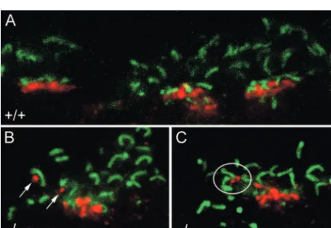
layer (IPL) (Fig. 1A). The processes in the inner IPL in Figure 1A
belong to NK3R-immmunoreactive amacrine cells (Haverkamp et al., 2003). There was no difference between wild-type and
CNGA3⫺/⫺retinas except for small NK3R-labeled dendrites
ex-tending into the outer part of the outer plexiform layer (Fig. 1A, arrows). To determine potential contact sites of these dendrites, we double labeled vibratome sections with antibodies against NK3R and the cytomatrix protein bassoon. Bassoon labels the photoreceptor ribbons in both cone pedicles and rod spherules (Brandsta¨tter et al., 1999). They show a horseshoe-shaped struc-ture in rod spherules (Fig. 1B–F), whereas they are clustered in a row in cone pedicles (Fig. 1B, frame). We found in all
CNGA3⫺/⫺mice tested (10 mice between pw3.2 and postnatal
month 12) clear examples of dendrites extending into the outer OPL (Fig. 1C–F). In contrast, this was almost never the case in age-matched wild-type mice (Fig. 1B). Figure 1C–Fshows exam-ples in which the tips of outgrowing bipolar cell dendrites are in close relationship to bassoon-labeled ribbons, indicating that they are in contact with rod spherules.
OFF cone bipolar cells in theCNGA3ⴚ/ⴚmouse contact rod spherules
At the ultrastructural level, ON and OFF bipolar cells can be clearly distinguished with respect to their contacts with cone pedicles. OFF cone bipolar cells make flat contacts at the cone pedicle base, whereas ON cone bipolar cells make invaginating contacts at the ribbons (Kolb and Nelson, 1995). In the case of the NK3R-immunolabeled OFF cone bipolar cells, we found many examples of flat contacts at the cone pedicle base ofCNGA3⫺/⫺
mice (Fig. 2A). In addition, labeled dendrites contacting rod spherules were seen frequently (Fig. 2B,C). To demonstrate that
these contacts were flat, noninvaginating synaptic contacts, we performed serial sectioning and followed the labeled dendrites in 8 –10 sections (Fig. 2C,D). Altogether, eightCNGA3⫺/⫺mice of
different age have been investigated, and in all cases, numerous ectopic synapses at rod spherules have been found. In contrast, almost no ectopic contacts were found in wild-type mouse reti-nas. For quantification, we counted the number of flat contacts at rod spherules from single sections of threeCNGA3⫺/⫺and three wild-type animals (see Materials and Methods). In the case of the
CNGA3⫺/⫺tissue, 207 of 1282 rod spherules made ectopic syn-apses with NK3R-labeled dendrites (16%); in the case of the wild-type tissue, only 6 of 867 rod spherules were possibly contacted by NK3R-labeled dendrites (0.67%).
We now asked whether the presence of these ectopic synapses depends on the activity of rod photoreceptors. To this end, we looked for ectopic synapses in retinas of CNGA3⫺/⫺Rho⫺/⫺
mice, in which both cones and rods are nonfunctional. In all double-mutant mice, we investigated (four mice between post-natal week 3.3 and 6.6), the NK3R-labeled OFF cone bipolar cells did not contact rod spherules. Apparently, cone bipolar cells form ectopic synapses with functional rods but not with rods sending no light-driven output onto second-order neurons, sug-gesting that the formation of ectopic synapses in the OPL requires a functional presynaptic photoreceptor.
GluR5 expression at ectopic synapses in the
CNGA3ⴚ/ⴚmouse
Connections between rods and OFF cone bipolar cells have been described in rodent and rabbit retina, and the synaptic nature of theses contacts was confirmed by the presence of the GluR1 and GluR2 receptors (Hack et al., 1999; Li et al., 2004). To proof the functional relevance of the ectopic bipolar cell synapses in the Figure 1. Confocal images of NK3R-immunoreactive bipolar cells from vertical sections ofCNGA3⫺/⫺and wild-type retinas.A,
CNGA3⫺/⫺retina, we studied the localization of the glutamate receptor subunit GluR5. GluR5 has been shown to be expressed by OFF cone bipolar cell dendrites at their contacts with cone pedicles (Haverkamp et al., 2001); however, it was not expressed at the dendritic tips of NK3R-immunoreactive bipolar cells (Haverkamp et al., 2003). We double labeled sections for GluR5 and bassoon and found a clear difference between wild-type and
CNGA3⫺/⫺ retina (Fig. 3). In the wild-type retina, GluR5 was exclusively aggre-gated in postsynaptic clusters at the cone pedicle base (Fig. 3A) [Haverkamp et al. (2003), their Fig. 8B–D]. In contrast, in theCNGA3⫺/⫺retina, clear examples of
GluR5 puncta were found at the positions where OFF bipolar cell dendrites contact rod spherules (compare Figs. 3B,C, 1C– F). The localization of GluR5 puncta at ectopic contact sides strongly indicates that the rod– cone bipolar cell synapses are functional. In addition, it shows that OFF cone bipolar cells other than NK3R-positive cells are engaged in ectopic syn-apses with rod spherules.
Plasticity of ON cone bipolar cells in the
CNGA3ⴚ/ⴚmouse
[image:4.594.44.286.425.592.2]Direct synaptic contacts of ON cone bipo-lar cells with rods have not been described so far. This could be because of a technical problem, because immunocytochemical markers for specific ON cone bipolar cell types are not available, and chances of an-alyzing all ON types in a sufficient number by intracellular dye injection are low. Therefore, we concentrated on a trans-genic mouse line with strong GFP expres-sion in a single type of ON cone bipolar cell. GFP expression was also present in rod bipolar cells but was significantly weaker than in ON cone bipolar cells (GUS–GFP mouse) (Huang et al., 2003). The labeled ON cone bipolar cells resemble those termed type 7 by Ghosh et al. (2004). Figure 4 shows the dendritic trees of two type 7 cells double labeled with antibodies against GluR5 and GFP. It can be seen that all the bipolar dendrites terminate at clusters of GluR5 puncta, which represent individual cone pedicles; none of the dendrites terminates at a rod spherule (Fig. 4B,D). This was the case for all type 7 cells we examined (n⫽36). Their dendrites contacted all the cone pedicles (between 6 and 10 per cell) within their dendritic field. None of the cells made a potential contact with a rod spherule.
Because ON cone bipolar cells resembling type 7 in the GUS– GFP mouse are exclusively connected to cones, we injected this bipolar cell type in theCNGA3⫺/⫺mouse retina. In addition, we also studied the presynaptic contacts of type 5 ON cone bipolar cells. Figure 5Ashows a projection of a type 5 ON cone bipolar cell with its axon terminating in stratum 3 of the IPL. When double-labeled with antibodies against kinesin and mGluR6, the presynaptic and postsynaptic components of the photoreceptor to bipolar cell synapse can be visualized. In single optical sections, cone pedicles can be easily identified according to the row-like clustering of the synaptic ribbon marker kinesin. As expected, all type 5 cells examined (n⫽6) contacted cone pedicles (Fig. 5B). However, this cell type also extended dendrites into close prox-imity of rod spherules, suggesting potential synaptic contact sites (Fig. 5C). We observed putative ectopic synapses between type 5 ON cone bipolar cells and rod spherules in all cells injected (n⫽6). Injection and double-labeling of type 7 ON cone bipolar cells in theCNGA3⫺/⫺mouse retina (Fig. 5D) also provided evidence
for ectopic contacts with rod spherules. Again, the clustering of Figure 3. Confocal images of vertical sections through the outer plexiform layer of wild-type
andCNGA3⫺/⫺retina double labeled for GluR5 (red) and bassoon (green).A, Wild-type retina with strong punctate immunofluorescence of GluR5 at three cone pedicles.B,C, GluR5 immu-noreactivity in the OPL ofCNGA3⫺/⫺mice. Four examples of red GluR5 puncta (arrows inB, circle inC) at the locations where OFF bipolar cell dendrites contact rod spherules (compare with Fig. 1C–F). Scale bar: (inC) 5m.
kinesin and mGluR6 immunoreactivity is indicative for cone pedicles, and dendrites of type 7 ON cone bipolar cells clearly make synapses with cones (Fig. 5E). However, individual contact sites (in contrast to the cone pedicle clusters) suggest also putative synapses with rod spherules (Fig. 5F). Dendrites extending from type 7 ON cone bipolar cells to rod spherules were observed in six of seven injected cells (86%).
Cone contacts of rod bipolar cells in theRhoⴚ/ⴚmouse
We have shown so far that cone bipolar cells react to the loss of light-driven output by establishing synaptic contacts to rod spherules. We now asked whether rod bipolar cells do also show a corresponding behavior when the rod photoreceptor output is missing. It has been shown that rod bipolar cells form ectopic synapses with cones in diverse photoreceptor degeneration mod-els (Peng et al., 2000, 2003), and one would expect to find the same in theRho⫺/⫺mouse.
To analyze their synaptic contacts at the light microscopic level, rod bipolar cells were labeled with antibodies against PKC␣ (Haverkamp and Wa¨ssle, 2000) and photoreceptor ribbon syn-apses with antibodies against CtBP2 (tom Dieck et al., 2005) (Fig. 6). We did not find any PKC␣-labeled dendrites closely associ-ated with cone pedicles in the wild-type retina (Fig. 6B), and we found hardly any examples of rod bipolar cell dendrites making potential contact with cone pedicles in theRho⫺/⫺retina (Fig. 6C). Analyzing the contacts of rod bipolar cell dendrites at the ultrastructural level was much more promising. We found a number of examples of PKC␣-labeled dendrites at cone pedicles in theRho⫺/⫺mouse (Fig. 7). In the wild-type retina, rod bipolar cells make invaginating synaptic connections with rod spherules (Fig. 7A) but not with cone pedicles (Peng et al., 2000). In the
Rho⫺/⫺mouse, most of the rod bipolar cell dendrites, which were
followed through a series of sections at the cone pedicle base, made invaginating synaptic contact (Fig. 7B–E). This result is in contrast to the findings in rhodopsin transgenic pigs and RCS rats, in which nearly all of the ectopic cone–rod bipolar cell syn-apses had flat, noninvaginating characteristics (Peng et al., 2000, 2003).
When looking for ectopic rod bipolar cell synapses in the
CNGA3⫺/⫺Rho⫺/⫺mouse, the success rate, again, was extremely low. We found only one positive example in three animals, indi-cating that rod bipolar cells form ectopic synapses with func-tional cones but not with those lacking cone function.
Discussion
In the wild-type mouse retina, most of the cone bipolar cells do not form connections with rods. We analyzed the dendritic con-nections made by cone bipolar cells in the retina ofCNGA3 -deficient mice, in which the cone light input is specifically switched off by genetic deletion of the A subunit of the cone CNG channel. Interestingly, we found that most of the cone bipolar cells in these mice show synaptic plasticity and form ectopic syn-apses with rods.
The demonstration of ectopic synapses between rod bipolar cells and cones in the OPL of RCS rats has been interpreted as a process of synaptic rewiring during retinal degeneration (Peng et al., 2003). Our results demonstrate that also cone bipolar cell dendrites have the capability to make alternative connections when the preferred contacts are out of function. Hence, the rules that govern synaptic partnering between rods and rod bipolar cells and between cones and cone bipolar cells are not absolute.
The molecular events that mediate the formation of normal rod and cone synapses during retinal development are poorly understood; however, an inherent molecular flexibility for form-ing synaptic connections may provide an adaptive advantage for the visual system. Molecular flexibility in forming synaptic con-tacts at the photoreceptor terminals have been explored in retinas in which the cones are genetically eliminated (Soucy et al., 1998) or in retinas in which the rods fail to form and all photoreceptors are cone-like (Nrl⫺/⫺mouse) (Strettoi et al., 2004; Daniele et al., 2005). The direct connections between rods and cone bipolar cells in the “coneless” mouse can be interpreted as a reaction caused by the genetic manipulation, such that cone bipolar cells search for and make inappropriate contacts with rods in the ab-sence of the normal synaptic target. The same holds true for the
Nrl⫺/⫺mouse, in which rod bipolar cells form synaptic connec-tions with cone-like cells that presumably were supposed to fur-ther develop into rods.
Although a large number of cones inCNGA3⫺/⫺mice
degen-erate in the first postnatal months, the ultrastructure of the re-maining ones appears normal (Michalakis et al., 2005). OFF cone bipolar cells make the usual flat contacts at the cone pedicle base (Fig. 2A,B); ON cone bipolar cells and horizontal cells make invaginating contacts at the ribbon synapses [Michalakis et al. (2005), their Fig. 7]. In this study, we found several clear exam-ples of OFF cone bipolar cells making flat contacts with rod spherules. ON cone bipolar cells also made putative contact with rod spherules, but EM reconstruction will be necessary to show whether these contacts are flat or invaginating ones. Most of the ectopic rod bipolar cell contacts we found in theRho⫺/⫺mouse were invaginating contacts, which shows that bipolar cells keep their synaptic features at ectopic synapses in theCNGA3⫺/⫺and
Rho⫺/⫺mouse.
We have no explanation for the contrary findings of Peng and his colleagues (2000, 2003). They showed in different retinal de-generation models that most of the ectopic cone–rod bipolar cell synapses had flat, noninvaginating characteristics.
Interestingly, we found no evidence of cone bipolar cell sprouting into the ONL (NK3R immunostaining inCNGA3⫺/⫺
andCNGA3⫺/⫺Rho⫺/⫺mice; data not shown), whereas rod bi-polar cell and horizontal cell sprouting has been demonstrated in several animal models. Horizontal and rod bipolar cell processes Figure 4. ON cone bipolar cells (type 7) in the GUS–GFP mouse retina.A,C, Horizontal view
grow into the ONL and form ectopic synapses with photorecep-tors as a result of photoreceptor degeneration (Claes et al., 2004), after retinal detachment (Lewis et al., 1998), or in mutant mice deficient for bassoon (Dick et al., 2003) or CaBP4 (Haeseleer et al., 2004), which are important proteins for ribbon synapse for-mation or transmitter release. The molecular mechanisms medi-ating neurite outgrowth and the formation of ectopic synapses in these diverse animal models are unknown.
We found ectopic bipolar synapses in
CNGA3⫺/⫺andRho⫺/⫺single knock-out
mice but not inCNGA3⫺/⫺Rho⫺/⫺ dou-ble knock-out mice, which clearly shows that the formation of ectopic synapses be-tween rod and cone bipolar cells requires functional rods and between cone and rod bipolar cells requires functional cones. Obviously, heterologous gap junctions be-tween rods and cones (Smith et al., 1986) cannot substitute for the endogenous light-driven input mediated by CNG channels or activation of rhodopsin. This is in line with the observed degeneration of rods and cones in the respective transgenic models. In addition, we found no altered expression of connexin36 (Cx36) in trans-genic animals when compared with wild-type mice (data not shown). Therefore, it seems unlikely that upregulation of Cx36 would compensate for defects in the visual transduction cascade. It is possible that the lateral spread of the electrical signal from the network of functional rods into the in-operable cone system (and vice versa) is not sufficient, because the signal is too small to mimic by itself the endogenous photoreceptor response. Although it seems reasonable to assume, we currently do not know whether or not Cx36-mediated gap junctions are functional in the transgenic animals. Because gap junc-tional channels respond in a very sensitive manner to changes in the concentration of various intracellular metabolites, the en-tire loss of the signal transduction cascade could also have profound functional im-plications for the electrical coupling be-tween rods and cones.
The expression of GluR5 at locations where OFF bipolar cells contact rod spher-ules (Fig. 3B,C) indicates that the ectopic rod– cone bipolar cell synapses are func-tional. If this is the case, the rods could provide input to both the rod-mediated and cone-mediated signaling pathways. A study of the functional properties of the ectopic synapses will require a detailed physiological analysis at the cellular level.
Three different pathways responsible for the transmission of rod signals have been postulated (Vo¨lgyi et al., 2004) (for review, see Wa¨ssle, 2004), and the gap junction protein Cx36 is essential for two of them (Deans et al., 2002). InCx36⫺/⫺
retinas, the coupling between AII amacrine and ON cone bipolar cells (primary rod pathway with the highest sensitivity) and be-tween rods and cones (secondary rod pathway with intermediate sensitivity) is lost (Gu¨ldenagel et al., 2001). Responses of low-intermediate sensitivity OFF ganglion cells survive in the
Cx36⫺/⫺mouse retina (Vo¨lgyi et al., 2004), and they are carried by the tertiary pathway (direct contact of OFF bipolar cells with rods). Although the rod-mediated electroretinogram of Figure 5. Confocal images of type 5 and type 7 ON cone bipolar cells in theCNGA3⫺/⫺mouse retina.A, Overall morphology of
a type 5 ON cone bipolar cell as visualized by projecting az-stack of optical images and superimposed onto the differential interference contrast image of the corresponding vertical section. The horizontal lines indicate strata S1 through S5 in the IPL.B, Double-labeling immunocytochemistry with antibodies against mGluR6 (red) and kinesin (blue) reveals a contact site with a cone pedicle (CP; circle).C, In a different optical section, a putative synapse with a rod spherule (RS; circle) is visible.D, Overall morphology of a type 7 ON cone bipolar cell.E, In a single optical section, a dendrite of this bipolar cells makes synaptic contacts with two cone pedicles (circles).F, In a different section, three contact sites with rod spherules (circles) are clearly visible. Double-labeling immunocytochemistry same as inBandC. Scale bars: (inA)A,D, 20m;B,C, 12m;E,F, 10m.
CNGA3⫺/⫺ mice shows no anomalous features (Biel et al., 1999), the generation ofCNGA3⫺/⫺Cx36⫺/⫺double knock-out mice combined with single unit (Deans et al., 2002) or multineuron (Meister et al., 1994) recordings might be a way to study the functional properties of the ectopic rod– cone bipolar cell synapses in more detail. Given that functional ectopic syn-apses between ON cone bipolar cells and rods do form in CNGA3⫺/⫺Cx36⫺/⫺
mice, we would expect to find low-intermediate sensitivity ON ganglion cells, which do not exist in the normal ON sys-tem (Vo¨lgyi et al., 2004). Furthermore, the data of multielectrode recordings from coneless mice strongly suggest the exis-tence of functional rod– cone bipolar cell contacts (Soucy et al., 1998). In wild-type retina, the APB-resistant OFF pathway re-lies mainly on electrical coupling between rods and cones, whereas in the coneless mouse retina, the APB-resistant responses can be explained if rods connect directly to OFF bipolar cells. The fact that all ganglion cells with OFF responses were ABP resis-tant indicates that not only the type 3 OFF bipolar cell makes functional synapses with rods (Tsukamoto et al., 2001) in the
coneless mouse, but also the other OFF bipolar cell types. This would be comparable with our model in which the cone bipolar cells react after the missing cone input and form ectopic and most likely functional synapses with rods.
References
Biel M, Seeliger M, Pfeifer A, Kohler K, Gerstner A, Ludwig A, Jaissle G, Fauser S, Zrenner E, Hofmann F (1999) Selective loss of cone function in mice lacking the cyclic nucleotide-gated channel CNG3. Proc Natl Acad Sci USA 96:7553–7557.
Brandsta¨tter JH, Fletcher EL, Garner CC, Gundelfinger ED, Wa¨ssle H (1999) Differential expression of the presynaptic cytomatrix protein bassoon among ribbon synapses in the mammalian retina. Eur J Neurosci 11:3683–3693.
Claes E, Seeliger M, Michalakis S, Biel M, Humphries P, Haverkamp S (2004)
Morphological characterization of the retina of the CNGA3⫺/⫺Rho⫺/⫺
mutant mouse lacking functional cones and rods. Invest Ophthalmol Vis Sci 45:2039 –2048.
Dacheux RF, Raviola E (1986) The rod pathway in the rabbit retina: a depo-larizing bipolar and amacrine cell. J Neurosci 6:331–345.
Daniele LL, Lillo C, Lyubarsky AL, Nikonov SS, Philp N, Mears AJ, Swaroop A, Williams DS, Pugh Jr EN (2005) Cone-like morphological, molecu-lar, and electrophysiological features of the photoreceptors of the Nrl knockout mouse. Invest Ophthalmol Vis Sci 46:2156 –2167.
Deans MR, Vo¨lgyi B, Goodenough DA, Bloomfield SA, Paul DL (2002) Connexin36 is essential for transmission of rod-mediated visual signals in the mammalian retina. Neuron 36:703–712.
DeVries SH (2000) Bipolar cells use kainate and AMPA receptors to filter visual information into separate channels. Neuron 28:847– 856. Dick O, tom Dieck S, Altrock WD, Ammermuller J, Weiler R, Garner CC,
Gundelfinger ED, Brandstatter JH (2003) The presynaptic active zone protein bassoon is essential for photoreceptor ribbon synapse formation in the retina. Neuron 37:775–786.
Ding YQ, Shigemoto R, Takada M, Ohishi H, Nakanishi S, Mizuno N (1996) Localization of the neuromedin K receptor (NK3) in the central nervous system of the rat. J Comp Neurol 364:290 –310.
Fyk-Kolodziej B, Qin P, Pourcho RG (2003) Identification of a cone bipolar
cell in cat retina which has input from both rod and cone photoreceptors. J Comp Neurol 464:104 –113.
Ghosh KK, Bujan S, Haverkamp S, Feigenspan A, Wa¨ssle H (2004) Types of bipolar cells in the mouse retina. J Comp Neurol 469:70 – 82.
Gu¨ldenagel M, Ammermuller J, Feigenspan A, Teubner B, Degen J, Sohl G, Willecke K, Weiler R (2001) Visual transmission deficits in mice with targeted disruption of the gap junction gene connexin36. J Neurosci 21:6036 – 6044.
Hack I, Peichl L, Brandstatter JH (1999) An alternative pathway for rod signals in the rodent retina: rod photoreceptors, cone bipolar cells, and the localization of glutamate receptors. Proc Natl Acad Sci USA 96:14130 –14135.
Haeseleer F, Imanishi Y, Maeda T, Possin DE, Maeda A, Lee A, Rieke F,
Palczewski K (2004) Essential role of Ca2⫹-binding protein 4, a Cav1.4
channel regulator, in photoreceptor synaptic function. Nat Neurosci 7:1079 –1087.
Haverkamp S, Wa¨ssle H (2000) Immunocytochemical analysis of the mouse retina. J Comp Neurol 424:1–23.
Haverkamp S, Gru¨nert U, Wa¨ssle H (2001) Localization of kainate receptors at the cone pedicles of the primate retina. J Comp Neurol 436:471– 486. Haverkamp S, Ghosh KK, Hirano AA, Wa¨ssle H (2003)
Immunocytochem-ical description of five bipolar cell types of the mouse retina. J Comp Neurol 455:463– 476.
Huang L, Max M, Margolskee RF, Su H, Masland RH, Euler T (2003) G protein subunit G gamma 13 is coexpressed with G alpha o, G beta 3, and G beta 4 in retinal ON bipolar cells. J Comp Neurol 455:1–10.
Humphries MM, Rancourt D, Farrar GJ, Kenna P, Hazel M, Bush RA, Sieving PA, Sheils DM, McNally N, Creighton P, Erven A, Boros A, Gulya K, Capecchi MR, Humphries P (1997) Retinopathy induced in mice by targeted disruption of the rhodopsin gene. Nat Genet 15:216 –219. Jaissle GB, May CA, Reinhard J, Kohler K, Fauser S, Lutjen-Drecoll E, Zrenner
E, Seeliger MW (2001) Evaluation of the rhodopsin knockout mouse as a model of pure cone function. Invest Ophthalmol Vis Sci 42:506 –513. Kolb H, Nelson R (1995) The organization of photoreceptor to bipolar cell
synapses in the outer plexiform layer. In: Neurobiology and clinical as-pects of the outer retina (Djamgoz MBA, Archer SN, Vallerga S, eds), pp 273–296. London: Chapmann and Hall.
Lewis GP, Linberg KA, Fisher SK (1998) Neurite outgrowth from bipolar
and horizontal cells after experimental retinal detachment. Invest Oph-thalmol Vis Sci 39:424 – 434.
Li W, Keung JW, Massey SC (2004) Direct synaptic connections between rods and OFF cone bipolar cells in the rabbit retina. J Comp Neurol 474:1–12.
Meister M, Pine J, Baylor DA (1994) Multi-neuronal signals from the retina: acquisition and analysis. J Neurosci Methods 51:95–106.
Michalakis S, Geiger H, Haverkamp S, Hofmann F, Gerstner A, Biel M (2005) Impaired opsin targeting and cone photoreceptor migration in the retina of mice lacking the cyclic nucleotide-gated channel CNGA3. Invest Ophthalmol Vis Sci 46:1516 –1524.
Nakajima Y, Iwakabe H, Akazawa C, Nawa H, Shigemoto R, Mizuno N, Nakanishi S (1993) Molecular characterization of a novel retinal metabotropic glutamate receptor mGluR6 with a high agonist selectivity
forL-2-amino-4-phosphonobutyrate. J Biol Chem 268:11868 –11873.
Nomura A, Shigemoto R, Nakamura Y, Okamoto N, Mizuno N, Nakanishi S (1994) Developmentally regulated postsynaptic localization of a metabo-tropic glutamate receptor in rat rod bipolar cells. Cell 77:361–369. Peng YW, Hao Y, Petters RM, Wong F (2000) Ectopic synaptogenesis in the
mammalian retina caused by rod photoreceptor-specific mutations. Nat Neurosci 3:1121–1127.
Peng YW, Senda T, Hao Y, Matsuno K, Wong F (2003) Ectopic synaptogen-esis during retinal degeneration in the royal college of surgeons rat. Neu-roscience 119:813– 820.
Pignatelli V, Strettoi E (2004) Bipolar cells of the mouse retina: a gene gun, morphological study. J Comp Neurol 476:254 –266.
Protti DA, Flores-Herr N, Li W, Massey SC, Wa¨ssle H (2005) Light signaling in scotopic conditions in the rabbit, mouse and rat retina: a physiological and anatomical study. J Neurophysiol 93:3479 –3488.
Smith RG, Freed MA, Sterling P (1986) Microcircuitry of the dark-adapted cat retina: functional architecture of the rod– cone network. J Neurosci 6:3505–3517.
Soucy E, Wang Y, Nirenberg S, Nathans J, Meister M (1998) A novel signal-ing pathway from rod photoreceptors to ganglion cells in mammalian retina. Neuron 21:481– 493.
Strettoi E, Mears AJ, Swaroop A (2004) Recruitment of the rod pathway by cones in the absence of rods. J Neurosci 24:7576 –7582.
tom Dieck S, Altrock WD, Kessels MM, Qualmann B, Regus H, Brauner D, Fejtova A, Bracko O, Gundelfinger ED, Brandstatter JH (2005) Molec-ular dissection of the photoreceptor ribbon synapse: physical interaction of Bassoon and RIBEYE is essential for the assembly of the ribbon com-plex. J Cell Biol 168:825– 836.
Tsukamoto Y, Morigiwa K, Ueda M, Sterling P (2001) Microcircuits for night vision in mouse retina. J Neurosci 21:8616 – 8623.
Vardi N, Duvoisin R, Wu G, Sterling P (2000) Localization of mGluR6 to dendrites of ON bipolar cells in primate retina. J Comp Neurol 423:402– 412.
Vo¨lgyi B, Deans MR, Paul DL, Bloomfield SA (2004) Convergence and seg-regation of the multiple rod pathways in mammalian retina. J Neurosci 24:11182–11192.