D
E
V
E
LO
P
M
E
N
T
INTRODUCTION
Preimplantation development is a mammalian-specific occurrence and involves a number of important events. Both compaction and blastocyst formation are the best-studied, represent morphologically dynamic changes and are important for successful implantation (Wang and Dey, 2006). Although these conspicuous events have been extensively studied, genes or mechanisms regulating early cell division stages before compaction have remained unclear. Recently, global gene expression profiles during preimplantation development have been examined, and two principal transient waves of de novo transcription have been identified (Hamatani et al., 2004; Wang et al., 2004a). However, the possible link between global gene expression profiles and developmental stages has not been addressed.
The mitogen-activated protein kinase (MAPK) cascades are highly conserved and have central roles in diverse cellular functions (Sturgill and Wu, 1991; Nishida and Gotoh, 1993; Robinson and Cobb, 1997; Lewis et al., 1998; Pearson et al., 2001; Pouyssegur and Lenormand, 2003; Chang and Karin, 2001). In mammals, MAPK members include extracellular signal-regulated kinases 1 and 2 (ERK1/2; MAPK3 and MAPK1, respectively), cJun N-terminal kinase (JNK), p38 and ERK5 (MAPK7). Each member of the MAPK family is activated in response to various extracellular stimuli and regulates various biological processes, mainly through regulating gene expression. Recent studies have shown that JNK and
p38 are involved in cavity formation during preimplantation development (Natale et al., 2004; Maekawa et al., 2005). However, the ERK pathway inhibitor U0126 (Favata et al., 1998) has no apparent effect on cavity formation when added at the eight-cell stage (Maekawa et al., 2005), suggesting that ERK might not have a role in mouse preimplantation development. We, however, considered the possibility that ERK function is required for the early cell division stages before compaction. In this study, we describe an essential role of ERK function in two-cell to eight-cell-stage embryos, and suggest a loose parallelism between the gene expression programs and the developmental stages before compaction.
MATERIALS AND METHODS Embryo collection and culture
Two-cell-stage embryos were flushed from oviducts of ICR mice (Japan SLC) using M2 medium (Nagy et al., 2003). In the experiments shown in Fig. 4, females were superovulated with pregnant mare serum gonadotropin (PMSG) and human chorionic gonadotropin (hCG). Embryos were cultured in KSOM culture media (Chemicon) at 37°C in 5% CO2. U0124 was purchased from Calbiochem, U0126 was from Promega and actinomycin D was from Sigma. In some experiments, the zona pellucida was removed by the method using acid tyrode. Embryos were transferred in acid tyrode (Sigma) at room temperature and observed continuously under the stereomicroscope. As soon as the zona was dissolved, embryos were collected and transferred back to in KSOM culture media.
Immunohistochemistry
Prior to fixation, the zona pellucida was removed. Embryos were fixed overnight in 4% paraformaldehyde in PBS at 4°C and washed in 2% BSA in PBS. The fixed embryos were permeabilized and blocked by incubation overnight in 2% BSA in PBS plus 0.2% Triton X-100 at 4°C. The embryos were then washed and incubated with anti-phospho-p44/42 MAPK (Thr202/Tyr204) E10 monoclonal antibody (Cell Signaling) (⫻100), anti-phospho-ELK1 (Ser383) antibody (Cell Signaling; ⫻400), anti-cyclin B1 (Santa Cruz; ⫻50), or anti-phospho-histone H3 antibody (Upstate; ⫻500)
Requirement for ERK MAP kinase in mouse preimplantation
development
Momoko Maekawa1,*, Takuya Yamamoto1,*, Michiaki Kohno2, Masatoshi Takeichi3and Eisuke Nishida1,†
Preimplantation development is a crucial step for successful implantation and pregnancy. Although both compaction and blastocyst formation have been extensively studied, mechanisms regulating the early cell division stages before compaction have remained unclear. Here, we show that extracellular signal regulated kinase (ERK) mitogen-activated protein (MAP) kinase function is required for early embryonic cell division before compaction. Our analysis demonstrates that inhibition of ERK activation in late two-cell-stage embryos leads to a reversible arrest in the G2 phase at the four-cell two-cell-stage. The G2-arrested four-cell-two-cell-stage embryos showed weakened cell-cell adhesion as compared with control embryos. Remarkably, microarray analyses showed that most of the
programmed changes of upregulated and downregulated gene expression during the four- to eight-cell stages proceeded normally in four-cell-stage-arrested embryos that were subsequently released to resume development; however, the expression profiles of a proportion of genes in these embryos closely paralleled the stages of embryonic rather than normal development. These parallel genes included the genes encoding intercellular adhesion molecules, whose expression appeared to be positively regulated by the ERK pathway. We also show that, whereas ERK inactivation in eight-cell-stage embryos did not lead to cell division arrest, it did cause this arrest when cadherin-mediated cell-cell adhesion was disrupted. These results demonstrate an essential role of ERK function in two-cell to eight-cell-stage embryos, and suggest a loose parallelism between the gene expression programs and the developmental stages before compaction.
KEY WORDS: Preimplantation development, MAP kinase, Adhesion, Mouse
Development 134, 2751-2759 (2007) doi:10.1242/dev.003756
1Department of Cell and Developmental Biology, Graduate School of Biostudies,
Kyoto University, Sakyo-ku, Kyoto 606-8502, Japan. 2Laboratory of Cell Regulation,
Department of Pharmaceutical Sciences, Graduate School of Biomedical Sciences, Nagasaki University, 1-14, Bunkyo-machi, Nagasaki 852-8521, Japan.3RIKEN Center
for Developmental Biology, Chuo-ku, Kobe 650-0047, Japan.
*These authors contributed equally to this work
†Author for correspondence (e-mail: [email protected])
D
E
V
E
LO
P
M
E
N
T
in 2% BSA in PBS for 16 hours at 4°C. Embryos were washed and incubated with anti-rabbit or anti-mouse IgG secondary antibodies and Hoechst (10g/ml) in 2% BSA in PBS for 16 hours at 4°C. To detect BrdU-positive embryos, embryos were incubated for 1 hour at 37°C with anti-BrdU monoclonal antibody (Becton Dickinson) and DNase I followed by incubation with anti-mouse IgG secondary antibody and Hoechst. Fluorescence images were viewed with a Bio-Rad confocal microscope (Radiance 2000) or a DeltaVision Image Restoration Microscope (Applied Precision Instruments, Olympus and Seki Technotron) with softWoRx software. In the images of Fig. 3B (Hoechst and p-Histone H3), deconvolving images were performed by using softWoRx.
Microarray experiments
For microarray analysis, we performed two independent experiments. For each microarray experiment, we collected two sets of 40 embryos from six kinds of pools: control embryos at day 1.5 (cont. 1.5), day 2.5 (cont. 2.5) and day 3.5 (cont. 3.5); U0126-treated embryos at day 2.5 (U2.5); and embryos released from the U0126-induced arrest, collected at day 3.5 (U3.5) and at day 4.5 (U4.5), as shown in Fig. 5B. Each stage embryos were collected and stored at –80°C. Total RNA was isolated by following the manual of ISOGEN (Nippon Gene). A total of 80 embryos were used for each array. Synthesis of cDNA, in vitro transcription and biotin labeling of cRNA, and hybridization to the Mouse Genome 430 2.0 array (Affymetrix) were performed according to Affymetrix protocols (Two-Cycle Target Labeling Assays). Hybridized arrays were scanned using an Affymetrix GeneChip Scanner. Scanned chip images were analyzed with GeneChip Operating Software v. 1.2 (GCOS).
Microarray data analysis
The Affymetrix output (CEL files) was imported into GeneSpring 7.3 (Agilent Technologies) microarray analysis software for statistical analysis and presentation of the condition tree, the expression profiles and the average expression profiles. Probe intensities were normalized, and expression signals of all genes (probe sets) were calculated using GCRMA (GC robust multi-array analysis, as implemented in GeneSpring software). Differentially expressed genes and ERK-regulated genes were identified by fold-changes and statistical analysis. Statistical analysis was performed by one-way ANOVA with a Benjamini and Hochberg False Discovery Rate (BH-FDR=0.05) for multiple testing correction followed by Tukey’s post-hoc tests (GeneSpring). The microarray data have been submitted to the Gene Expression Omnibus (GEO) public database at NCBI, and the accession number is GSE7309.
RESULTS
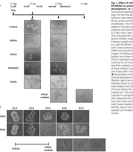
To examine the effect of ERK pathway inhibition on early cell division cycles, we added U0126 at the late two-cell stage. Rather surprisingly, the inhibition of the ERK pathway resulted in the arrest of cell division at the four-cell stage (Fig. 1A, U0126). At 2 days after U0126 addition, when control embryos entered the morula or blastocyst stage, U0126-treated embryos remained at the four-cell stage (Fig. 1A). An inactive analog of U0126, U0124, had no effect (Fig. 1A, U0124). Moreover, another MEK inhibitor, PD98059, also had the same effect as U0126, and the effects of PD98059 and U0126 were dose-dependent (data not shown). U0126 and PD98059 are known to inhibit not only the ERK1/2 pathway but also the ERK5 pathway (Kamakura et al., 1999; Mody et al., 2001). To see which pathway is important for progression passed the four-cell stage, we used PD184352, which selectively inhibits the ERK1/2 pathway (Mody et al., 2001; Squires et al., 2002; Tanimura et al., 2003). PD184352, like U0126, induced cell division arrest at the four-cell stage. The effect of PD184352 was also dose-dependent; the drug at 5 M, which does not inhibit the ERK5 pathway (Mody et al., 2001; Squires et al., 2002), significantly induced cell division arrest (Fig. 1A). Therefore, we conclude that the ERK1/2 pathway is required for progression of early cell division cycles. In addition, we found that the ERK1/2-pathway-inhibited embryos showed a
weakened adhesion between blastomeres, especially at later time points, as clearly seen in the photographs shown in Fig. 1B. To confirm this, we investigated the ability of one embryo to adhere to another embryo. When two-cell-stage embryos were placed very close to one another, control four- or eight-cell-stage embryos often aggregated into large aggregates within a day, but the ERK1/2-inhibited embryos (four-cell stage) remained unaggregated (data not shown), confirming the weakened adhesiveness in ERK1/2-inhibited blastomeres.
We then examined whether the inhibitor-induced developmental arrest was reversible or not. Late two-cell-stage embryos were treated with U0126 for 24 hours, and then U0126 was washed out and embryos were cultured in a drug-free medium. After the drug washout, embryos started to develop again and, 24 and 48 hours after the washout, they became morphologically normal eight-cell embryos and morula or blastocyst embryos, respectively (Fig. 1B). These results indicate that treatment with ERK pathway inhibitor induces a reversible developmental arrest during early cell division cycles.
To examine whether the ERK1/2 pathway is activated during mouse preimplantation development, embryos were stained with anti-phospho-ERK antibody and anti-phospho-ELK1 antibody. ELK1 is a well-known substrate of ERK1/2. The obtained immunofluorescence images showed that both phospho-ERK and phospho-ELK1 began to appear between the two-cell and four-cell stages, and remained until blastocyst stages (Fig. 2A). This result is in good agreement with previously reported data on embryonic day (E)2.5 and E3.5 embryos (Wang et al., 2004b). Treatment with U0126 for 1 hour markedly decreased the staining intensities of phospho-ERK1/2 and phospho-ELK1 (Fig. 2B). Furthermore, our kinase assay showed that the ability of the lysates obtained from four-cell-stage embryos to phosphorylate ELK1 in vitro was significantly reduced by U0126 treatment of the embryos. These results show that the ERK1/2 pathway is activated in mouse preimplantation development, and that U0126 is able to inhibit the ERK pathway in this system.
four-cell-D
E
V
E
LO
P
M
E
N
T
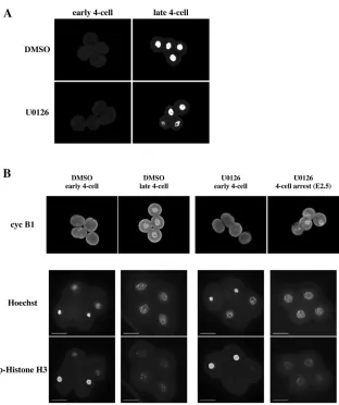
arrested embryos showed the same, dotted phospho-histone H3 staining pattern (Fig. 3B), suggesting that they did not enter M phase. Next, we performed Hoechst staining. In early four-cell or eight-cell-stage embryos, some blastomeres showed chromosome condensation (Fig. 3B, and data not shown). However, in U0126-treated, four-cell-arrested embryos, chromosome condensation was not observed (Fig. 3B). Taken together, these results demonstrate that U0126-induced developmental arrest at the four-cell-stage occurs at G2 arrest, just before M phase.
Next, we added U0126 to early two-cell-stage embryos to determine whether it induces arrest at the two-cell stage or the four-cell stage. As a result, the embryos did not proceed to the four-four-cell stage, and were arrested in the two-cell stage (Fig. 4A). Then, we performed the same experiments as above to determine a cell cycle
phase in which the U0126-treated, two-cell-stage embryos were arrested. The results from BrdU incorporation (Fig. 4B), and cyclin B1 and phospho-histone H3 staining (Fig. 4C), experiments demonstrate that the U0126-induced developmental arrest at the two-cell stage also occurs at the G2 phase, just before M phase. Therefore, it is likely that ERK activity is essential for cells to enter M phase in very early embryonic cell cycles before compaction during mouse preimplantation development.
[image:3.612.60.506.54.564.2]Because the ERK pathway is known to regulate gene expression, we examined the effect of inhibition of transcription on the early cell cycles. In agreement with a previous study with another inhibitor, ␣-amanitin (Clarke et al., 1992), treatment of late two-cell-stage embryos with the transcription inhibitor actinomycin D completely blocked development between the two-cell and four-cell
D
E
V
E
LO
P
M
E
N
T
stages (Fig. 5A). Therefore, normal cell division to the eight-cell stage requires de novo synthesis of mRNAs. To identify those genes whose expression levels are regulated by the ERK pathway during the early cell division stages, we performed the genome-wide analysis by using Affymetrix GeneChip oligonucleotide
[image:4.612.51.375.60.285.2]microarrays, which contain about 30,000 genes (about 45,000 probe sets). For this analysis, we collected embryos at six points, as follows: control embryos at day 1.5 (cont. 1.5, two-cell stage), day 2.5 (cont. 2.5, four- to eight-cell) and day 3.5 (cont. 3.5, morula to blastocyst); U0126-treated embryos at day 2.5 (U2.5, four-cell
Fig. 2. ERK and ELK1 are phosphorylated during mouse preimplantation development. (A) Phosphorylated ERK (p-ERK, upper) and phosphorylated ELK1 (p-ElK-1, lower) were detected using anti-phospho-ERK antibody and anti-phospho-ELK1 antibody, respectively. Embryos (from left to right) at the two-cell, four-cell, eight-four-cell, morula or blastocyst stages were fixed and stained. Fluorescence was viewed with a confocal microscope. (B) Phosphorylation of ERK or ELK1 with or without U0126. Four-cell (top) and two-cell (bottom) stage embryos were treated with U0126 for 1 hour, and then the embryos were fixed and stained with either phospho-ERK antibody (four cell) or anti-phospho-ELK1 antibody (two cell). Fluorescence was viewed with a confocal microscope.
[image:4.612.52.364.367.739.2]D
E
V
E
LO
P
M
E
N
T
arrested); and embryos released from the U0126-induced arrest were collected at day 3.5 (U3.5, eight-cell) and at day 4.5 (U4.5, morula to blastocyst), as shown in Fig. 5B. We identified 6863 probe sets whose expression levels in cont. 2.5 or cont. 3.5 were increased or decreased by more than threefold as compared to those in cont. 1.5 with statistical significance in replicate experiments (see Fig. 5C). These probe sets included genes encoding NANOG and
CDX2, which are known to be crucial for cell-lineage segregation during preimplantation development (Wang and Dey, 2006). Then, to analyze the gene expression program in the U0126-treated and released embryos, samples at six points were clustered according to their relative distances by using the above-mentioned 6863 probe sets (Fig. 5D). The obtained hierarchical clustering data showed that the gene expression profile in U3.5 embryos is most similar to
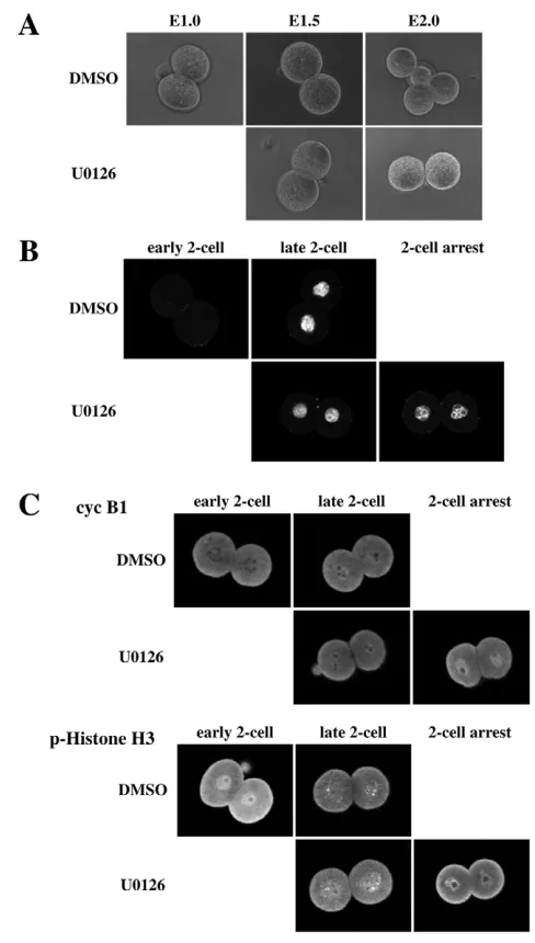
[image:5.612.49.293.152.582.2]Fig. 4. ERK inactivation in early two-cell-stage embryos induces developmental arrest in the G2 phase, just before M phase in the two-cell stage.(A) Early two-cell-stage embryos were treated with 20M U0126 or DMSO, and embryos were observed from E1.0 to E2.0. The zona pellucida was removed before inhibitor treatment. (B) BrdU incorporation into the nucleus was investigated. Early two-cell-stage embryos were cultured in the presence of BrdU and U0126 or BrdU and DMSO (control), fixed, and stained with anti-BrdU antibody. Fluorescence was viewed at the stages indicated with a confocal microscope. (C) Cyclin B1 accumulation in the nucleus and phosphorylation of histone H3 were examined. Early two-cell-stage embryos were cultured in the presence of U0126 or DMSO. Embryos were fixed and stained with anti-cyclin B1 or anti-phospho-histone H3 antibody. Fluorescence was viewed with a confocal microscope at the stages indicated.
Fig. 5. The transcriptional program during mouse
preimplantation development.(A) Actinomycin D completely blocked development. Two-cell-stage embryos were treated with actinomycin D (0.04 M or 0.4 M), and compared with control embryos (0 M) at 24 (E2.5) and 48 (E3.5) hours after treatment. (B) Schedule of inhibitor treatment and of the microarray experiment. Solid and broken lines indicate the duration of U0126 and DMSO (vehicle) treatment, respectively. The inhibitor was added at the late two-cell stage, and embryos were collected for microarray
[image:5.612.318.560.156.517.2]D
E
V
E
LO
P
M
E
N
T
that in cont. 3.5, although U3.5 embryos (at the eight-cell stage) and cont. 3.5 embryos (morula or blastocyst) are in different stages of development. Similarly, the gene expression profile in U2.5 embryos is most similar to that in cont. 2.5. These results show that most of the programmed changes of upregulated and downregulated gene expression during the four- to eight-cell stages proceeded normally in the four-cell stage-arrested embryos, and therefore suggest that the gene expression program does not necessarily parallel the stages of embryonic development during the early cell division stages. Moreover, the results show that the ERK pathway regulates only a portion of gene expression programs, and suggest that the ERK-regulated genes might be involved in the cell cycle arrest.
To identify genes whose expression is regulated by the ERK pathway, we first defined ERK-dependent genes as genes whose change in expression level was reduced by more than half after the 24 hours of U0126 treatment. In those genes whose expression level in cont. 2.5 was increased or decreased by more than threefold as compared to that in cont. 1.5, we identified 420 and 109 probe sets as ERK-dependent, upregulated and downregulated genes, respectively. In addition, there were a number of genes whose expression level was not significantly changed from cont. 1.5 to cont. 2.5, but was changed by ERK inactivation; 173 and 64 probe sets were increased and decreased, respectively, more than threefold by U0126 treatment. These 237 probe sets were the second type of dependent genes. Both the first and second types of ERK-dependent genes were analyzed with respect to their gene ontology
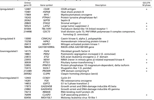
(GO-annotation) as given in NetAffyx (http://www.affymetrix.com/ analysis/index.affx). The ERK-dependent genes were assigned to GO biological process categories, and 30 genes were found to belong to the ‘cell cycle’ category (Table 1), of which ten genes belonged to the ‘M phase’ category (Table 1). Six out of ten genes were in the first type of ERK-dependent genes (downregulated). The concentration of the ‘M phase’ category genes to the ERK-dependent genes (downregulated) was statistically significant (P=0.00569). Although the concentration of the ‘M phase’ category genes in the dependent genes might result from ERK-inhibition-induced G2 arrest, it is also possible that ERK regulation of these ‘M phase’ genes would be important for progression to M phase in the early embryonic cell division stages.
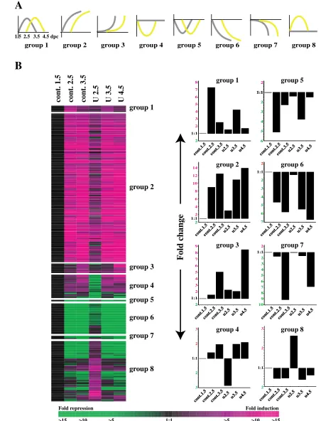
[image:6.612.52.569.383.720.2]To identify genes whose expression profiles closely parallel the stages of embryonic development when arrested in the four-cell G2 phase and released to resume development, we further analyzed the ERK-dependent genes. Thus, genes whose expression levels in cont. 1.5, cont. 2.5 and cont. 3.5 (Fig. 6A, gray) were similar to those in U2.5, U3.5 and U4.5, respectively (Fig. 6A, yellow), were classified into eight groups (Fig. 6A). In Fig. 6B, the number of genes in each group, the expression pattern of each gene (left) and the average expression profiles for genes (right) in groups 1-8 are shown (see Table S1 in the supplementary material for a complete list of genes in groups 1-8). GenMAPP/MAPPFinder was used to examine the biological context (Doniger et al., 2003) and, for each group, pathways with the permute p-value less than 0.01 were searched for. As a result, two pathways were found to have a significant coherence
Table 1. ERK-dependent genes that belong to the ‘cell cycle’ category
Entrez GO:279:
gene ID Gene symbol Description M phase
Upregulated I 12487 CD28 CD28 antigen +
15481 HSPA8 Heat shock protein 8
17869 MYC Myelocytomatosis oncogene
19243 PTP4A1 Protein tyrosine phosphatase 4a1
20362 SEPT8 Septin 8
20843 STAG2 Stromal antigen 2 +
50523 LATS2 Large tumor suppressor 2 +
81004 TBL1XR1 Transducin (beta)-like 1X-linked receptor 1 +
214498 CDC73 Vcell division cycle 73, PAF1/RNA polymerase II complex component,
homolog (S. cerevisiae)
Upregulated II 13000 CSNK2A2 Casein kinase II, alpha 2, polypeptide
15258 HIPK2 Homeodomain interacting protein kinase 2
26413 MAPK1 Mitogen activated protein kinase 1
98828 G431001I09Rik RIKEN cDNA G431001I09 gene
Downregulated I 14175 FGF4 Fibroblast growth factor 4
18861 PMS2 Postmeiotic segregation increased 2 (S. cerevisiae) +
23872 ETS2 E26 avian leukemia oncogene 2, 3⬘domain
23955 NEK4 NIMA (never in mitosis gene a)-related expressed kinase 4 +
30939 PTTG1 Pituitary tumor-transforming 1 +
53892 PPM1D Protein phosphatase 1D magnesium-dependent, delta isoform
72415 SGOL1 Shugoshin-like 1 (S. pombe) +
213389 PRDM9 VPR domain containing 9 +
269582 CLSPN Claspin homolog (Xenopus laevis)
Downregulated II 12443 CCND1 Cyclin D1
14281 FOS FBJ osteosarcoma oncogene
14853 GSPT2 G1 to S phase transition 2
17873 GADD45B Growth arrest and DNA-damage-inducible 45 beta
23882 GADD45G Growth arrest and DNA-damage-inducible 45 gamma
74213 RBM26 RNA binding motif protein 26
76499 CLASP2 CLIP associating protein 2 +
83456 MOV10L1 Moloney leukemia virus 10-like 1
D
E
V
E
LO
P
M
E
N
T
cont.1.5cont.2.5cont.3.5u2.5 u3.5 u4.5 cont.1.5cont.2.5cont.3.5u2.5 u3.5 u4.5
2
1:1
2 4 6 8 10 12 14
2
1:1
2 3 4 5 6 8 9
7
7 6 5 4 3 2
1:1
2
10 9 8 7 6 5 4 3 2
1:1
2
F
old change
2
1:1
2 3 4 5 6 7
8 group 5
group 2 group 6
group 3 group 7
group 4 group 8
6 5 4 3 2
1:1 1:1
2
cont.1.5cont.2.5cont.3.5u2.5 u3.5 u4.5 cont.1.5cont.2.5cont.3.5u2.5 u3.5 u4.5
cont.1.5cont.2.5cont.3.5u2.5 u3.5 u4.5 cont.1.5cont.2.5cont.3.5u2.5 u3.5 u4.5
group 3 group 4
group 2 group 5 group 6 group 7 group 8
cont.1.5cont.2.5cont.3.5u2.5 u3.5 u4.5
cont.1.5cont.2.5cont.3.5u2.5 u3.5 u4.5 cont.1.5cont.1.5cont.2.5cont.2.5cont.3.5cont.3.5u2.5 u3.5 u4.5u2.5 u3.5 u4.5
group 1
cont.1.5cont.2.5cont.3.5u2.5 u3.5 u4.5 cont.1.5cont.2.5cont.3.5u2.5 u3.5 u4.5
A
B
group 1
1.5 2.5 3.5 4.5 dpc
3 2
1:1
2 3
2
1:1
2 3
Fold repression Fold induction
>15 >10 >5 1:1 >5 >10 >15
cont. 1.5
cont. 2.5
cont. 3.5
U 2.5
U 3.5
U 4.5
group 2 group 1
group 3
group 4
group 5
group 6
group 7
group 8
cont.1.5cont.2.5cont.3.5u2.5 u3.5 u4.5 cont.1.5cont.2.5cont.3.5u2.5 u3.5 u4.5 cont.1.5cont.2.5cont.3.5u2.5 u3.5 u4.5
[image:7.612.56.508.61.653.2]cont.1.5cont.2.5cont.3.5u2.5 u3.5 u4.5
D
E
V
E
LO
P
M
E
N
T
indicator in group 2: one is Mm_mRNA_processing_binding_ Reactome (permute p-value=0.003) and the other is cell junction (permute p-value=0.006). The ‘cell junction’ pathway is intriguing, because our present result has shown that cell-cell adhesion is weakened in ERK-pathway-inhibited embryos, as described before. Then, we searched for genes that belong to the ‘cell junction’ category in all eight groups, and identified five genes, as shown in Fig. 7A. Because these five genes belong to group 2 or group 3, their expression is positively regulated by the ERK pathway. These genes encode products that participate in (i) tight junctions, which contribute to the epithelial barrier function by regulating the free diffusion of solutes between adjacent cells (CLDN4, OCLN) (Furuse and Tsukita, 2006); (ii) adherens junctions [ECAD (also known as CDH1 – Mouse Genome Informatics)], which mediate cell-cell adhesion, connect actin filaments to the cell surface and produce cytoskeleton-regulated cell communication (Takeichi, 1988); (iii) gap junctions [CX43 (also known as GJA1 – Mouse Genome Informatics)], which provide an intercellular communication pathway directly connecting adjacent cell cytoplasms (Evans et al., 2006); and (iv) desmosomes (DSG2), which connect intermediate filaments to the cell surface and mediate strong cell-cell adhesion (Kottke et al., 2006). The ERK-pathway-dependent upregulation of these genes might contribute to integrity and to strengthened cell-cell adhesion in four-cell to eight-cell-stage embryos. It is also possible that enhanced cell-cell adhesion could facilitate progression to the M phase during early cell division cycles (see below). Our analyses thus strongly suggest the importance of
cell adhesion in the early cell division stages before compaction, in addition to the established role of cadherin in compaction (Takeichi, 1988).
[image:8.612.54.372.378.742.2]These analyses suggested the possibility that inhibition of cell-cell adhesion, similar to inhibition of the ERK pathway, might induce cell cycle arrest in four-cell-stage embryos. To test this possibility, we added ECCD-1, the monoclonal antibody that inhibits Ca2+ -dependent cell-cell adhesion (Yoshida-Noro et al., 1984), to late two-cell-stage embryos. However, developmental arrest at the four-cell stage did not take place (data not shown). It is possible that inhibition of other types of cell-cell adhesion would also be needed to induce cell cycle arrest. However, we have found that ECCD-1 treatment potentiates the sensitivity of embryos to U0126 treatment. Thus, consistent with our previous results (Maekawa et al., 2005), U0126 addition in eight-cell-stage embryos did not significantly affect subsequent developmental processes (Fig. 7B). Similarly, ECCD-1 alone did not inhibit cell division or blastocyst formation when added to eight-cell stage embryos (Fig. 7B), although it did induce defects in compaction (data not shown). These observations are consistent with the previous report that embryos cultured in the presence of ECCD-1 remained uncompacted at the morula stage, but the morphology of the embryos became undistinguishable from that of control embryos at the blastocyst stage (Shirayoshi et al., 1983). However, when both U0126 and ECCD-1 were added, cell division arrest between the eight-cell to the 16-cell stages occurred (Fig. 7B). These results suggest that cadherin-mediated cell-cell adhesion should facilitate cell cycle progression.
D
E
V
E
LO
P
M
E
N
T
DISCUSSION
Preimplantation development involves a number of biologically significant events, such as compaction and blastocyst formation, which represent morphologically dynamic changes. Although both compaction and blastocyst formation have been well examined, molecular mechanisms regulating the early cell division stages following these events remain unclear. In this study, we have shown that the ERK pathway is activated in the early cell division stages during mouse preimplantation development, and has an essential role in the G2/M transition during the cell cycle progression of 2-cell to 8-2-cell-stage embryos. This role of ERK MAPK is different from the one in mammalian cultured cells, in which ERK activity is involved in the cell cycle progression from G0/G1 to S phase (Lewis et al., 1998; Pearson et al., 2001; Pouyssegur and Lenormand, 2003). In addition, because it is also known that the ERK pathway plays an important role in producing M phase arrest in unfertilized vertebrate eggs (Gotoh and Nishida, 1995; Brunet and Maro, 2005), our present finding suggests that the role of the ERK pathway might alter after fertilization. Elucidating a molecular basis for the different roles of ERK MAPK in cell cycle progression in different situations should be performed in future studies.
Our microarray experiments have identified a set of cell cycle-related genes whose expression levels are increased or decreased by the inhibition of the ERK pathway (Table 1). These genes would be good candidates for transcriptional targets of the ERK pathway, which could regulate the G2/M transition during early cell division cycles. Moreover, our microarray analysis has demonstrated that the expression programs of most genes do not parallel the developmental stages before compaction, and that the expression programs of a subset of genes, particularly adhesion-related genes, correlate well with the cell cycle progression and/or the developmental stages. Clarifying the biological significance of the loose parallelism between the gene expression programs and the developmental stages, and the regulatory mechanisms of the expression of adhesion molecules, will provide new insights into preimplantation development.
We thank members of our laboratory for their technical assistance and helpful discussion. This work was supported by grants from the Ministry of Education, Culture, Sports, Science and Technology of Japan (to E.N.).
Supplementary material
Supplementary material for this article is available at http://dev.biologists.org/cgi/content/full/134/15/2751/DC1
References
Brunet, S. and Maro, B. (2005). Cytoskeleton and cell cycle control during meiotic maturation of the mouse oocyte: integrating time and space. Reproduction130, 801-811.
Chang, L. and Karin, M.(2001). Mammalian MAP kinase signalling cascades. Nature410, 37-40.
Clarke, H. J., Oblin, C. and Bustin, M.(1992). Developmental regulation of chromatin composition during mouse embryogenesis: somatic histone H1 is first detectable at the 4-cell stage. Development115, 791-799.
Doniger, S. W., Salomonis, N., Dahlquist, K. D., Vranizan, K., Lawlor, S. C. and Conklin, B. R.(2003). MAPPFinder: using Gene Ontology and GenMAPP to create a global gene-expression profile from microarray data. Genome Biol. 4, R7.
Evans, W. H., De Vuyst, E. and Leybaert, L.(2006). The gap junction cellular
internet: connexin hemichannels enter the signalling limelight. Biochem. J. 397, 1-14.
Favata, M. F., Horiuchi, K. Y., Manos, E. J., Daulerio, A. J., Stradley, D. A., Feeser, W. S., Van Dyk, D. E., Pitts, W. J., Earl, R. A., Hobbs, F. et al.(1998). Identification of a novel inhibitor of mitogen-activated protein kinase kinase. J. Biol. Chem. 273, 18623-18632.
Furuse, M. and Tsukita, S.(2006). Claudins in occluding junctions of humans and flies. Trends Cell Biol. 16, 181-188.
Gotoh, Y. and Nishida, E.(1995). The MAP kinase cascade: its role in Xenopus oocytes, eggs and embryos. Prog. Cell Cycle Res.1, 287-297.
Hamatani, T., Carter, M. G., Sharov, A. A. and Ko, M. S.(2004). Dynamics of global gene expression changes during mouse preimplantation development. Dev. Cell6, 117-131.
Kamakura, S., Moriguchi, T. and Nishida, E.(1999). Activation of the protein kinase ERK5/BMK1 by receptor tyrosine kinases. Identification and
characterization of a signaling pathway to the nucleus. J. Biol. Chem. 274, 26563-26571.
Kottke, M. D., Delva, E. and Kowalczyk, A. P.(2006). The desmosome: cell science lessons from human diseases. J. Cell Sci. 119, 797-806.
Lewis, T. S., Shapiro, P. S. and Ahn, N. G.(1998). Signal transduction through MAP kinase cascades. Adv. Cancer Res. 74, 49-139.
Maekawa, M., Yamamoto, T., Tanoue, T., Yuasa, Y., Chisaka, O. and Nishida, E.(2005). Requirement of the MAP kinase signaling pathways for mouse preimplantation development. Development132, 1773-1783.
Mody, N., Leitch, J., Armstrong, C., Dixon, J. and Cohen, P.(2001). Effects of MAP kinase cascade inhibitors on the MKK5/ERK5 pathway. FEBS Lett. 502, 21-24.
Nagy, A., Gertsentein, M., Vintersten, K. and Behringer, R.(2003). Manipulating the Mouse Embryo. New York: Cold Spring Harbor Laboratory Press.
Natale, D. R., Paliga, A. J. M., Beier, F., D’Souza, S. L. A. and Watson, A. J.
(2004). P38 MAPK signaling during murine preimplantation development. Dev. Biol. 268, 76-88.
Nishida, E. and Gotoh, Y.(1993). The MAP kinase cascade is essential for diverse signal transduction pathways. Trends Biochem. Sci. 18, 128-131.
Pearson, G., Robinson, F., Beers Gibson, T., Xu, B. E., Karandikar, M., Berman, K. and Cobb, M. H.(2001). Mitogen-activated protein (MAP) kinase pathways: regulation and physiological functions. Endocr. Rev. 22, 153-183.
Pouyssegur, J. and Lenormand, P.(2003). Fidelity and spatio-temporal control in MAP kinase (ERKs) signalling. Eur. J. Biochem. 270, 3291-3299.
Robinson, M. J. and Cobb, M. H.(1997). Mitogen-activated protein kinase pathways. Curr. Opin. Cell Biol. 9, 180-186.
Shirayoshi, Y., Okada, T. S. and Takeichi, M.(1983). The calcium-dependent cell-cell adhesion system regulates inner cell mass formation and cell surface polarization in early mouse development. Cell35, 631-638.
Squires, M. S., Nixon, P. M. and Cook, S. J.(2002). Cell-cycle arrest by PD184352 requires inhibition of extracellular signal-regulated kinases (ERK) 1/2 but not ERK5/BMK1. Biochem. J. 366, 673-680.
Sturgill, T. W. and Wu, J.(1991). Recent progress in characterization of protein kinase cascades for phosphorylation of ribosomal protein S6. Biochim. Biophys. Acta1092, 350-357.
Takeichi, M.(1988). The cadherins: cell-cell adhesion molecules controlling animal morphogenesis. Development102, 639-655.
Tanimura, S., Asato, K., Fujishiro, S. H. and Kohno, M.(2003). Specific blockade of the ERK pathway inhibits the invasiveness of tumor cells: down-regulation of matrix metalloproteinase-3/-9/-14 and CD44. Biochem. Biophys. Res. Commun. 304, 801-806.
Wang, H. and Dey, S. K.(2006). Roadmap to embryo implantation: clues from mouse models. Nat. Rev. Genet. 7, 185-199.
Wang, Q. T., Piotrowska, K., Ciemerych, M. A., Milenkovic, L., Scott, M. P., Davis, R. W. and Zernicka-Goetz, M.(2004a). A genome-wide study of gene activity reveals developmental signaling pathways in the preimplantation mouse embryo. Dev. Cell6, 133-144.
Wang, Y., Wang, F., Sun, T., Trostinskaia, A., Wygle, D., Puscheck, E. and Rappolee, D. A.(2004b). Entire mitogen activated protein kinase (MAPK) pathway is present in preimplantation mouse embryos. Dev. Dyn. 231, 72-87.