Laurence J. Sages' Edwin D. Cacayorin' George R. Petro' Tarakad S. Ramachandran2
Received June 18, 1987; accepted after revision November 26,1987.
Presented at the annual meeting of the American Society of Neuroradiology, New York, May 1987.
1 Department of Radiology, Neuroradiology Sec-tion, SUNY Health Science Center at Syracuse, 750 E. Adams St., Syracuse, NY 13210. Address reprint requests to L. J. Soges.
21000 E. Genesee St., Syracuse, NY 13210.
AJNR 9:425-429, May/June 1988 0195-6108/88/0903-0425
II:! American Society of Neuroradiology
Migraine:
Evaluation
by
MR
Twenty-four patients clinically diagnosed as having migraine (17 of the classic or common type and seven of the complicated type) were evaluated on a O.S-T or O.6-T superconductive MR imaging unit with the objective of detecting associated parenchymal lesions. Thirteen (54%) of the patients had normal MR studies. Eleven (46%) of the patients (seven with classic or common and four with complicated migraine) showed well-defined lesions with prolonged T2 signal intenSity. The lesions associated with classic migraine were focal and predominantly distributed in the periventricular white maHer, bilateral in four and unilateral in three. In the group with complicated migraine, larger cortical abnormalities similar to infarcts were seen in three patients and multiple bilateral focal white maHer lesions were seen in one. Almost all the lesions were evident only on T2-weighted studies; a few exhibited hypointense signal intensity on T1-weighted studies. The focal periventricular white maHer lesions were not necessarily associated with neurologic deficits, but the cortical lesions were.
Our study indicates that parenchymal changes are frequently associated with migraine and that MR may well be the screening and diagnostic method of choice for their detection and evaluation.
Headache is a very common problem, affecting the majority of individuals in Western cultures. Of these, approximately 15-20% suffer from migraine type headaches [1). Classic migraine, the most common type, is clinically recognized as a form of severe, usually unilateral recurrent throbbing headache, which is preceded by visual symptoms (the "aura"), and responds to ergotamine [2]. Similar headaches without visual symptoms are referred to as common migraine. Migraine associated with neurologic deficits, which are usually transient, is referred to as complicated migraine, a much less common entity [2).
The reported findings in migraine from numerous imaging techniques (CT, angiography, pOSitron emission tomography) have been variable (see Discussion). MR imaging has emerged as a diagnostic tool with superb sensitivity in detecting early and subtle alterations in brain parenchyma, prompting us to use this method to evaluate patients with migraine.
Materials and Methods
Twenty-four patients, eight males and sixteen females, ages 15-55 years, clinically diagnosed as having migraine, had MR studies of the brain on a Picker 1.0-T superconductive MR unit' operating at 0.5 T (21 patients) or a Technicare 0.6-T superconductive MR unitt (three patients). One patient was scanned on both units. Seventeen had clinically determined classic or common migraine, and seven had complicated migraine. All patients had MR studies of the brain using 2500/100 (TRITE) T2-weighted 10-mm spin-echo images in the axial and coronal planes and 600/40 T1-weighted 5-mm spin-echo images in the sagittal plane. Images had 256 x 256 matrixes and were obtained from a 30-cm field of view.
One patient was scanned within 24 hr of onset of symptoms of dysphasia and had follow -up scans 3 weeks and 4 months later; the other patients were studied at random. The
426 SOGES ET AL. AJNR:9, May/June 1988
location and signal intensity of lesions were noted on both T1-and T2-weighted studies. Attempts were also made to correlate the parenchymal changes neurologically.
Results
Seven of the 17 patients with common or classic migraine and four of the seven patients with complicated migraine had abnormal MR findings (see Table 1). The findings consisted of focal regions of increased signal intensity on T2-weighted studies (Fig. 1). In the patients with classic or common mi-graine, the lesions were seen exclusively within white matter (unilaterally in three and bilaterally in four), while three of the patients with complicated migraine had lesions involving the cerebral or cerebellar cortex unilaterally. In a patient who had an MR within 24 hr of onset of dysphasia, T2-weighted images showed regions of increased signal intensity in the left cen-trum semiovale and insular cortex, as well as small foci of
TABLE 1: Distribution of Patients by Age and Gender
Under 40 years old
Over 40 years old
Total
• Neurologic deficits.
A
Positive Study Age 28 31 38 38 39 41 43 44 44 47 51 11 Gender F F" F F Ma M F" F F F" F Negative StudyAge Gender
18 M
20 M"
27 F
27 F
30 F
32 F
34 F
35 M
36 F
40 F"
42 M"
44 M
55 M
13
B
increased signal intensity in the left cerebral white matter (Fig. 2). CT was normal. This patient was treated with nifedipine and recovered. A follow-up MR study obtained 3 weeks later showed an increase in the size of the lesions in the insular cortex and frontal operculum despite the resolution of the neurologic deficits. Of the other six patients with complicated migraine, one had multiple tiny foci of increased signal in the cerebral white matter bilaterally, one had increased signal in the cerebral cortex consistent with an infarct (Fig. 3), and one had involvement of the cerebellar cortex (Fig. 4). The remain-ing three had normal MR findremain-ings. The cortical abnormalities appropriately correlated with the patients' neurologic deficits, which all resolved within a few days after the migrainous attacks. The smaller white matter lesions seen in patients with classic or common migraine could not be accounted for neurologically.
Discussion
Complex and variable etiologic considerations and obser-vations have evolved from numerous studies in the past on patients with migraine. The most widely accepted hypothetical basis for migraine headache is that initial vasoconstriction of extra- and intracranial arteries takes place, leading to ischemia (clinically manifested by the "aura" and neurologic deficits), followed by reactive vasodilatation and hyperemia resulting in the development of headache [3-5]. Numerous factors are thought to account for the vascular events, including the presence of circulating vasoactive substances such as sero-tonin [6] and histamine [7], sensitivity to ingested substances such as nitrites [8], and inherent abnormalities of the auto-nomic nervous system in patients with migraine [9].
Investigators using positron emission tomography have demonstrated decreased regional glucose metabolism in the preheadache phase, which rose but remained below resting levels during the headache phase [10]. Oxygen inhalation studies have revealed decreased blood flow and diminished oxygen extraction in regions of migraine infarction as dem-onstrated on CT [11]. Xenon-133 blood studies have sub -stantiated the occurrence of decreased cortical blood flow
[image:2.612.54.299.322.513.2] [image:2.612.54.393.550.731.2]A and B, MR study performed within 24 hr of the attack shows focal regions of increased sig-nal in left centrum semiovale (arrow, A), frontal white matter, and insular cortex (arrows, B). There is also diffusely increased signal involving
the sylvian and frontal cortex (arrowheads, B).
e, Follow-up MR image 3 weeks later shows
that the abnormality in the sylvian and frontal cortex is more pronounced (arrowheads) despite clinical resolution of symptoms.
D, 4 months later, cortical abnormality is still
faintly visible in sylvian region (arrow). Lesion in centrum semiovale was unchanged.
Fig. 3.-A and B, 43-year-old woman with
classic migraine who presented with aphasia
and right hemiparesis. MR image obtained
sev-eral days later demonstrates increased signal in left cerebral cortex (arrows, A and B) centered around insula (arrowheads, A).
A
B
c
D
[image:3.612.226.560.49.468.2] [image:3.612.226.559.502.729.2]428 SOGES ET AL. AJNR:9, May/June 1988
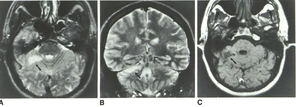
Fig. 4.-39-year-old man who had several episodes of severe headache associated with right-sided weakness, ataxia, and slurred speech. He was
diagnosed as having vertebrobasilar migraine.
A and B, MR obtained several months after last episode shows abnormally increased signal in right cerebellar hemisphere on T2-weighted images
(arrow, A and B).
C, T1-weighted image shows abnormality as a region of decreased signal (arrow).
and postischemic hyperemia with increased cerebral blood flow on the symptomatic side in classic migraine [12, 13]. Angiography has occasionally demonstrated vascular spasm or dilatation, but in most cases has been normal [14-16].
The frequency of parenchymal abnormalities associated
with migraine is indeterminate. Reported CT observations on patients with migraine have ranged from all normal in a large
series of 435 patients [17] to findings of cerebral edema and
infarctions [14, 16, 18, 19]. The most consistent finding was
an increased incidence of atrophy [20-22].
In our series, MR showed parenchymal abnormalities in
41 % of patients with classic or common migraine and in 57%
of patients with complicated migraine. These consisted mainly
of focal lesions with prolonged T2 signal intensity that were
distributed exclusively in white matter in those with common
or classic migraine. These small lesions were not associated with overt neurologic dysfunctions. Cortical lesions
resem-bling infarcts were seen in some patients with complicated
migraine in regions appropriately correlating with the patients'
neurologic deficits.
Any process that increases the water content of a tissue,
thereby prolonging its T2 relaxation time, will result in in-creased signal intensity in the affected region on MR images
with long TR and TE (T2-weighted) pulse sequences. In white
matter, increased water content may be due to either loss of
the hydrophobic myelin (demyelination) or increased interstitial
water (edema, hydrocephalus) [23].
Patchy foci of increased signal intensity in white matter is a fairly common normal finding in the elderly, in whom an incidence of 20-30% has been reported [24]. However, they are rarely found in normal individuals under 45 years old [25, 26]. These lesions have been attributed to ischemic demyeli-nation and lacunar infarcts [25-28]. Kirkpatrick and Hayman [29] examined pathologic preparations of 15 brains of clinically healthy subjects ages 52-72 years to determine the
fre-quency and nature of white matter lesions. They found white
matter lesions in 12 (80%) of the 15. These included atrophic
periventricular demyelination in eight, two white matter in-farcts, four vascular malformations (three telangiectases and one capillary hemangioma), and three small ventricular diver-ticula.
The white matter lesions encountered in patients with mi-graine resemble those of multiple sclerosis and the periven-tricular white matter lesions of small vessel atherosclerotic disease (subcortical arteriosclerotic encephalopathy). How-ever, the dominant clinical feature in our patient population is
headache, which is not described as a common presenting
manifestation of multiple sclerosis [30] and arteriosclerotic cerebrovascular disease [28, 31, 32]. Furthermore, even
lim-iting our observations to patients under 40 years old to reduce
the overlap with age-related white matter lesions, we still find an incidence of 27% in patients with the classic or common
type of migraine and 67% for those with complicated migraine.
We could not offer histologic correlation for the visualized
lesions nor can we infer their cause. However, our study
demonstrated a high frequency of white matter and cortical
changes in patients with migraine and indicates that MR may
well be the diagnostic technique of choice for detecting them ..
REFERENCES
1. Waters WE, O'Connor PJ. Prevalence of migraine. J Neurol Neurosurg
Psychiatry 1975;38:613-616
2. Rosenberg RN, ed. The clinical neurosciences. New York: Churchill living-stone. 1983: 1:382-1:403
3. Blau IN. Migraine: a vasomotor instability of the meningeal circulation.
Lancet 1978;2:1136-1139
4. Simard D, Paulson OB. Cerebral vasomotor paralysis during migraine attack. Arch Neuro/1973;29:207-209
[image:4.612.57.559.85.267.2]6. Somerville BW. Platelet-bound and free serotonin levels in jugular and forearm venous blood during migraine. Neurology (Minneap) 1976;26:
41-45
7. Anthony M, Lance JW. Migraine neuralgia-blood histamine levels and clinical response to H, and H2 receptor blockade. In: Greene R, ed. Current concepts in migraine research. New York: Raven, 1978: 149-151
8. Henderson WR, Raskin NH. "Hot-dog" headache: individual susceptibility
to nitrite. Lancet 1972;2: 1162-1163
9. Morley S. Migraine: a generalized vasomotor dysfunction? A critical review of evidence. Headache 1977;17:71-74
10. Sachs H, Russel JAG, Christman DR. Positron emission tomographic
studies on induced migraine. Lancet 1984;25:465 (Itr)
11. Bousser MG, Baron JC, Iba-Zizen MT. Migrainous cerebral infarction: a
tomographic study of cerebral blood flow and oxygen extraction fraction with the oxygen-15 inhalation techniques. Stroke 1980;11 :145-153
12. Lauritzen M, Olesen J. Regional cerebral blood flow during migraine attacks by xenon-133 inhalation and emission tomography. Brain 1984;107:
447-461
13. Fumihiko S, Meyer JS. Regional cerebral hemodynamics during migraine
and cluster headaches measured by the '33Xe inhalation method. Head
-ache 1978;18:122-132
14. Dorfmann LJ, Marshall WH, Engmann DR. Cerebral infarction and migraine:
clinical and radiologic correlations. Neurology 1979;29: 317 -322
15. Masuzawa T, Shinoda S, Furuse M. Cerebral angiographic changes on examination of a patient with migraine. Neuroradiology 1983;24:277-281
16. Pereira Monteiro JM, Leite Carmeiro A, Bastos Lima AT. Migraine and cerebral infarction: three case studies. Headache 1985;25:429-433
17. Cuetter AC, Aita JF. CT scanning in classic migraine. Headache
1983;23:195 (Itr)
18. Mathews NT, Meyer JS, Welch KMA. Abnormal CT scans in migraine.
Headache 1977;16:272-279
19. Bousser MG, Baron JC, Chiras J. Ischemic strokes and migraine.
Neuro-radiology 1985;27:583-587
20. Sargeant JD, Lawson RC, Sol bach P. Use of CT scans in an outpatient
headache population: an evaluation. Headache 1979;19:388-390
21. du Boulay GH, Ruiz JS, Rose FC. CT changes associated with migraine.
AJNR 1983;4:472-473
22. Sargeant JD, Lawson RC. Computered tomographic scans for headache.
JAMA 1980;244:133 (Itr)
23. Zimmerman RD, Fleming CA, Lee BCP, St.-Louis LA, Deck MD. Periven -tricular hyperintensity as seen by magnetic resonance: prevalence and significance. AJNR 1986;7: 13-20
24. Bradley WG, Waluck V, Brant-Zawadzki M, Yadley RA, Wycoff RR. Patchy,
periventricular white matter lesions in the elderly: common observation
during NMR imaging. Noninvasive Med Imaging 1984;1 :35-41
25. Ajax EG, de Leon MJ, Kalmin A, Rosner L, Goodgold A, Chase N.
Leucoencephalopathy in normal and pathological aging: 2. MRI of brain
lucencies. AJNR 1986;7: 567 -570
26. Brant-Zawadzki M, Fein G, VanDyke C, Kiernan R, Davenport L, de Groot
J. MRI imaging of the aging brain: patchy white matter lesions and dementia. AJNR 1985;6:675-682
27. Gerard G, Weisberg L. MRI peri ventricular lesions in adults. Neurology
1986;36:998-1001
28. Roman GC. Senile dementia of the Binswanger type. JAMA
1987;258: 1782-1788
29. Kirkpatrick JB, Hayman LA. White matter lesions in MRI imaging of clinically
healthy brains of elderly subjects: possible pathological basis. Radiology
1987;162:509-511
30. Rosenberg RN, ed. The clinical neurosciences. New York: Churchill livi ng-stone, 1983:1:618-1:662
31. Caplan LR, Schoene WC. Clinical features of subcortical arteriosclerotic
encephalopathy (Binswanger disease). Neurology 1978;28: 1206-1215
32. Babikian V, Ropper AH. Binswanger's disease. a review. Stroke