The following handle holds various files of this Leiden University dissertation:
http://hdl.handle.net/1887/61147
Author: Meinders, I.H.
Migraine and brain changes
© copyright Inge Meinders, Maastricht 2018
Printing: Datawyse | Universitaire Pers Maastricht
ISBN 978 94 6159 792 2
UNIVERSITAIRE
PERS MAASTRICHT
U
P
Migraine and brain changes
PROEFSCHRIFT
ter verkrijging van
de graad van Doctor aan de Universiteit Leiden,
op gezag van Rector Magnificus prof.mr. C.J.J.M. Stolker,
volgens besluit van het College voor Promoties
te verdedigen op donderdag 15 februari 2018
klokke 13:45 uur
door
Inge Hester Meinders
PROMOTORES
Prof. dr. M.A. van Buchem
Prof. dr. M.D. Ferrari
COPROMOTOR
Dr. M.C. Kruit
LEDEN PROMOTIECOMMISSIE
Dr. G.M. Terwindt
Prof. dr. A. de Roos
Dr. L.J. Launer, Laboratory of Epidemiology, Demography and Biometry, NIH, Bethesda, MD
Prof. C.I. de Zeeuw, Department of Neuroscience, Erasmus MC Rotterdam, the Netherlands
Dr. F.R. Rosendaal
Contents
Chapter 1 General outline and introduction
7
Chapter 2 Structural brain changes in migraine
15
Chapter 3 Iron accumulation in migraine
39
Chapter 4 Iron in deep brain nuclei in migraine? CAMERA follow-up MRI
findings
49
Chapter 5 Volumetric brain changes in migraineurs from the general
population
61
Chapter 6 Systemic right-to-left shunts, ischemic brain lesions, and
persistent migraine activity
77
Chapter 7 Summary
93
Chapter 8 Nederlandse samenvatting en conclusies
97
Chapter 9 Curriculum Vitae
101
Chapter
1
This thesis reports on brain changes in migraine and its possible causes and
conse-quences.
MIGRAINE
Migraine is a common, chronic, multifactorial neurovascular brain disorder, affecting
15% of adults. Migraine is the 3rd most common and 7th most disabling disease in the
world
1, and is responsible for the highest socio-economic burden of any brain disorders
in Europe
1, 2. It is characterized by recurrent attacks of disabling headache and
associat-ed features of autonomic nervous system dysfunction (migraine without aura, MO). Up
to one third of patients also have neurological aura symptoms (migraine with aura,
MA).
3, 4For a long time, migraine was believed to be just an episodic disorder without
permanent effects on health, other than being disabling during the attack. However, in
the last three decades, associations have been reported between migraine and
cere-brovascular diseases as well as cardiovascular diseases.
MIGRAINE AND BRAIN CHANGES
Contrary to being just a harmless condition, in the past decades it has become clear that
suffering from migraine may have permanent negative consequences to the brain.
Ac-cumulating evidence shows that migraine is linked to the risk of having migrainous
in-farction, ischemic and hemorrhagic stroke, and white matter changes in the brain.
Migrainous infarction, defined as an ischemic stroke developing during migraine
with aura, is a rare complication of migraine. In this situation a migraine attack and the
occurrence of brain damage are directly linked, which suggests a shared
pathophysio-logical pathway. But, although this phenomenon has already first been described long
ago, the exact pathophysiology is still not yet fully understood.
5, 6An increased risk of clinical ischemic stroke in migraine patients, irrespective of
mi-graine attacks, has been shown by several epidemiological studies. This increased risk
notably affects younger women with migraine, in particular those with higher attack
frequency and MA.
7-9In addition to clinical infarction, silent cerebral infarcts are also more prevalent
among migraineurs as was shown by the CAMERA-1 study (the baseline part of the
CAMERA-study).
10Silent infarcts are defined by the presence of a brain parenchymal
defect of vascular origin, confirmed by CT or MRI, in the absence of neurological
symp-toms and signs and a history of clinical stroke or TIA.
mi-graine diagnosis, etc.) and presented conflicting results, leading to difficulties in
inter-pretation.
13The CAMERA-1 study was designed to overcome these problems, and was
the first study that provided clear answers. The study showed that migraineurs from the
general population were indeed at increased risk of posterior circulation territory
in-farcts, and that the higher risk was most pronounced among migraine with aura
pa-tients with a higher attack frequency. Further, women with migraine were found to be
at significantly increased risk of deep (not periventricular) WMHs. This increased risk
was not explained by other cardiovascular risk factors. Similarly, infratentorial
hyperin-tense lesions (IHLs) were found to be more prevalent among migraineurs.
10, 14-16These
findings were subsequently confirmed by other groups
17-19The CAMERA-1 study also demonstrated that migraine patients have increased iron
deposition in multiple deep brain nuclei in line with clinic-based studies
20-25. The
under-lying mechanisms, however, remained elusive.
Because the CAMERA-1 study had a cross-sectional design, it was not possible to
draw any conclusion on a possible causal relationship between migraine attacks and
brain changes. Therefore, a follow-up study was needed to assess the development of
new brain changes or progression of existing brain changes over time.
In the present thesis I describe studies evaluating the incidence and evolution of
brain changes and associated migraine features in the CAMERA-cohort over a period of
9 years. In addition, possible causes and functional consequences were assessed.
In Chapter 2, I describe a study assessing whether migraineurs have a higher
inci-dence and greater volume-increase of WMHs, IHLs and infarcts over time compared to
a non-migraine control group. We also assessed possible cognitive consequences of
(progressive) WMLs. In chapters 3 and 4 we describe follow-up study on iron
accumula-tion in the basal ganglia of migraineurs. In chapter 5 we compared other types of
struc-tural brain changes between migraineurs and non-migraine controls by evaluating
over-all and regional gray and white matter structure using state-of-the-art voxel based
mor-phometry analyses.
POSSIBLE CAUSES
Now that we know that migraine is associated with several types of brain changes, it is
important to explore possible causes and consequences.
In the last two decades, several clinic based case-control studies demonstrated a
two to threefold increased prevalence of RLS in migraine with aura patients, as
meas-ured using Transcranial Doppler with air contrast.
27, 28A recent review reported that
PFO was more prevalent in migraine with aura patients (pooled OR 2.54; 95%CI
2.01-3.08)
29, but a population based study evaluating the presence of RLS in migraineurs and
non-migraine controls of the general population was lacking up to that moment.
Therefore, I describe in Chapter 6 the prevalence of RLS in migraineurs and controls
of the population-based CAMERA-cohort. RLS has been linked to increased risk of stroke
in several studies, especially among younger persons.
30In addition, among young
is-chemic stroke patients, those with a RLS are believed to be at higher risk of having
mi-graine with aura.
31It has been suggested that emboli, passing from the venous systemic
circulation through a RLS to the systemic arterial circulation may both trigger migraine
aura attacks and may cause (sub)clinical ischemic infarcts, thus providing a possible
explanation for a link between migraine (aura), RLS and stroke. Supporting evidence for
this theory was found in a recent study, showing that injecting emboli into the carotid
artery of mice caused cortical spreading depression, linking emboli to migraine aura.
32In chapter 6 I also describe the association in the CAMERA-cohort between RLS,
ischem-ic brain lesions and persisting migraine activity.
POSSIBLE CONSEQUENCES
It is of great importance to migraine patients to know if brain changes might have
tional consequences. In the study described in chapter 2, we assessed cognitive
func-tion in migraineurs with and without brain lesions.
CEREBELLAR FUNCTION
Cognitive function
Clearly, the diagnosis of migraine is not as harmless as was thought before. As described
above, migraine is linked to the presence and progression of white matter
hyperintensi-ties. In general, in the elderly, WMH have been linked to worse cognitive performance
38-41. Moreover, several papers have described cognitive dysfunction among migraineurs
42-49. Migraine patients often complain about cognitive difficulties during or directly after
their migraine attacks
46, 50In one of our own studies, migraineurs had no impaired
at-tentional or working-memory functioning in the two days after an attack. They did,
however, show impairments in the processing of global visual features compared with
controls, both between and immediately after an attack.
51Little is known about the effect of WMH on cognitive function in migraineurs. Recently,
the Epidemiology of Vascular Aging study has evaluated this complex relation and
demonstrated no evidence that migraine in itself, or in combination with structural
brain lesions, results in cognitive impairment.
17Chapter 2 of this thesis describes
cogni-tive functioning of the CAMERA-participants with and without migraine and whether
high WMH load might be associated with reduced cognitive performance.
AIM OF THIS THESIS
REFERENCES
1. Steiner TJ, Stovner LJ, Birbeck GL. Migraine: the seventh disabler. J Headache Pain 2013;14:1.
2. Lipton RB, Bigal ME, Diamond M, Freitag F, Reed ML, Stewart WF. Migraine prevalence, disease burden, and the need for preventive therapy. Neurology 2007;68:343-349.
3. Classification and diagnostic criteria for headache disorders, cranial neuralgias and facial pain. Headache Classification Committee of the International Headache Society. Cephalalgia 1988;8 Suppl 7:1-96. 4. Ferrari MD. Migraine. Lancet 1998;351:1043-1051.
5. Laurell K, Lundstrom E. Migrainous infarction: aspects on risk factors and therapy. Curr Pain Headache Rep 2012;16:255-260.
6. Elliott D. Migraine and stroke: current perspectives. Neurol Res 2008;30:801-812.
7. Etminan M, Takkouche B, Isorna FC, Samii A. Risk of ischaemic stroke in people with migraine: systematic review and meta-analysis of observational studies. Bmj 2005;330:63.
8. Chang CL, Donaghy M, Poulter N. Migraine and stroke in young women: case-control study. The World Health Organisation Collaborative Study of Cardiovascular Disease and Steroid Hormone Contraception. Bmj 1999;318:13-18.
9. Tzourio C, Iglesias S, Hubert JB, et al. Migraine and risk of ischaemic stroke: a case-control study. Bmj 1993;307:289-292.
10. Kruit MC, van Buchem MA, Hofman PA, et al. Migraine as a risk factor for subclinical brain lesions. Jama 2004;291:427-434.
11. Kaplan RD, Solomon GD, Diamond S, Freitag FG. The role of MRI in the evaluation of a migraine population: preliminary data. Headache 1987;27:315-318.
12. Soges LJ, Cacayorin ED, Petro GR, Ramachandran TS. Migraine: evaluation by MR. Ajnr: American Journal of Neuroradiology 1988;9:425-429.
13. Palm-Meinders ea. Migraine and brain lesions. In: Borsook, ed. The migraine brain. New York2011: 123-137. 14. Kruit MC, Launer LJ, Ferrari MD, van Buchem MA. Brain stem and cerebellar hyperintense lesions in
migraine. Stroke 2006;37:1109-1112.
15. Kruit MC, van Buchem MA, Launer LJ, Terwindt GM, Ferrari MD. Migraine is associated with an increased risk of deep white matter lesions, subclinical posterior circulation infarcts and brain iron accumulation: the population-based MRI CAMERA study. Cephalalgia 2010;30:129-136.
16. Kruit MC, Launer LJ, van Buchem MA, Terwindt GM, Ferrari MD. MRI findings in migraine. RevNeurol(Paris) 2005;161:661-665.
17. Kurth T, Mohamed S, Maillard P, et al. Headache, migraine, and structural brain lesions and function: population based Epidemiology of Vascular Ageing-MRI study. Bmj 2011;342:c7357.
18. Swartz RH, Kern RZ. Migraine is associated with magnetic resonance imaging white matter abnormalities: a meta-analysis. ArchNeurol 2004;61:1366-1368.
19. Scher AI, Gudmundsson LS, Sigurdsson S, et al. Migraine headache in middle age and late-life brain infarcts. Jama 2009;301:2563-2570.
20. Kruit MC, van Buchem MA, Overbosch J, Ferrari MD, Launer LJ. Iron deposits in migraine: Red nucleus and putamen involved? Cephalalgia 2002;22:571-571.
21. Kruit MC, Launer LJ, Overbosch J, van Buchem MA, Ferrari MD. Iron accumulation in deep brain nuclei in migraine: a population-based magnetic resonance imaging study. Cephalalgia 2009;29:351-359. 22. Welch KM. Iron in the migraine brain; a resilient hypothesis. Cephalalgia 2008.
23. Welch KM, Nagesh V, Aurora SK, Gelman N. Periaqueductal gray matter dysfunction in migraine: cause or the burden of illness? Headache 2001;41:629-637.
24. Tepper SJ, Lowe MJ, Beall E, et al. Iron Deposition in Pain-Regulatory Nuclei in Episodic Migraine and Chronic Daily Headache by MRI. Headache 2011.
26. Sarisoy S, Aydin OF, Sungur M, et al. The relationship between migraine and right-to-left shunt in children. Eur J Pediatr 2011;170:365-370.
27. Anzola GP, Magoni M, Guindani M, Rozzini L, Dalla VG. Potential source of cerebral embolism in migraine with aura: a transcranial Doppler study. Neurology 1999;52:1622-1625.
28. Tembl J, Lago A, Sevilla T, Solis P, Vilchez J. Migraine, patent foramen ovale and migraine triggers. J Headache Pain 2007;8:7-12.
29. Schwedt TJ, Demaerschalk BM, Dodick DW. Patent foramen ovale and migraine: a quantitative systematic review. Cephalalgia 2008;28:531-540.
30. Serena J, Jimenez-Nieto M, Silva Y, Castellanos M. Patent foramen ovale in cerebral infarction. Curr Cardiol Rev 2010;6:162-174.
31. Pezzini A, Grassi M, Lodigiani C, et al. Predictors of migraine subtypes in young adults with ischemic stroke: the italian project on stroke in young adults. Stroke 2011;42:17-21.
32. Nozari A, Dilekoz E, Sukhotinsky I, et al. Microemboli may link spreading depression, migraine aura, and patent foramen ovale. AnnNeurol 2010;67:221-229.
33. Sandor PS, Mascia A, Seidel L, De P, V, Schoenen J. Subclinical cerebellar impairment in the common types of migraine: a three-dimensional analysis of reaching movements. AnnNeurol 2001;49:668-672. 34. Harno H, Hirvonen T, Kaunisto MA, et al. Subclinical vestibulocerebellar dysfunction in migraine with and
without aura. Neurology 2003;61:1748-1752.
35. Akdal G, Donmez B, Ozturk V, Angin S. Is balance normal in migraineurs without history of vertigo? Headache 2009;49:419-425.
36. Gerwig M, Rauschen L, Gaul C, Katsarava Z, Timmann D. Subclinical cerebellar dysfunction in patients with migraine: evidence from eyeblink conditioning. Cephalalgia 2014;34:904-913.
37. Koppen H, Boele HJ, Palm-Meinders IH, et al. Cerebellar function and ischemic brain lesions in migraine patients from the general population. Cephalalgia 2017;37:177-190.
38. Longstreth WT, Jr., Manolio TA, Arnold A, et al. Clinical correlates of white matter findings on cranial magnetic resonance imaging of 3301 elderly people. The Cardiovascular Health Study. Stroke 1996;27:1274-1282.
39. Prins ND, van Dijk EJ, den Heijer T, et al. Cerebral small-vessel disease and decline in information processing speed, executive function and memory. Brain 2005;128:2034-2041.
40. Vermeer SE, Prins ND, den Heijer T, Hofman A, Koudstaal PJ, Breteler MM. Silent brain infarcts and the risk of dementia and cognitive decline. NEnglJMed 2003;348:1215-1222.
41. Debette S, Markus HS. The clinical importance of white matter hyperintensities on brain magnetic resonance imaging: systematic review and meta-analysis. Bmj 2010;341:c3666.
42. Ardila A, Sanchez E. Neuropsychologic symptoms in the migraine syndrome. Cephalalgia 1988;8:67-70. 43. Farmer K, Cady R, Bleiberg J, Reeves D. A pilot study to measure cognitive efficiency during migraine.
Headache 2000;40:657-661.
44. Hooker WD, Raskin NH. Neuropsychologic alterations in classic and common migraine. ArchNeurol 1986;43:709-712.
45. Le Pira F, Zappala G, Giuffrida S, et al. Memory disturbances in migraine with and without aura: a strategy problem? Cephalalgia 2000;20:475-478.
46. Mulder EJ, Linssen WH, Passchier J, Orlebeke JF, de Geus EJ. Interictal and postictal cognitive changes in migraine. Cephalalgia 1999;19:557-565.
47. O'Bryant SE, Marcus DA, Rains JC, Penzien DB. The neuropsychology of recurrent headache. Headache 2006;46:1364-1376.
48. Martins IP, Sa Ce. Loss of topographic memory and prosopagnosia during migraine aura. Cephalalgia 1999;19:841-843.
49. Waldie KE, Hausmann M, Milne BJ, Poulton R. Migraine and cognitive function: a life-course study. Neurology 2002;59:904-908.
Chapter
2
Structural brain changes in migraine
Inge H. Palm-Meinders, MD
*Hille Koppen, MD
*Gisela M. Terwindt, MD, PhD
Lenore J. Launer, PhD
Junya Konishi, MD
Juliette M. E. Moonen, BSc,
Jacobus T. N. Bakkers, MD,
Paul A. M. Hofman, MD, PhD
Baldur van Lew, MSc
Huub A. M. Middelkoop, PhD
Mark A. van Buchem, MD, PhD
Michel D. Ferrari, MD, PhD
**Mark C. Kruit, MD, PhD
**ABSTRACT
Background. Migraine affects up to 15% of the general population.
1-3One-third of
pa-tients with migraine have associated symptoms of neurological aura.
2,3Previous work in
the cross-sectional community-based Cerebral Abnormalities in Migraine, an
Epidemio-logical Risk Analysis (CAMERA-1) study demonstrated a higher prevalence and greater
volume of magnetic resonance imaging (MRI)–measured deep white matter
hyperin-tensities, infratentorial hyperinhyperin-tensities, and posterior circulation territory infarctlike
lesions in participants with migraine.
4-6A higher volume of deep white matter
hyperin-tensities
7and increased prevalence of posterior circulation territory infarctlike lesions
has also been demonstrated in women with migraine with aura
8and the prevalence of
deep white matter hyperintensities was increased among patients with migraine
identi-fied from neurology clinics.
9Context. A previous cross-sectional study showed an association of migraine with a
higher prevalence of magnetic resonance imaging (MRI)–measured ischemic lesions in
the brain.
Objective. To determine whether women or men with migraine (with and without aura)
have a higher incidence of brain lesions 9 years after initial MRI, whether migraine
fre-quency was associated with progression of brain lesions, and whether progression of
brain lesions was associated with cognitive decline.
Design, Setting, and Participants. In a follow-up of the 2000 Cerebral Abnormalities in
Migraine, an Epidemiological Risk Analysis cohort, a prospective population-based
ob-servational study of Dutch participants with migraine and an age and sex-matched
con-trol group, 203 of the 295 baseline participants in the migraine group and 83 of 140 in
the control group underwent MRI scan in 2009 to identify progression of MRI-measured
brain lesions. Comparisons were adjusted for age, sex, hypertension, diabetes, and
educational level. The participants in the migraine group were a mean 57 years (range,
43-72 years), and 71% were women. Those in the control group were a mean 55 years
(range, 44-71 years), and 69% were women.
Main outcome measures. Progression of MRI-measured cerebral deep white matter
hyperintensities, infratentorial hyperintensities, and posterior circulation territory
in-farctlike lesions. Change in cognition was also measured.
non-high deep white matter hyperintensity load with change in cognitive scores (−3.7 in
the migraine group vs 1.4 in the control group; 95% CI, −4.4 to 0.2; adjusted P = .07).
Conclusions. In a community-based cohort followed up after 9 years, women with
mi-graine had a higher incidence of deep white matter hyperintensities but did not have
significantly higher progression of other MRI-measured brain changes. There was no
association of migraine with progression of any MRI-measured brain lesions in men.
INTRODUCTION
White matter hyperintensities, infratentorial hyperintensities, and posterior circulation
territory infarctlike lesions are believed to be of ischemic origin. In particular, white
matter hyperintensities are associated with atherosclerotic disease risk factors,
9in-creased risk of ischemic stroke,
10-12and cognitive decline.
13The associations of migraine
with these MRI-measured lesions and clinical ischemic stroke
7,14are consistent with the
hypothesis that recurring migraine headaches may be associated with cerebral ischemia
and that migraine-associated cerebral ischemia may be attack related. In the current
study, we report associations of migraine and migraine subtype with the progression of
MRI-measured cerebral ischemic lesions at the 9-year follow-up of the original CAMERA
study population. In exploratory analyses, we report associations of migraine frequency,
total number of migraine attacks during follow-up, and presence of current migraine
headache symptoms with progression of brain lesions. In additional exploratory
anal-yses, we determined whether progression of brain lesions was associated with cognitive
decline and whether the presence of migraine headache influenced any association of
brain lesion progression with cognitive decline.
METHODS
Study Population and Procedures
The original participants of the CAMERA-1 study included 295 well characterized
indi-viduals with migraine
3and 140 age and sex-matched controls who were randomly
se-lected from a community-based study of the general population.
1The MRI scans were
2000. The interview was structured so that participants could recount their history of
migraine using personal benchmarks (e.g. pregnancy) for when a different pattern
started and stopped. These benchmarks were used to define periods. Information was
collected on migraine prophylaxis and treatment. All non-imaging data were collected
blinded to diagnosis and MRI findings. To avoid introduction of false-positive differences
due to upgraded MRI techniques, we used the same scanners and protocols that were
used for CAMERA-1.
4The protocol was approved by the local medical ethics
commit-tees. All participants gave written informed consent.
Outcome Measures
Primary outcome measure of this study was change in number and volume of
MRI-measured deep white matter hyperintensities in individuals with migraine vs controls
during follow-up. In addition, progression of posterior circulation territory infarctlike
lesions as well as infratentorial hyperintensities was evaluated.
Results of automatic segmentation of white matter lesions (QBrain 1.1 software)
were, if necessary, corrected manually in a conservative manner by 1 rater, in
anony-mized baseline and follow-up scans separately, blinded for scan order and diagnosis.
Reproducibility data include (random, n = 40 of participants reanalyzed): 1.0T-scanner:
p, 0.999 (
P
< .001) and 1.5T-scanner: p, 0.963 (
P
< .001). Periventricular white matter
hyperintensities were attached to the lateral ventricle; other supratentorial
hyperinten-sities were deep white matter hyperintenhyperinten-sities, which were calculated by number, total,
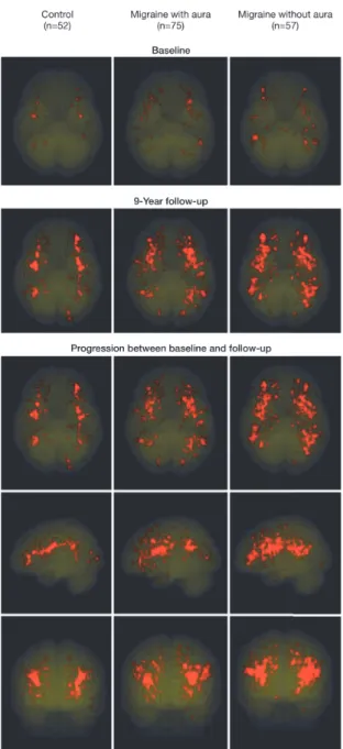
and mean volume for each participant. Geographical location was evaluated by
normal-izing the individual MRI scans with segmented lesions to standard Montreal
Neurologi-cal Institute–space, and projecting the lesions (weighted for group size) of all
partici-pants per diagnostic group in a transparent 3-dimensional map (glass brain).
Infratentorial hyperintensities were hyperintense on T2- and
proton-density-weighted and not hypointense on fluid attenuated inversion recovery images. Presence
and progression of lesions was diagnosis, by comparing baseline and follow-up scans
side by side. Reproducibility data (random, n = 40 [14%]; baseline, K = 0.908;
P
= .09 and
follow-up, K = 1.000;
P
< .001). Lesion progression was defined as an increase in size,
number, or both (Figure 1).
Infarctlike lesions were nonmass parenchymal defects with a vascular distribution,
isointense to cerebrospinal fluid signal on all sequences, and, when supratentorial,
surrounded by a hyperintense rim on FLAIR images.
4Virchow-Robin spaces were
ex-cluded based on typical location, shape, and absence of a hyperintense rim. In the basal
ganglia, only parenchymal defects larger than 3 mm in diameter were considered in
order to exclude nonspecific lesions. Location and vascular territory of new and
preex-isting infarcts were read by 2 neuroradiologists, who were blinded to diagnosis (K= 0.87,
final diagnosis in the 9 cases in which the 2 raters disagreed. An exploratory outcome
measure of this study was the changes in cognition related to white matter
hyperinten-sities at baseline and at follow-up. Similarly, the change in cognition between baseline
and follow-up was evaluated as function of baseline and follow-up lesion volume as well
as lesion volume change. For each participant, normalized test scores (Z scores of
sepa-rate tests in domains of memory, executive function, attention, visuospatial ability, and
speed) were summed to achieve a total composite cognitive score for each time point.
Change in raw test scores (follow-up minus baseline) were normalized by Z
scores. The
tests, evaluating cognitive performance in the domains of memory, concentration, and
attention, executive functioning, psychomotor, and processing speed, organization, fine
motor skills, fluid intelligence, and visuospatial skills, consisted of the 15-word Verbal
Learning Test
15; abbreviated Stroop test,
16consisting of 3 subtasks; verbal Fluency which
is a modified version of the Symbol Digit Modalities Test; and the Purdue pegboard
test.
19In follow-up investigation, the Block Design Test from the Wechsler Adult
Intelli-gence Scale III test battery
20was added. Further details on cognition testing are
provid-ed in eTable 3 (available at http://www.jama.com).
Covariates and Definitions
Sociodemographic and medical history characteristics were assessed by interview.
Edu-cational level was dichotomized into low, primary school or less than voEdu-cational
educa-tion, and high, more than higher vocational or professional educaeduca-tion, college, or
uni-versity. A diagnosis of diabetes or hypertension was based on patient report of a
physi-cian’s diagnosis.
Statistical Analysis
using linear regression models, adjusting for age, sex, and educational level (model 1)
and additionally for migraine (model 2) to assess the effect of migraine diagnosis. Data
were analyzed using the statistical software package for social sciences (SPSS, version
17.0. for Windows).
RESULTS
Study Population
A total of 411 of 435 (95%) of baseline participants were successfully recontacted; 14
participants had moved, 4 were lost to civil registry information, and 6 had died (eTable 1).
Two hundred eighty-six participants (66%) underwent follow-up MRI scan (114
mi-graine with aura, 89 mimi-graine without aura, 83 controls). Mean follow-up was 8.5 years
(range, 7.9-9.2; SD, 0.24 years). Reasons for nonparticipation were no interest (n = 51),
inability to visit the research center (n = 30), claustrophobia (n = 8), and
non-neurological illness (n = 36). There was no association between responder rate and
diagnosis of migraine (response rate in both migraine groups was 203 of 296 (69%) vs
83 of 139 (60%) in the control group (P = .07). Compared with nonparticipants,
partici-pants were younger at baseline (48 vs 50 years; P = .01), more often reported high
edu-cational level (52% vs 40%; P = .01), smoked fewer pack years (8 vs 14 years; P <.001;
eTable 1), had a similar prevalence of posterior circulation territory infarctlike lesions
(4%), brain infarcts (6% vs 9%; P = .24), and a high load of deep white matter
hyperin-tensities (based on semi quantitative measures at baseline; 19% vs 22%; P
= .44). At
follow-up, participants in the migraine group were slightly older than those in the
con-trol group (57 vs 55 years; P = .03) and had a higher prevalence of diabetes (9% vs 2%; P
= .05; TABLE 1).
Deep White Matter Hyperintensities
progression (adjusted odds ratio [OR], 2.1; 95% CI, 1.0-4.1;
P
= .04; TABLE 3). Similarly,
women in the migraine group had a higher incidence of high progression than women
in the control group (23% vs 9%;
P
= .03; Table 2). Hypertension was not associated with
a higher incidence of white matter hyperintensity progression (
P
= .06). Interaction
terms for hypertension (
P
= .90) and diabetes (
P
= .60) were not significant. Further
exploratory analyses showed no association of the number of migraine attacks,
mi-graine attack duration, mimi-graine frequency, type of attack, or mimi-graine therapy with
lesion progression (
e
Table 2).
Figure 1. Brain Magnetic Resonance T2-Weighted images at baseline and follow-up from three representative participants showing progression of infratentorial hyperintensities
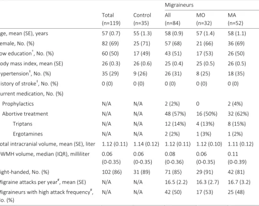
Table 1. Follow-up Characteristics of Study Participants
Migraine
Characteristic Total
(n=286)
Controls (n=83)
Migraine (n=203)
No Aura (n=89)
Aura (n=114) Age, mean (SD), y 57 (7.7) 55 (7.3) 57 (7.8)f 58 (7.5) 57 (8.0)
Women 202 (71) 57 (69) 145 (71) 64 (72) 81 (71)
Maastricht research center 128 (45) 38 (46) 90 (44) 35 (40) 55 (48)
Low educationa 137 (48) 38 (46) 99 (49) 46 (52) 53 (47)
BMI, mean (SD) 26 (4.1) 26 (3.8) 26 (4.3) 25 (4.3) 26 (4.2)
Hypertensionb 97 (34) 24 (29) 73 (36) 32 (36) 41 (36)
Use of antihypertensive medicationc 79 (28) 19 (23) 60 (30) 28 (32) 32 (28)
Blood pressure, mean (SD), mm Hgb
Systolic 151 (21) 152 (19) 151 (21) 148 (20) 154 (22)
Diastolic 94 (11) 94 (12) 94 (11) 92 (10) 96 (12)
Diabetes (self-reported) 20 (7) 2 (2) 18 (9)g 9 (10) 9 (8)
History of stroked 8 (3) 0 (0) 8 (4) 2 (2) 6 (5)
History of transient ischemic attack 12 (4) 2 (2) 10 (5) 5 (6) 5 (4)
Smoking
Ever 193 (68) 58 (70) 135 (67) 58 (65) 77 (68)
Current 67 (35) 19 (33) 48 (36) 22 (38) 26 (34)
Pack-years, mean (SD) 11 (15) 12(15) 11 (15) 13 (18) 10 (13)
Alcohol use
None during last 12 mo 42 (15) 10 (12) 32 (16) 18 (20) 14 (12)
≥3 U/d 29 (10) 11 (13) 18 (9) 6 (7) 12 (11)
Current use of migraine medicatione
Triptans 25 (12.3) 8 (9) 17 (14.9)
Ergotamines 5 (2.5) 1 (1.1) 4 (3.5)
Prophylactic drugs 7 (3.4) 1 (1.1) 6 (5.3)
Oral contraceptive use, women only
Current 16 (6) 6 (12) 10 (8) 3 (6) 7 (9)
≥ 15 y 71 (25) 24 (48) 47 (38) 21 (42) 26 (35)
Abbreviations: BMI: Body mass index calculated as weight in kilograms divided by height in meters squared. a: Low education indicates primary school or lower vocational education. b: Hypertension self-reported physician diagnosed. c: Use of antihypertensive medication by participants with hypertension, not used as migraine prophylaxis. Mean blood pressure indicates mean of two blood pressure measurements after tran-scranial Doppler examination with Valsalva maneuver. d: Ischemic or hemorrhage, self-reported. e: current use of migraine medication defined as use in the year. f: compared with controls: P=.03. Unless indicated otherwise, differences were not significant (P >0.05). g: Compared with controls P=.05
had a higher incidence of 10 or more new lesions among 43 of 145 participants (30%) vs
5 of 57 in the control group (9%) (adjusted OR, 3.5; 95% CI, 1.3-9.6;
P
= 01). Among
women with migraine, deep white matter hyperintensities were more diffusely
distrib-uted in the deep white matter than among controls (FIGURE 2).
Periventricular White Matter Hyperintensities
Progression of periventricular white matter hyperintensities did not differ between
participants with migraine and controls. There was no association of sex, aura status, or
migraine frequency with progression.
Infratentorial Hyperintensities
The prevalence of infratentorial hyperintensities at follow-up was 21% among women
with migraine and 4% among controls (adjusted OR, 6.5; 95% CI, 1.5-28.3;
P
= .01; Table
3). Progression of infratentorial hyperintensities was not significantly higher among
women with migraine (15%) than women in the control group (2%; adjusted OR, 7.7;
95% CI, 1.0-59.5;
P
=.05; Table 3). There was no relationship between migraine aura and
Ta bl e 2. P re va le nc e an d Pr og re ss io n of In fa rc ts a nd D ee p W hi te M at te r a nd Infra tentoria l Hy perintens ities Mi gra ine H ea da ch e Con tr ols
(n = 83)
Mi gra ine H ea da ch e
(n = 203)
P Va lu e a W ith ou t A ur a
(n = 89)
W
ith
A
ur
a
(n = 114)
P
Va
lu
e
a
Deep white matter hyperintens
itie
s
Men, No. (%)
26 (31) 58 (2 9) .7 25 (2 8) 33 (2 9) >. 99 Lesio n v ol ume, me
dian (IQR), mL
Ba se lin e 0. 04 ( 0. 00-0. 20) 0. 02 ( 0. 00-0. 07) .1 8 0. 01 ( 0. 00-0. 08) 0. 02 ( 0. 00-0. 09) .7 6 9-y Fo llo w -u p 0. 14 (0 .0 1-0. 67 ) 0. 06 (0 .0 8-0. 34 ) .3 6 0. 05 (0 .0 0-0. 15) 0. 11 ( 0. 01-0. 42) .47 Di ffe re nc e 0. 08 ( 0. 01-0. 43 ) 0. 04 ( 0. 00-0. 29 ) .3 1 0. 04 ( 0. 00-0. 10) 0. 08 ( 0. 01-0. 31) .47 Lesio n pro gressi on
, No. (%)
b 2 1 (8 1) 4 0 (6 9) .30 1 5 (6 0) 2 5 (7 6) .26 Hig h pr ogre ss ion, No. (%) c 6 (2 3) 1 2 (2 1) .78 5 (2 0) 7 (2 1) >. 99 New les ion s Media n ( IQ R) 3 (1-11) 3 (0-8) .6 4 3 (0-5) 4 (0-10) .5 0 ≥1 0, No. (%) 8 (3 1) 1 2 (2 1) .41 4 (1 6) 8 (2 4) .53 Me an v ol ume , me dia n (IQ R) 0. 03 ( 0. 01-0. 05 ) 0. 02 ( 0. 01-0. 04 ) .2 3 0. 02 ( 0. 01-0. 07) 0. 02 ( 0. 01-0. 04) .65 Wome
n, No. (%)
57 (69) d 14 5 (7 1) .67 6 4 (7 2) 8 1 (7 1) >. 99 Lesio n v ol ume, me
dian (IQR), mL
Ba se lin e 0. 00 ( 0. 00-0. 04) 0. 02 ( 0. 00-0. 09) .009 0. 03 ( 0. 00-0. 12) 0. 01 ( 0. 00-0. 06) .0 8 9-y Fo llo w -u p 0. 04 (0 .0 0-0. 19 ) 0. 09 (0 .0 2-0. 34 ) .0 4 0. 16 (0 .0 2-0. 43) 0. 05 ( 0. 01-0. 28) .03 Di ffe re nc e 0. 02 ( 0. 00-0. 14 ) 0. 05 ( 0. 01-0. 27 ) .0 4 0. 11 ( 0. 01-0. 36) 0. 04 ( 0. 00-0. 15) .04 Lesio n pro gressi on
, No. (%)
Mi gra ine H ea da ch e Con tr ols
(n = 83)
Mi gra ine H ea da ch e
(n = 203)
P Va lu e a W ith ou t A ur a
(n = 89)
W
ith
A
ur
a
(n = 114)
P Va lu e a In fr at en to ria l h yp er in te ns iti es , N o. (% ) M en Prevalence 3 (1 2) 9 (1 6) .7 5 4 (1 6) 5 (1 5) >. 99 Pr og re ss io n b 1 (4) 5 (9) .66 2 (8) 3 (9) >. 99 Wome n Prevalence 2 (4) 30 (2 1) .002 18 (2 8) 12 (1 5) .0 6 Pr og re ss io n b 1 (2) 21 ( 15) .01 13 ( 20) 8 (1 0) .1 0 Po st er io r c irc ul at io n te rr ito ry in fa rc tli ke le si on s, N o. (% ) e Ba se lin e 3 (4) 11 (5) .7 6 2(2) 9 (8) .1 2 9-y Fo llow-up 3 (4) 18 ( 9) .1 4 6 (7) 12 ( 11) .46 N ew le sion 0 10 (5) .07 5 (6) 5 (4) .75 Anterior c irc ula tio
n or ba
sa l ga ng lia infa rc tlik e lesi on s (non-p osteri or c irc ula tion territory ), No. (%) Ba se lin e 8 (1 0) 15 (7) .6 3 6 (7) 9 (8) .7 9 9-y Fo llow-up 11 ( 13) 20 ( 10) .41 12 ( 11) 8 (9) .82 Ne w le sion s 3 (4) 5 (3) .6 9 2 (2) 3 (3) >. 99 Ab br ev ia tio n: IQ R, in te rq ua rt ile ra ng e, a P va lu es a re fo r di ffe re nc es b et we en th e co nt ro l g ro up a nd th e m igra ine gr oup an d bet w een tho se in the migra ine grou p w ith and w ith ou t a ur a. b Pr og re ssi on o f d eep wh ite m atter hyperintensitie s is define d as an in cr eas e i n vo lu m e af ter 9 year s ( A be tw een fo llow -u p an d bas eline >0 .01 mL); progress ion of infra tentoria l h yperintens iti es is defined as an in crease in s ize , n um be r, or b ot h,
c Hig
Table 3. Risk of Deep White Matter and Infratentorial Hyperintensities in Women by Migraine Statusa
Controls (n=57)
Migraine (n=145)
P Value Migraine Without Aura (n=64)
Migraine With Aura (n=81)
P Value Deep white matter
hyperintensities
Progression, No. (%)b 33 (60)e 112 (77) 53 (83) 59 (73)
OR (95% Cl) 1 [Reference] 2.1 (1.0-4.1)f .04 2.9 (1.2-6.7)f 1.7 (0.8-3.5) .23
High progression, No. (%)c 5 (9) e 33 (23) 19 (30) 14 (17)
OR (95% Cl) 1 [Reference] 2.3 (0.8-6.4) .12 3.3 (1.1-9.9)f 1.6 (0.5-5.0) .12
High increase in number, No. (%)d 5 (9)
e 43 (30) 25 (39) 18 (22)
OR (95% Cl) 1 [Reference] 3.5 (1.3-9.6)f .01 5.3 (1.8-15.4)f 2.4 (0.8-7.0) .04
Infratentorial hyperintensities
Prevalence, No. (%)
2 (4) 30 (21) 18 (28) 12 (15)
OR (95% Cl) 1 [Reference] 6.5 (1.5-28.3)f .01 9.6 (2.1-44.1)f 4.4 (0.9-20.5) .07
Progression, No. (%)b 1 (2) 21 (15) 13 (20) 8 (10)
OR (95% Cl) 1 [Reference] 7.7 (1.0-59.5) .05 11.5 (1.4-92.9)f 5.0 (0.6-41.7) .10
Abbreviation: OR, odds ratio. aOR (95% CI) are adjusted for age, education, hypertension, and diabetes. b
Pro-gression is defined as an increase in volume after 9 years (delta between follow-up and baseline > 0.01 mL); progression of infratentorial hyperintensities is defined as an increase in size, number, or both. cHigh
progres-sion is defined as the upper 20th percentile of progresprogres-sion distribution. dHigh increase in number of lesions is
defined as 10 or more new lesions, which reflects the upper 20th percentile of the distribution of lesions count. eFor analyses of deep white matter hyperintensity progression, 2 women in the control group were
excluded (leaving n = 55), because of missing baseline volumes due to software failures during lesion segmen-tations. Visual comparison revealed no progression between baseline and follow-up for these 2 women.
fCompared with controls: P < .05
Infarcts and Infarctlike Lesions
Cognitive Changes
There were no differences in cognitive functioning between groups at follow-up (mean
composite Z
score, migraine group, 1.2 vs control group, 0; adjusted
P = .90; 95% CI,
−2.0 to 2.0). At follow-up, deep white matter hyperintensity load was not associated
with cognitive performance (mean composite Z
score high load, −3.7 vs low load, 1.4;
adjusted P = .07; 95% CI, −4.4 to 0.2; men and women were analyzed together, see also
eTable3 for original clinical scores of the separate subtest domains). Presence of
mi-graine did not influence this association (adjusted P = .30; 95% CI, −2.0 to 2.1).
Individu-als with a high deep white matter hyperintensity load at baseline did not experience
greater change in cognitive function at the 9-year follow-up than those without a high
load at baseline (mean composite Z score, −0.5 vs 0.2; adjusted P = .4; 95% CI, -1.7 to
0.7). Similarly, there were no significant differences between groups with respect to
tests of individual cognitive domains (eTable3).
COMMENT
We prospectively evaluated associations of migraine with structure and function of the
brain at the 9-year follow-up. Among men, we found no association of migraine with
progression of MRI-measured brain lesions. Women in the migraine group had a higher
prevalence and a greater increase of deep white matter hyperintensities than women in
the control group. Although migraine was associated with a higher prevalence of
in-fratentorial hyperintensities at follow up, there were no significant associations of
mi-graine with progression of infratentorial hyperintensities or posterior circulation
territo-ry infarct like lesions among women. In addition, the number of migraines, frequency of
migraines, migraine severity, type of migraine, and migraine therapy were not
associat-ed with lesion progression. Increase in deep white matter hyperintensity volume was
not significantly associated with poorer cognitive performance at follow-up.
This study has several strengths, including the longitudinal study design, length of
follow-up, the relatively well characterized cohort, use of standardized International
Headache Society criteria-based diagnosis of migraine by headache experts, and
sensi-tive and reproducible methods of MRI reading. The sensisensi-tive MRI techniques used
al-lowed for a more detailed analysis of the brain, in particular the cerebellum.
non-responders, additional imputation analyses to support the sensitivity of the current
results could not be performed. An additional study limitation is that confidence
inter-vals are wide (Table 3).
The number of migraine attacks, frequency of migraines, migraine severity, type of
migraine headaches, and migraine therapy were not associated with lesion progression.
In contrast, our baseline data showed that more frequent migraine headaches were
associated with a higher prevalence of MRI findings.
4However, our findings at baseline regarding frequency-related difference in MRI
findings was most pronounced among those in the migraine group who were 50 years
or younger and less so in older patients. Thus, with increasing age of the study
popula-tion, when attacks generally diminish,
1other migraine disease-related conditions
lead-ing to white matter hyperintensities are possibly increaslead-ing, complicatlead-ing the detection
of migraine attack-related associations. A similar, age-dependent mechanism is also
seen for the risk of stroke in participants with migraine, which is increased in young
patients only.
14,21At older age, other risk factors such as hypertension may obscure or
overcome any potential role of migraine. In the present case, we hypothesize there are
at least 2 different types of vascular mechanisms that may cause structural brain
changes in migraine: one, which is primarily related to attacks and mainly present at
younger age, and another, which is probably ongoing as part of having the disease
mi-graine. The observation of migrainous stroke, with stroke occurring during a migraine
attack, would support the hypothesis that ischemia may occur during attacks.
22Howev-er, our finding that migraine was not significantly associated with progression of all
evaluated types of brain lesions at the 9-year follow-up raises questions about the role
of cerebral ischemia over time in people with migraine.
21,23Possible explanations for an association of migraine headache with structural brain
changes include a chronic procoagulatory or proinflammatory state due to endothelial
dysfunction
24,25or elevated homocysteine levels,
26,27or recurrent paradoxical (micro-)
emboli due to right-to-left shunts.
28Increased incidence of brain lesions among people
with migraine headaches and atherosclerotic risk factors such as hypertension,
diabe-tes, or other cardiovascular risk factors is also possible, but we did not identify any
sig-nificant interactions for hypertension or diabetes. A relation with headache in general
7cannot be excluded. Finally, sex differences seem to play an important role because
progression of deep white matter hyperintensities was only found in women. This
find-ing is in line with results from another study
8and consistent with the higher risk of brain
infarcts in women with migraine.
14Our sample size was too small for a proper analysis
Figure 2. Geographical Location of All Individual Deep White Matter Hyperintensities Projected on Transparent 3-Dimensional Maps After Normalization of the Individual Magnetic Resonance Scans With Segmented Le-sions to Standard Montreal Neurological Institute Space
White matter hyperintensities have been associated with cognitive deficits in the
el-derly
29,30and some studies found evidence for worse cognitive performance in
individu-als with migraine.
31-34We tested memory, speed, and attention
35in all participants at
baseline and follow-up and found no significant association between deep white matter
hyperintensity volume and cognitive dysfunction. Most prior studies were conducted in
older participants with larger deep white matter intensity volumes; this cohort is rather
young with relatively little volume.
7In summary, in a community-based cohort followed up for 9 years, migraine was
as-sociated only with a higher incidence of deep white matter brain changes among
wom-en. There were no significant associations of migraine with progression of other brain
lesions among women, and there were no associations of migraine headache with
pro-gression of any brain lesions among men. These findings raise questions about the role
of migraine headaches with progression of cerebral vascular changes. The functional
implications of MRI brain lesions in women with migraine and their possible relation
with ischemia and ischemic stroke warrant further research.
This work was supported by grants 1R01NS061382-01 from the National Institutes
of Health, 2007B016 from the Netherlands Heart Foundation, and 903-52-291, VICI
918-56-602, and Spinoza 2009 from the Netherlands Organisation for Scientific
Re-search, and 907-00-217 and Vidi 917-11-319 from the Intramural Research Program at
the National Institute on Aging.
Role of the Sponsor: None of the funding bodies had any role in the design and
con-duct of the study; collection, management, analysis, and interpretation of the data; and
preparation, review, or approval of the manuscript. Disclaimer: The contents of this
article are solely the responsibility of the authors and do not necessarily represent the
official view of the National Institutes of Health or the Netherlands Heart Foundation.
Online-Only Material: Author Video Interview and eTables 1 through 3 are available at
http://www.jama.com.
O
N
LINE
-ONLY M
ATER
IAL
eT ab le 1 . B as el in e ch ar ac te ris tic s of fo llo w -u p pa rt ic ip an ts a nd n on -p ar tici pants Char acterist ic at ba seline (C AMER A-1) CAMERA-1 (n =4 35 ) CAMERA-2 T ota l g roup CAMER A-2 M igra in eurs CAMERA-2 C ontro ls N on -p ar tic ip an ts (n =1 49 ) Pa rt ic ip an ts (n =2 86 ) N on -p ar tic ip an ts (n=93) Pa rt ic ip an ts (n =2 03 ) N on -p ar tic ip an ts (n=56) Parti ci pants (n=83) De m og ra ph ic s Age a t CAM ERA-1, me an (SD ), y 48 ( 7. 8) 50 ( 7. 9) $ 48 (7. 7) 49 (8. 0) 48 (7. 8) 51 (7. 5) $$ 46 (7. 2) Fe m al e 31 7 (7 3% ) 11 5 (7 7% ) 20 2 (7 1% ) 73 (7 9% ) 14 4 (7 1% ) 42 (7 5% ) 58 (6 9%) �o w e duc a� on� 22 7 (5 2%) 90 ( 60 % ) $ 13 7 (4 8% ) 57 (6 1%) 98 (4 9%) 33 (5 9%) 39 (4 6%) Ph ys ic al a nd la b ex am Bod y m ass in de x, m ea n (S D) 25 (4. 2) 25 (4. 4) 25 (4. 0) 26 (4. 7) $ 25 (4. 1) 24 (3. 8) 24 (3. 6) Blo od press ure, mean (SD), mm H
g Sy stol ic Dias tolic 13 4 (1 8) 91 (1 0) 13 6 (1 8) 92 (1 0) 13 4 (1 8) 91 (1 0) 13 6 (1 8) 93 (1 1) $ 13 3 (1 7) 91 (9) 13 5 (1 8) 90 (8) 13 5 (1 8) 91 (1 0) Hy pe rt ensi on * 16 7 (3 8%) 58 ( 39 % ) 10 9 (3 8%) 43 ( 46 % ) 79 ( 39 % ) 15 (27 %) 30 ( 36 % ) Di ab et es 9 (2 % ) 2 (1 % ) 7 (2 % ) 0 4 (2 % ) 2 (4 % ) 3 (4 % ) Hig h ri sk c ho le st er ol* 65 ( 15 % ) 27 ( 18 % ) 38 ( 13 % ) 17 ( 18 % ) 29 ( 14 % ) 10 (1 8%) 9 (1 1% ) Medical hist ory Smok ing
Ever Pack-y
ears
, mean (SD)
28 7 (6 6%) 10 (1 3) 10 4 (7 0%) 14 (1 6) $ 18 3 (6 4%) 8 (1 1) 64 (6 7%) 13 (1 6) $ 12 5 (6 2%) 8 (1 1) 40 (7 1%) 14 (1 7) 58 (6 9%) 10 (1 2) Hig h a lc oho l c on su mpt ion* 44 ( 10 % ) 14 ( 9%) 30 ( 11 % ) 6 (7%) 16 ( 8% ) 8 (1 4% ) 14 (1 7%) >1 5 yr s of o ra l c on tr ac ep tiv e us e (women only) 77 /3 17 (2 4% ) 22 /1 15 (1 9% ) 55 /2 02 (2 7% ) 15 /7 3 (2 1% ) 38 /1 44 (2 6% ) 7/ 42 (1 7% ) 17/ 58 (2 9% ) U nl es s in di ca te d ot he rw ise , d iff er en ce s w er e no t s ig ni fic an t ( P >. 05) Co m pa re d w ith p ar tic ip an ts
$ P
<.
05
,
$$ P
Ca
us
es
o
f d
ea
th
d
ur
ing follow-up period
Six
ba
sel
in
e
parti
ci
pant
s
had
died
duri
ng
fol
low-u
p
pe
riod:
one
du
e
to
m
al
ig
na
nt
n
eo
pl
as
m
o
f o
va
ry
, o
ne
h
ad
e
m
ph
ys
em
a,
o
ne
c
er
ebra
l
infarct
ion,
on
e
acu
te
pe
ritoni
tis
wit
h
sep
tic
shoc
k,
an
d
two
un
kn
own
c
au
se
s
of
d
ea
th
. O
nl
y
em
ph
ys
em
a
pat
ie
nt
w
as
c
on
tr
ol
p
ar
tic
ip
an
t,
ot
he
rs
w
er
e
m
ig
ra
in
eu
rs
.
eT ab le 2 . Migra ine cha ra cteristics in r
ela tion t o MR I out come mea sures a s co mpa
red to c
ontr ol s (Fema le s o nly ) H ig h DW MH loa d at FU Pr og re ss io n of DW MH loa d Hig h pr ogre ss ion of DW MH loa d Pro gress io n of I H Dura tion of mi gra ine < 2 9 yrs ≥ 2 9 yrs 1. 8 [ 0. 6-4. 9] 1. 2 [ 0. 4-3. 3] P= .3 2. 9 [1 .3 -6 .7 ] 1. 5 [ 0. 7-3. 2] P= .0 6 3. 3 [1 .0 -9 .9 ] 1. 6 [ 0. 5-5. 0] P= .08 11. 2 [1. 4-90. 1] 4. 9 [0 .6 -4 1. 8] P= .1 lo w 2 5% high 2 5% 0. 5 [ 0. 1-1. 6] 0. 7 [ 0. 3-2. 0] P= .8 0. 9 [0 .3 -2 .4 ] 0. 3 [ 0. 1-1. 0] P= .0 4 0. 7 [0 .2 -2 .2 ] 0. 6 [ 0. 2-1. 8] P= .5 2. 1 [0 .7 -6 .4 ] 0. 5 [ 0. 1-1. 9] P= .2 median (
IQR) of th
ose w ith le sio ns medi an ( IQ R) o f thos e w ithout 23 ( 11-39) 20 (0 -3 4) P*=. 2 22 ( 7-34) 15 (0 -3 6) P* =. 1 23 (1 6-38 ) 20 (0 -3 4) P* =. 1 24 (1 5-37 ) 20 (0 -3 4) P*=. 2 Mi graine subtype w ith ou t a ur a w ith a ur a 1. 8 [ 0. 7-5. 1] 1. 2 [ 0. 4-3. 2] P= .3 2. 9 [1 .2 -6 .7 ] 1. 7 [ 0. 8-3. 5] P= .2 3. 3 [1 .1 -9 .9 ] 1. 6 [ 0. 5-5. 0] P= .1 11. 5 [1. 4-92. 9] 5. 0 [0 .6 -4 1. 7] P= .1 Number o
f headache attacks
<median (lifetime) ≥median (lifetime)
1. 7 [ 0. 6-4. 7] 1. 3 [ 0. 5-3. 5] P= .5 1. 9 [0 .9 -4 .2 ] 2. 2 [ 1. 0-4. 9] P= .9 2. 8 [0 .9 -8 .7 ] 1. 9 [ 0. 6-5. 8] P= .4 8. 9 [1 .1 -7 3. 3] 6. 7 [0 .8 -5 5. 0] P= .5 median (
IQR) of th
ose w ith le sio ns medi an ( IQ R) o f thos e w ithout 27 1 (1 07-64 6) 26 4 (1 68-48 0) P*=. 9 27 4 (1 23-57 5) 25 2 (1 74-35 3) P*=. 4 26 3 (1 05-58 1) 27 4 (1 73-49 6) P*=. 6 28 4 (1 00-92 3) 26 4 (1 59-49 6) P*=. 9 <m edi an (F U o nl y) ≥m edi an (F U o nl y) 1. 9 [ 0. 7-5. 5] 1. 2 [ 0. 4-3. 3] P= .3 2. 1 [0 .9 -4 .7 ] 2. 2 [ 1. 0-4. 5] P= .8 3. 2 [1 .0 -9 .9 ] 1. 9 [ 0. 6-5. 6] P= .2 12. 3 [1. 5-101. 5] 5. 3 [0 .6 -4 3. 8] P= .1
Mean attacks per month,
H ig h DW MH loa d at FU Pr og re ss io n of DW MH loa d Hig h pr ogre ss ion of DW MH loa d Pro gress io n of I H H eadache acti vi ty s tatus in ac tiv e at b as el in e
active at bas
eline 3. 4 [1 .1 -1 0. 7] 1. 1 [ 0. 4-2. 8] P= .0 3 3. 1 [1 .0 -9 .5 ] 1. 9 [ 0. 9-3. 8] P= .4 5. 3 [1 .5 -1 8. 1] 1. 7 [ 0. 6-5. 1] P= .03 10. 2 [1. 1-93. 1] 7. 0 [0 .9 -5 5. 6] P= .6 ina ctiv e durin g 9 y ea r FU acti ve duri ng 9 year FU 1. 8 [ 0. 6-5. 0] 1. 2 [ 0. 4-3. 4] P= .5 2. 9 [1 .0 -5 .4 ] 1. 9 [ 0. 9-4. 1] P= .9 2. 6 [0 .8 -8 .1 ] 2. 1 [ 0. 7-6. 3] P= .7 5. 7 [0 .7 -4 7. 4] 10. 6 [1. 3-87. 0] P= .2 N um be r o f a ur a at ta ck s
<median (lifetime) ≥median (lifetime)
1. 1 [ 0. 3-4. 0] 0. 9 [ 0. 3-3. 3] P= .8 1. 7 [0 .7 -4 .1 ] 1. 7 [ 0. 7-4. 3] P= .8 1. 6 [0 .4 -6 .2 ] 1. 3 [ 0. 3-5. 0] P= .8 5. 7 [0 .6 -5 7. 1] 4. 6 [0 .5 -4 5. 9] P= .8 median (
IQR) of th
ose w ith le sio ns medi an ( IQ R) o f thos e w ithout 15 8 (7 3-411) 15 0 (4 9-262) P*=. 2 15 8 (6 8-283) 14 9 (4 3-281) P*=. 5 15 8 (7 3-411) 15 0 (4 9-262) P*=. 2 11 3 (2 6-265) 15 4 (6 5-292) P*=. 5 <media n a t fol low-up ≥media n a t fol low-up 1. 1 [ 0. 2-5. 3] 1. 1 [ 0. 3-4. 6] P= .7 1. 5 [0 .5 -4 .2 ] 2. 2 [ 0. 7-6. 7] P= .9 1. 6 [0 .3 -8 .7 ] 1. 5 [ 0. 3-6. 8] P= .7 6. 0 [0 .5 -7 7. 7] 3. 2 [0 .2 -4 8. 1] P= .6 Au ra a ct iv ity s ta tu s in ac tiv e at b as el in e
active at bas
eline 2. 0 [ 0. 5-7. 8] 0. 8 [ 0. 2-2. 5] P= .2 2. 0 [0 .6 -6 .2 ] 1. 6 [ 0. 7-3. 5] P= .8 2. 7 [0 .6 -1 1. 8] 1. 1 [ 0. 3-3. 8] P= .2 5. 9 [0 .5 -6 8. 5] 5. 4 [0 .6 -4 8. 2] P= .9 in ac tiv e at fo llo w -u p ac tiv e at fo llo w -u p 0. 3 [ 0. 1-2. 0] 1. 7 [ 0. 6-4. 8] P= .0 6 1. 5 [0 .5 -4 .3 ] 2. 3 [ 1. 0-5. 0] P= .4 0. 5 [0 .1 -3 .0 ] 2. 6 [ 0. 8-8. 3] P= .0 5 6. 8 [0 .6 -7 1. 4] 7. 8 [0 .9 -6 4. 3] P= .8
Treatment no t
riptan
s ever used
tri ptans ever us ed 1. 4 [ 0. 6-3. 7] 1. 5 [ 0. 4-6. 0] P= .8 1. 8 [0 .9 -3 .7 ] 4. 3 [1 .1 -1 6. 6] P= .2 2. 2 [0 .8 -6 .3 ] 2. 9 [0 .7 -1 1. 8] P= .5 8. 9 [1 .1 -6 9. 3] 2. 5 [0 .1 -4 2. 5] P= .2 OR with [95% CI] f or com parison w ith contro ls; c ontro
ls as a re
ference gr oup P-values between migra ine subgr ou ps a dj us ted fo r a ge , h yp er te ns io n, diab
etes, education; P
*-val
ues by
M
ann
Whitney U test
DWMH=Deep whit e matte r h yp er in te ns iti es IH =Infra tentor ia l h yperintensities Progressi on of DW MH defined as a n in crease in D W M H v olume after 9 y ea rs (Δ C AM 2-CA M1 ≥0. 01 m l); p rogr es sion of IHs de fin ed a s an in crease in size and /or num ber of IH s; h igh pro gressi
on of D
WM
H de
fin
ed as the up
eT ab le 3 . M ea n Z-sc or es o f c og ni tiv e pe rf or m an ce in d iff er en t d om ai ns b y de ep w hi te m at te r h yp er in te ns ity lo ad (D W M H ) non-h igh D WM H high DW M H P [9 5% C I] (model 1) P [9 5% C I] (model 2) N Mea n (SD) N Mea n (SD) Co gn iti ve fu nc tio n at b as el in e Me m ory : im me dia te re ca ll 21 9 0. 0 (2. 7) 57 -0. 0 (2. 5) 0. 5 [-0. 5 to 1. 0] 0. 5 [-0. 5 t o 1. 0] M em or y: d el ay ed re ca ll 21 9 0. 0 (1 .0 ) 57 -0 .1 (0 .9 ) 0. 8 [-0. 2 to 0. 3] 0. 8 [-0. 2 t o 0. 3] Conc en tr at io n, a tt ent ion 21 6 0. 0 (2. 6) 57 -0. 2 (2. 5) 0. 4 [-1. 0 to 0. 4] 0. 4 [-1. 0 t o 0. 4] Pr oc essi ng spe ed 21 9 0. 1 (1. 0) 57 -0. 3 (1. 0) 0. 3 [-0. 4 t o 0. 1] 0. 3 [-0 .4 to 0 .1 ] Vi su o-spa tia l, mot or sk ill s 18 7 0. 2 (3. 6) 50 -0. 8 (2. 9) 0. 4 [-1. 7 to 0. 6] 0. 4 [-1. 7 to 0. 6] Ex ec ut iv e f un ct io n 21 6 0. 1 (1 .6 ) 57 -0 .3 (1 .5 ) 0. 6 [-0. 5 to 0 .3 ] 0. 6 [-0 .5 to 0 .3 ] Co gn iti ve fu nc tio n at fo llo w -u p Me m ory : im me dia te re ca ll 22 3 0. 2 (2. 6) 53 -0. 7 (2. 7) 0. 2 [-1. 2 to 0. 3] 0. 2 [-1. 2 t o 0. 3] Me m ory : de la ye d r ec all 22 3 0. 1 (1. 0) 53 -0. 3 (1. 0) 0. 06 [-0. 6 t o 0. 0] 0. 06 [-0. 6 t o 0. 0] Co nc en tr at io n, a tt en tio n 22 3 0. 1 (2. 8) 51 -0 .6 (2. 4) 0. 7 [-0 .6 to 0 .9 ] 0. 8 [-0 .6 to 0 .9 ] Proces sing s peed 22 2 0. 1 (1. 0) 53 -0 .4 (0. 9) 0. 1 [-0 .5 to 0 .0 ] 0. 1 [-0 .5 to 0 .0 ] Visu o-spa tia l, mot or sk ill s 21 9 0. 4 (4. 3) 53 -1 .7 (4. 0) 0. 09 [-2. 1 t o 0. 2] 0. 09 [-2. 1 t o 0. 2] Exec utive fun ction 22 3 0. 3 (1. 7) 51 -0 .1 (1. 7) 0. 3 [-0 .2 to 0 .7 ] 0. 3 [-0 .2 to 0 .7 ] Flui d i ntell igence 16 6 0. 1 (1. 0) 30 -0 .2 (1. 0) 0. 4 [-0 .5 to 0 .2 ] 0. 4 [-0 .5 to 0 .2 ] O ve ra ll co gn iti ve p er fo rm an ce CAMERA-1 (base line) 18 4 0. 3 (8. 9) 50 -1 .8 (8. 2) 0. 8 [-2 .9 to 2 .3 ] 0. 8 [-2 .9 to 2 .3 ] CA M ER A-2 (fol lo w -u p) 21 8 1. 4 (9. 2) 51 -3 .7 (8. 9) 0. 07 [-4. 5 t o 0. 2] 0. 07 [-4. 5 t o 0. 2] Hig h DW M H defin
ed as the up
per q ui ntile of DW MH di stributi on. Mode l 1: Adju sted
for age, gende
r, le
vel of educatio
n; M ode l 2: Adj us ted for a ge, ge nder, le vel of educatio
n, and m
igraine dia gn os is Z-scores i ndicate b
y how ma
ny sta nd ar d de vi at io ns a n ob se rv at io n is ab ov
e or bel
Assessment of cognitive performance
Cognitive performance was evaluated by validated, widely used, cognitive tests in a
fixed order. The test battery, administered by four trained medical students, was the
same for both time points (test protocol and methods were the same for baseline and
follow-up) and included the 15 word Verbal Learning Test (Rey, 1985); abbreviated
Stroop test (Stroop, 1935) consisting of three subtasks; verbal Fluency test (Miller,
1984); Letter Digit Substitution Test (Van der E, 2006), which is a modified version of
the Symbol Digit Modalities Test; and Purdue pegboard test (Tiffin, 1948). In follow-up
investigation, the Block Design Test from the WAIS-III test battery (Wechsler, 1981) was
added. Higher score indicates better cognitive performance. The results of these tests
were normalized by calculation of Z-scores based on total sample means and standard
deviations, and added up per cognitive domain. The composite cognitive score was
calculated for baseline as well as follow-up time point by adding up the separate
do-main Z-scores.
Cognitive domains
Memory function was composed of immediate recall and delayed recall after 20
minutes. The reading subtasks of the Stroop test measured concentration and attention
ability. Executive function was scored by the interference task of the Stroop test and the
word fluency task. The Letter Digit Substitution Test evaluated psychomotor speed,
processing speed, and organization. Fine motor skills, motor speed, and visuo-spatial
ability were evaluated by the Purdue pegboard. The Block Design Test measured fluid
intelligence and visuo-spatial skills.
Statistical analysis
REFERENCES
1. Launer LJ, Terwindt GM, Ferrari MD. The prevalence and characteristics of migraine in a populationbased cohort: the GEM study. Neurology. 1999; 53(3):537-542.
2. Ferrari MD. Migraine. Lancet. 1998;351(9108): 1043-1051.
3. Headache Classification Committee of the International Headache Society. Classification and diagnostic criteria for headache disorders, cranial neuralgias and facial pain. Cephalalgia. 1988;8(suppl 7): 1-96. 4. Kruit MC, van Buchem MA, Hofman PA, et al. Migraine as a risk factor for subclinical brain lesions. JAMA.
2004;291(4):427-434.
5. Kruit MC, Launer LJ, Ferrari MD, van Buchem MA. Infarcts in the posterior circulation territory in mi-graine: the population-based MRI CAMERA study. Brain. 2005;128(Pt 9):2068-2077.
6. Kruit MC, Launer LJ, Ferrari MD, van Buchem MA. Brain stem and cerebellar hyperintense lesions in migraine. Stroke. 2006;37(4):1109-1112.
7. Kurth T, Mohamed S, Maillard P, et al. Headache, migraine, and structural brain lesions and function: population based Epidemiology of Vascular AgeingMRI study. BMJ. 2011;342:c7357.
8. Scher AI, Gudmundsson LS, Sigurdsson S, et al. Migraine headache in middle age and late-life brain infarcts. JAMA. 2009;301(24):2563-2570.
9. Dufouil C, de Kersaint-Gilly A, Besanc¸ on V, et al. Longitudinal study of blood pressure and white matter hyperintensities: the EVA MRI Cohort. Neurology. 2001;56(7):921-926.
10. Debette S, Markus HS. The clinical importance of white matter hyperintensities on brain magnetic reso-nance imaging: systematic review and meta-analysis. BMJ. 2010;341:c3666.
11. Buyck JF, Dufouil C, Mazoyer B, et al. Cerebral white matter lesions are associated with the risk of stroke but not with other vascular events: the 3-City Dijon Study. Stroke. 2009;40(7):2327-2331.
12. Vermeer SE, Hollander M, van Dijk EJ, Hofman A, Koudstaal PJ, Breteler MM; Rotterdam Scan Study. Silent brain infarcts and white matter lesions increase stroke risk in the general population: the Rotter-dam Scan Study. Stroke. 2003;34(5):1126-1129.
13. van der Flier WM, van Straaten EC, Barkhof F, et al. Small vessel disease and general cognitive function in nondisabled elderly: the LADIS study. Stroke. 2005; 36(10):2116-2120.
14. Schürks M, Rist PM, Bigal ME, Buring JE, Lipton RB, Kurth T. Migraine and cardiovascular disease: sys-tematic review and meta-analysis. BMJ. 2009; 339:b3914.
15. Rey A. L’examin Clinique en Psychologie. Paris, France: Presses Universitaires de France; 1985. 16. Stroop J. Studies of interference in serial verbal reactions. J Exp Psychol. 1935;18:643-662.
17. Miller E. Verbal fluency as a function of a measure of verbal intelligence and in relation to different types of cerebral pathology. Br J Clin Psychol. 1984; 23(pt 1):53-57.
18. Van der Elst W, Van Boxtel MP, Van Breukelen GJ, Jolles J. Normative data for the Animal, Profession and Letter M Naming verbal fluency tests for Dutch speaking participants and the effects of age, education, and sex. J Int Neuropsychol Soc. 2006;12(1):80-89.
19. Tiffin J, Asher EJ. The Purdue pegboard; norms and studies of reliability and validity. J Appl Psychol. 1948;32(3):234-247.
20. Wechsler D. Manual for the Wechsler Adult Intelligence Scale-Revised. New York, NY: Psychological Corp; 1981.
21. Kurth T, Diener HC. Current views of the risk of stroke for migraine with and migraine without aura. Curr Pain Headache Rep. 2006;10(3):214-220.
22. Wolf ME, Szabo K, Griebe M, et al. Clinical and MRI characteristics of acute migrainous infarction. Neu-rology. 2011;76(22):1911-1917.
23. Bigal ME, Kurth T, Santanello N, et al. Migraine and cardiovascular disease: a population-based study. Neurology. 2010;74(8):628-635.
25. Tietjen GE, Herial NA, White L, Utley C, Kosmyna JM, Khuder SA. Migraine and biomarkers of endothelial activation in young women. Stroke. 2009; 40(9):2977-2982.
26. Schürks M, Rist PM, Kurth T. MTHFR 677C>T and ACE D/I polymorphisms in migraine: a systematic review and meta-analysis. Headache. 2010;50(4):588-599.
27. Scher AI, Terwindt GM, Verschuren WM, et al. Migraine and MTHFR C677T genotype in a population-based sample. Ann Neurol. 2006;59(2):372-375.
28. Schwedt TJ, Demaerschalk BM, Dodick DW. Patent foramen ovale and migraine: a quantitative systemat-ic review. Cephalalgia. 2008;28(5):531-540.
29. Longstreth WT Jr, Manolio TA, Arnold A, et al. Clinical correlates of white matter findings on cranial magnetic resonance imaging of 3301 elderly people. The Cardiovascular Health Study. Stroke. 1996; 27(8):1274-1282.
30. Prins ND, van Dijk EJ, den Heijer T, et al. Cerebral small-vessel disease and decline in information pro-cessing speed, executive function and memory. Brain. 2005;128(Pt 9):2034-2041.
31. Le Pira F, Lanaia F, Zappalà G, et al. Relationship between clinical variables and cognitive performances in migraineurs with and without aura. Funct Neurol. 2004;19(2):101-105.
32. Meyer JS, Thornby J, Crawford K, Rauch GM. Reversible cognitive decline accompanies migraine and cluster headaches. Headache. 2000;40(8):638-646.
33. Mulder EJ, Linssen WH, Passchier J, Orlebeke JF, de Geus EJ. Interictal and postictal cognitive changes in migraine. Cephalalgia. 1999;19(6):557-565, discussion 541.
34. Waldie KE, Hausmann M, Milne BJ, Poulton R. Migraine and cognitive function: a life-course study.
Neurology. 2002;59(6):904-908.