Original Article
Correlation between soluble CD14 subtype, platelet
activating factor levels, and the severity of sepsis
Wen Yang, Yanbing Liang, Hao Tang, Zhenyu Li, Jingguo Wu, Lijin Zeng, Zhongfu Ma
Department of General Internal Medicine, The First Affiliated Hospital, Sun Yat-sen University, Guangzhou City, Guangdong Province, P. R. China
Received December 30, 2017; Accepted February 3, 2018; Epub March 15, 2018; Published March 30, 2018
Abstract: Objective: To investigate the correlation between soluble CD14 subtype (sCD14-ST), platelet activating
fac-tor (PAF) levels, and the severity of sepsis. Methods: Sixty-five septic patients admitted to our hospital from January
2015 to April 2017 were enrolled in this study. They were randomly assigned to the sepsis group (n=30), the severe sepsis group (n=20) or the septic shock group (n=15) in terms of the severity of disease. The plasma sCD14-ST and PAF levels, and the APACHE II scores were compared among the patients in the three groups. Correlations among the plasma sCD14-ST and PAF levels, and the APACHE II score were detected with the use of Pearson correlation analysis. In addition, the plasma sCD14-ST and PAF levels were measured in 50 healthy volunteers, and the value of the plasma sCD14-ST and PAF levels for diagnosis of sepsis was tested by the receiver operating characteristic (ROC) curves. Results: Plasma sCD14-ST and PAF levels, and the APACHE II scores were increased sequentially
and significantly among the sepsis group, the severe sepsis group, and the septic shock group (all P<0.05). There
were positive correlations of APACHE II scores with plasma sCD14-ST levels (r=0.805, P=0.003) and the PAF levels (r=0.712, P=0.014). The ROC curve analysis indicated that plasma sCD14-ST (P=0.015) and PAF (P=0.011) levels
were of value in differentiating patients with sepsis, and the differences were statistically significant. Conclusion:
The plasma sCD14-ST and PAF levels can be used as one of the markers for early sepsis diagnosis, and is favorable for assessing the severity in sepsis.
Keywords: Sepsis, severe sepsis, septic shock, soluble cluster of differentiation 14 subtype, platelet activating factor
Introduction
Sepsis, one of the common conditions encoun-tered in intensive care units, is severe and has high mortality in patients. Poorly controlled sepsis may progress into severe sepsis, septic shock, or multiple organ failure [1]. Accordingly, early diagnosis and identification of sepsis and timely formulation of reasonable protocols are of great significance for good prognosis. Recently, biomarkers used in combination or alone in clinical diagnosis of sepsis include calcitonin (PCT), interleukin and C-reactive pro-tein (CRP). However, the sensitivity and specific -ity of these biomarkers are not high or it takes a long time to get the results, therefore all these factors are unfavorable for early assess-ment of the disease [2]. The PCT level has been shown to be elevated in patients with non-sep-tic conditions such as large-area trauma, organ transplantation, or pancreatitis [3]. Blood
cul-ture is the gold standard for septic diagnosis, yet it takes 48 to 72 hours to get the result of a blood culture and it is time-consuming, and has a low positive rate [4]. It is noteworthy that it is of great clinical value to find a highly effi -cient and simple biomarker for early diagnosis of sepsis.
sCD14-ST and PAF levels in septic patients, with an aim to bring some insights into clinical treatment.
Materials and methods
Patients
Between January 2015 and April 2017, 65 sep-tic patients admitted to our hospital were recruited in this study. The enrolled patients included 42 male and 23 female patients, with a mean age of 58±7.9 years. In accordance with the severity of the disease, the patients were assigned to the sepsis group (n=30), the severe sepsis group (n=20) or the septic shock group (n=15). Patients older than 18 years old were eligible for enrollment if they met the new criteria for diagnosis of sepsis specified in the guidelines of ACCP/SCCM International Sepsis Definition Conference [9]. Sepsis was defined as the presence of clinically-confirmed infec -tion and systemic inflammatory response syn -drome (SIRS). Severe sepsis was defined as the presence of sepsis, organ dysfunction, hypo-perfusion, or sepsis-induced hypotension. Se- ptic shock was defined as sepsis-induced per -sistent hypotension, with a systolic blood pres-sure < 90 mmHg or over 40 mmHg lower than basic blood pressure. Patients were excluded if they had immunodeficiency disease, autoim -mune disease, granulocytisis (<0.5*109), tu- mor, or had received immunosuppressive ther-apy within 3 months before enrollment. Patients who were pregnant, hospitalized for less than 48 hours, or provided incomplete clinical data were also excluded from this study. The eligible patients provided written informed consent, and the Hospital Ethics Committee reviewed and approved the protocol of this study.
sCD14-ST and PAF determination
Before antibiotic therapy within 24 h after ad- mission, 5 mL of blood was collected by an endotoxin-free anticoagulation tube containing ethylenediaminetetraacetic acid (EDTA, antico-agulation agent) from the medial cubital vein of each patient, followed by plasma isolation from the collected blood at 2500 r/min for 10 min and detection of the plasma sCD14-ST levels with the use of a PATHFAST analyzer (Mitsubishi, Japan) for chemiluminescent immunoassay. The PAF levels of patients were detected by the enzyme-linked immunosorbent assay (ELISA),
strictly following the instructions on the ELISA kits (R&D Science, US).
Outcome measures
The sCD14-ST and PAF levels in plasma and the Acute Physiology and Chronic Health Evaluation (APACHE) II scores of patients were compared among the three study groups. The APACHE II scores included the acute physiological scores (0-48 points), healthy condition scores (2-5 points) and age (0-6 points). Scores range from 0 to 59, with higher score indicating more severe disease. Moreover, the correlations am- ong the sCD14-ST and PAF levels, and the APACHE II scores were analyzed among the three study groups.
Additionally, 50 healthy volunteers were assi- gned as normal controls, and their plasma sCD14-ST and PAF levels were measured. The receiver operating characteristic (ROC) curve was established according to the plasma sCD14-ST and PAF levels of healthy volunteers and septic patients, and the value of the plas-ma sCD14-ST and PAF levels in diagnosing sepsis was assessed based on the area under the curve (AUC).
Statistical analysis
All the data analyses were performed with SPSS software, version 21.0. Measurement data are presented as the mean ± standard deviation, with one-way analysis of variance (ANOVA) with post hoc Bonferroni’s test utilized for the comparisons across the three groups. Count data are described as percentages, with Chi-square tests for comparisons across the three groups, and Chi-square segmentation tests for comparisons between two groups. Correlations among the sCD14-ST and PAF lev-els, and the APACHEII scores were identified by Pearson correlation analysis. The ROC curve was established, and the ROC was utilized to evaluate the value of the sCD14-ST and PAF levels for diagnosing sepsis. A P value less than 0.05 were deemed as statistically significant.
Results
Patient characteristics at baseline
well-balanced among the three groups, so they were comparable (P>0.05, Table 1).
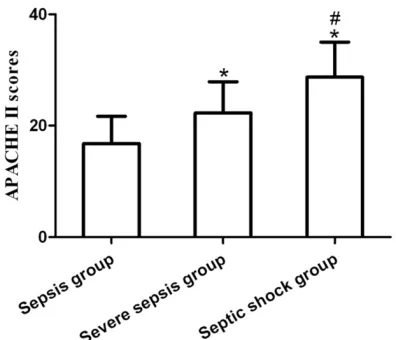
APACHE II scores
The APACHE II score was 16.8±4.9 in the sep-sis group, 22.3±5.6 in the severe sepsep-sis group and 28.7±6.3 in the septic shock group; the dif-ferences in the APACHE II scores were statisti-cally significant among the three groups (P<0.001), with the highest score in the septic shock group, the second highest score in the severe sepsis group, and the lowest score in the sepsis group (Figure 1).
Plasma sCD14-ST and PAF levels
The plasma sCD14-ST and PAF levels of patients were increased sequentially in the sepsis group, the severe sepsis group, and the septic shock group, with substantial differenc-es among the three groups (Both P<0.001; Table 2).
Correlations of the APACHE II scores with the sCD14-ST level and the PAF level
Pearson correlation analysis demonstrated that the sCD14-ST level was positively correlat-ed with the APACHE II score, and the PAF level was also positively correlated with the APACHE II score, with statistical differences (Both P<0.05), as reported in Table 3.
[image:3.612.89.523.86.165.2]Fifty healthy volunteers were assigned as nor-mal controls. According to the ROC curve analy-sis showing the value of the markers sCD14-ST and PAF for sepsis diagnosis, the AUC for sCD14-ST was 0.906, with the best cut-off value at 850.8 pg/mL. The AUC for PAF was 0.779, with the best cut-off value at 987.6 ng/L, suggesting that the markers sCD14-ST and PAF were of value in diagnosing sepsis and Table 1. Patient characteristics at baseline
Variable Case Age (year) M/F (n) BT (°C) RR (bpm) HR (bpm) LA (mmol/L) WBC (109/L)
SG 30 55.6±7.2 19/11 37.6±1.2 24.5±8.3 103.4±15.9 2.1±1.1 12.1±5.1
SSG 20 60.4±8.1 14/6 37.4±1.0 23.7±7.4 108.7±16.5 2.5±1.0 16.9±9.2
SSGR 15 59.7±7.8 9/6 37.8±0.8 24.8±8.7 109.4±16.9 2.7±1.2 17.3±6.8
F/χ2 0.339 0.415 0.132 0.015 0.119 0.198 0.480
P 0.725 0.813 0.879 0.986 0.889 0.826 0.640
[image:3.612.90.288.207.377.2]Note: M/F denotes male/female, BT body temperature, RR respiratory rate,HR heart rate, LA lactic acid,WBC white blood cell count,SG sepsis group,SSG severe sepsis group, and SSGR septic shock group.
Figure 1. Comparison of the APACHE II scores among the three groups. Compared to the sepsis group,
*P<0.001; Compared to the severe sepsis group, *P<0.001.
[image:3.612.91.289.476.558.2]shock group. Age, sex, white blood cell counts, lactic acid levels and vital signs were generally
Table 2. Comparison of the sCD14-ST and PAF levels among the three groups
Variable Case sCD14-ST (pg/ml) PAF (ng/L)
SG 30 725.6±216.3 1075.7±90.5
SSG 20 1412.8±635.7* 1256.3±107.6*
SSGR 15 2478.5±732.4*,# 1383.1±117.3*,#
F 28.105 29.358
P 0.000 0.000
Note: compared with the sepsis group, *P<0.001; com
[image:3.612.88.287.624.665.2]-pared with the severe sepsis group, #P<0.001.
Table 3. Correlations of the APACHE II scores with the sCD14-ST level and the PAF level
Marker Coefficient P
sCD14-ST 0.805 0.003
dramatically different in sepsis diagnosis (P<0.05), as illustrated in Table 4 and Figure 2.
Discussion
Sepsis is an infection-induced systemic inflam -matory response syndrome and one of the major causes of death in critically ill patients. Early differentiation of high-risk patients is favorable for clinicians to formulate timely treatment measures, and improve the recovery rate of patients with sepsis. PCT, CRP, and WBC are the primary biomarkers for evaluating the conditions and prognosis of septic patients. Among them, studies on PCT are the most and used most widely. Previous studies reveal that significant increases in PCT concentrations are observed in patients with severe trauma or undergoing operations [10, 11]. Of note, the prediction of prognosis, and differentiation and diagnosis of sepsis by PCT or other markers alone tend to result in missed diagnosis or mis-diagnosis. Therefore, it is of significance to find effective biomarkers for monitoring the patients with sepsis [12].
Soluble cluster of differentiation cell surface molecules disappear or newly-emerge in the process when hematopoietic stem cells are
[image:4.612.91.287.170.323.2]dif-ferentiated into different lineages, at differenti-ating stages, or in the course of activation. CD14 is a receptor for lipopolysaccharide-lipo-polysaccharide binding protein, and exists as soluble CD14 and membrane CD14 in the human body. Recent studies have indicated that sCD14-ST has certain advantages in the diagnosis and prognosis of sepsis [13, 14]. According to our current study, the sCD14-ST levels were elevated sequentially in the sepsis group, the severe sepsis group, and the septic shock group, and was remarkably different among the three groups, consistent with the results reported in previous studies [15, 16]. In another study, elevated sCD14-ST level is sig-nificantly better than PCT, WBC, and CRP at dif -ferentiating sepsis, which is attributed to the low specificity of PCT, WBC, and CRP, and the fact that their expression levels affected by multiple factors [17]. The results of Pearson correlation analysis revealed a positive correla-tion between the sCD14-ST and APACHE II scores. The ROC curve analysis demonstrated that the AUC showing the value of the sCD14-ST level in diagnosing sepsis was greater than 0.5, which was substantially different, suggest-ing that sCD14-ST level is valuable in the diag-nosis of patients with sepsis, and correlated with the disease progression in septic patients. PAF, a potent bioactive phospholipid, produced from leukocytes, platelets, endotheliocytes, and various cells and tissues in the lungs, liver, and kidneys [18, 19]. PAF also promotes the secretion of the inflammatory cytokines includ -ing TNF-1 and interleukin [20]. It is reported that the accumulation of PAF in the body fur-ther aggravates the inflammatory response, resulting in visceral injuries [21]. PAF has been shown to be associated with the severity of infectious diseases in patients in the ICUs [22]. The results of our current study indicated that the PAF levels were strikingly different among the three groups (P<0.05), with the highest PAF level in patients with septic shock and the low-est PAF level in those with sepsis. This might be explained by the fact that PAF is involved in sep-tic shock caused by gram-positive bacteria and endotoxin, and that the ischemia reperfusion injuries develop in the process of septic shock which stimulates synthesis of PAF [23, 24]. The results of the ROC curve analysis in our current study reveal that PAF is of great value in the differentiation and diagnosis of sepsis Table 4. ROC curve analysis for sepsis
diagno-sis by the markers sCD14-ST and PAF
Marker AUC SE P 95% CI
sCD14-ST (pg/ml) 0.906 0.058 0.015 0.459-0.681
PAF (ng/L) 0.779 0.064 0.011 0.561-0.907
Note: AUC denotes area under the curve, SE standard error, and CIconfidence interval.
and is positively correlated with the APACHE II score. It is noteworthy that the PAF level in sep-tic patients is a potential marker for assess-ment of the prognosis of patients.
In conclusion, sCD14-ST and PAF can be applied as markers for diagnosis of sepsis, which is conducive to evaluating the severity of sepsis. However, additional studies with large samples are needed for further investigation, with an aim to provide more experimental and theoretical evidence for clinical guidance of the treatment of sepsis.
Disclosure of conflict of interest
None.
Address correspondences to: Zhongfu Ma, Depar- tment of General Internal Medicine, The First
Affiliated Hospital, Sun Yat-sen University, No. 58,
Zhongshan Second Road, Guangzhou City 510080, Guangdong Province, P. R. China, Tel: +86-020-28823388, Fax: +86-020-+86-020-28823388, E-mail: [email protected]
References
[1] Emami-Razavi SH, Mohammadi A, Alibakhshi A, Jalali M and Ghajarzadeh M. Incidence of post-operative sepsis and role of charlson co-morbidity score for predicting postoperative sepsis. Acta Med Iran 2016; 54: 318-322. [2] Yaguchi A, Yuzawa J, Klein DJ, Takeda M and
Harada T. Combining intermediate levels of the Endotoxin Activity Assay (EAA) with other biomarkers in the assessment of patients with sepsis: results of an observational study. Crit Care 2012; 16: R88.
[3] Sager R, Kutz A, Mueller B and Schuetz P. Pro-calcitonin-guided diagnosis and antibiotic stewardship revisited. BMC Med 2017; 15: 15. [4] Dinc F, Akalin H, Ozakin C, Sinirtas M, Kebabci
N, Iscimen R, Kelebek Girgin N and Kahveci F. Comparison of blood culture and multiplex re-al-time PCR for the diagnosis of nosocomial sepsis. Minerva Anestesiol 2016; 82: 301-309.
[5] Yaegashi Y, Shirakawa K, Sato N, Suzuki Y, Ko-jika M, Imai S, Takahashi G, Miyata M, Furu-sako S and Endo S. Evaluation of a newly
iden-tified soluble CD14 subtype as a marker for
sepsis. J Infect Chemother 2005; 11: 234-238.
[6] Endo S, Suzuki Y, Takahashi G, Shozushima T, Ishikura H, Murai A, Nishida T, Irie Y, Miura M, Iguchi H, Fukui Y, Tanaka K, Nojima T and
Oka-mura Y. Usefulness of presepsin in the diagno-sis of sepdiagno-sis in a multicenter prospective study. J Infect Chemother 2012; 18: 891-897. [7] Yost CC, Weyrich AS and Zimmerman GA. The
platelet activating factor (PAF) signaling
cas-cade in systemic inflammatory responses. Bio -chimie 2010; 92: 692-697.
[8] Teixeira-da-Cunha MG, Gomes RN, Roehrs N, Bozza FA, Prescott SM, Stafforini D, Zimmer-man GA, Bozza PT and Castro-Faria-Neto HC. Bacterial clearance is improved in septic mice by platelet-activating factor-acetylhydrolase (PAF-AH) administration. PLoS One 2013; 8: e74567.
[9] Levy MM, Fink MP, Marshall JC, Abraham E, An-gus D, Cook D, Cohen J, Opal SM, Vincent JL and Ramsay G; SCCM/ESICM/ACCP/ATS/SIS. 2001 SCCM/ESICM/ACCP/ATS/SIS
interna-tional sepsis definitions conference. Crit Care
Med 2003; 31: 1250-1256.
[10] Hu L, Shi Q, Shi M, Liu R and Wang C. Diagnos-tic calue of PCT and CRP for detecting serious bacterial infections in patients with fever of un-known origin: a systematic review and meta-analysis. Appl Immunohistochem Mol Morphol 2017; 25: e61-e69.
[11] Andriolo BN, Andriolo RB, Salomao R and Atal-lah AN. Effectiveness and safety of procalcito-nin evaluation for reducing mortality in adults with sepsis, severe sepsis or septic shock. Co-chrane Database Syst Rev 2017; 1: Cd010959. [12] Giannakopoulos K, Hoffmann U, Ansari U,
Bertsch T, Borggrefe M, Akin I and Behnes M. The use of biomarkers in sepsis: a systematic review. Curr Pharm Biotechnol 2017; 18: 499-507.
[13] Matera G, Quirino A, Peronace C, Settembre P, Marano V, Loria MT, Marascio N, Galati L, Bar-reca GS, Giancotti A, Amantea B, Liberto MC and Foca A. Soluble CD14 subtype-a new bio-marker in predicting the outcome of critically Ill septic patients. Am J Med Sci 2017; 353: 543-551.
[14] de Guadiana Romualdo LG, Torrella PE, Acebes SR, Oton MDA, Sanchez RJ, Holgado AH, San-tos EJ and Freire AO. Diagnostic accuracy of presepsin (sCD14-ST) as a biomarker of infec-tion and sepsis in the emergency department. Clin Chim Acta 2017; 464: 6-11.
[15] Godnic M, Stubljar D, Skvarc M and Jukic T. Di-agnostic and prognostic value of sCD14-ST--presepsin for patients admitted to hospital in-tensive care unit (ICU). Wien Klin Wochenschr 2015; 127: 521-527.
new-borns with sepsis and SIRS. Clin Chim Acta 2015; 451: 65-70.
[17] Enguix-Armada A, Escobar-Conesa R, Garcia-De La Torre A and Garcia-De La Torre-Prados MV. Use-fulness of several biomarkers in the manage-ment of septic patients: C-reactive protein, procalcitonin, presepsin and mid-regional pro-adrenomedullin. Clin Chem Lab Med 2016; 54: 163-168.
[18] Mariano F, Bussolati B, Migliori M, Russo S, Tri-olo G and Camussi G. Platelet-activating factor synthesis by neutrophils, monocytes, and en-dothelial cells is modulated by nitric oxide pro-duction. Shock 2003; 19: 339-344.
[19] Corl CM, Contreras GA and Sordillo LM. Lipoxy-genase metabolites modulate vascular-derived platelet activating factor production following endotoxin challenge. Vet Immunol Immuno-pathol 2010; 136: 98-107.
[20] Han SJ, Ko HM, Choi JH, Seo KH, Lee HS, Choi EK, Choi IW, Lee HK and Im SY. Molecular mechanisms for lipopolysaccharide-induced biphasic activation of nuclear factor-kappa B (NF-kappa B). J Biol Chem 2002; 277: 44715-44721.
[21] Mazereeuw G, Herrmann N, Bennett SA, Swardfager W, Xu H, Valenzuela N, Fai S and Lanctot KL. Platelet activating factors in de-pression and coronary artery disease: a pot-
ential biomarker related to inflammatory
me-chanisms and neurodegeneration. Neurosci Biobehav Rev 2013; 37: 1611-1621.
[22] Poeze M, Froon AH, Ramsay G, Buurman WA and Greve JW. Decreased organ failure in pa-tients with severe SIRS and septic shock treat-ed with the platelet-activating factor antago-nist TCV-309: a prospective, multicenter, double-blind, randomized phase II trial. TCV-309 Septic Shock Study Group. Shock 2000; 14: 421-428.
[23] Iwase M, Yokota M, Kitaichi K, Wang L, Takagi K, Nagasaka T, Izawa H and Hasegawa T. Car-diac functional and structural alterations in-duced by endotoxin in rats: importance of platelet-activating factor. Crit Care Med 2001; 29: 609-617.