Original Article
Long non-coding RNA UPAT promotes cell proliferation
via regulating the miR-133a/IGF1R axis
in colorectal cancer
Xin Liu1, Wenxiao Wang1, Lei Zhang2, Chenggang Yang1
1Department of Gastrointestinal Surgery, Liaocheng People’s Hospital, Liaocheng 252000, Shandong Province,
China; 2Department of General Surgery, Liaocheng Dongchangfu People’s Hospital, Liaocheng, China
Received September 27, 2017; Accepted January 23, 2018; Epub April 15, 2018; Published April 30, 2018
Abstract: Many long noncoding RNAs (lncRNAs) are dysregulated in human cancers and play critical roles in tumor development and progression. It has been reported that long noncoding RNA UPAT promotes colon tumorigenesis by inhibiting degradation of UHRF1. However, the function and molecular mechanism of UPAT in colorectal cancer (CRC) needs to be further studied. In this study, we found that UPAT expression was inversely correlated to miR-133a expression in colorectal cancer tissues and cells. In addition, knockdown of UPAT inhibited cell proliferation and cell cycle progression of colorectal cancer. Further mechanistic studies revealed that UPAT could sponge endogenous miR-133a and inhibit its activity. Moreover, both UPAT knockdown and miR-133a overexpression in CRC cell lines led to cell proliferation and cell cycle progression was inhibited. We also found that insulin-like growth factor 1 receptor (IGF1R), a known target of miR-133a, was inhibited by both UPAT knockdown and miR-133a overexpression. Fur-thermore, tumor growth suppression was retarded with miR-133a down-regulated in UPAT knockdown of colorectal cancer cells xenografts. Taken together, our work provides the first evidence of a UPAT-miR-133a-IGF1R regulatory network in CRC and reveals that UPAT is a potential new oncogene for CRC.
Keywords: lncRNA UPAT, proliferation, miR-133a, IGF1R, colorectal cancer
Introduction
Colorectal cancer (CRC) is the third most pre- valent cancer type and the third leading cause of cancer-related deaths worldwide [1]. The oc- currence and progression of CRC is a multi-step process involving deregulation of multiple onco-genes and tumor suppressors [2]. Although great efforts have been made to understand the complicated pathogenesis of CRC and to improve its treatment, CRC remains a severe disease. Therefore, it is important to elucida- te the molecular mechanisms underlying CRC proliferation.
Long noncoding RNAs (lncRNAs, >200 nucleo-tides in length) have limited or no protein-cod-ing capacity [3, 4]. Previous studies have dem-onstrated that lncRNAs play important roles in diverse biological processes, including embry-onic development, cell growth and tumorigene-sis, by regulating gene expression at the
chro-matin organization, transcriptional, and post- transcriptional levels [5]. For example, UCA1 is up-regulated in CRC tissues and predicted po- or prognosis in two independent CRC cohorts [6]. The lncRNA H19 promotes cell proliferation by competitively binding to miR-200a and de- repressing β-Catenin expression in CRC [7]. However, the expression and function of lncRNA
UPAT in CRC remain unclear.
China). Written informed consent was obtained from the patients before samples collection. All experiments were approved by the Ethical Committee of the Nanjing Medical University Affiliated Cancer Hospital.
Tissue samples and cell lines
We collected 25 paired CRC tissues and the corresponding adjacent normal tissues. All tumor and paired adjacent normal tissues were confirmed by experienced pathologists. Infor- med written consent was obtained from all patients included in this study. The CRC cell lines CW-2, SW1116, and SW480 cells were purchased from Chinese Academy of Sciences (Shanghai, China). Cells were cultured in DMEM Medium (Life Technologies, USA) supplement-ed with 10% FBS (Life Technologies, USA) in a humidified 5% CO2 atmosphere at 37°C. Cell transfection
The sequences of miR-133a mimic, inhibitor, and UPAT siRNA were all synthesized by Ge- nePharma Company (Shanghai, China). Oligo- nucleotide and plasmid transfection were con-ducted by using the LipofectamineTM 2000 transfection reagent (Invitrogen, USA), follow- ed by the protocol recommended by the manu-facturer. After 48 h transfection, the SW480 cells were collected and used for further investigations.
Real-time PCR
Total RNA was isolated by TRIZOL Reagent (Invitrogen, USA) following the manufacturer’s instructions. After RNA extraction, RNA sam-ples were reversely transcribed by High Ca- pacity cDNA Reverse Transcription Kit (TAKARA, Japan). RT-PCR was performed using a SYBR premix Ex Taq kit (TAKARA, Japan) on the 7300 Real-time PCR system (Thermo Fisher Scienti- fic, USA) according to the manufacturer’s
proto-Cell lysates were lysed by RIPA buffer (Sigma-Aldrich, USA) with Complete Protease Inhibitor Cocktail (Roche, USA). Protein concentration was measured with the Bio-Rad protein assay kit. 50 µg protein extractions were separat- ed by SDS-polyacrylamide gel electrophoresis (SDS-PAGE), and then transferred to 0.22 µm nitrocellulose membranes (Sigma-Aldrich. USA). The membrane was blocked in 5% nonfat milk and incubated with diluted antibodies against IGF1R (Cell Signaling, USA) and GAPDH (Cell Signaling, USA), followed by incubation with a HRP-conjugated secondary antibody (Santa Cruz, USA). ECL chromogenic substrate was used to visualize the bands and the inten-sity of the bands was quantified by densitome-try (Quantity One software; Bio-Rad). GAPDH was used as control.
Cell Counting Kit-8 assay
Cell proliferation was monitored by the Cell Counting Kit-8 (CCK8) assay (Promega Cor- poration, USA) every 24 h following the manu-facturer’s protocol. In brief, the transfected cells were plated in 96-well plates (3 × 10⁴ cells/well) and then 10 µl of CCK8 solution was added and incubated for 2 h. Each solution was measured spectrophotometrically at 450 nm. Cell cycle analysis
First, cells were seeded in six-well plates at 2 × 10⁵ cells/well. Forty-eight hours after transfec-tion, cells were fixed in 70% ethanol and stain- ed with 20 μg/mL propidium iodide (PI). Next, cell cycle distribution was analyzed on a flow cytometer (FACSCalibur, BD Biosciences). The experiment was repeated at least three times. Luciferase assay
vec-Figure 1. UPAT and miR-133a are inversely expressed in clinical CRC tissues. A and B. Relative expression of UPAT and miR-133a in gastric cancer tissues was analyzed by real-time PCR and was normalized to normal tissues. C. A statistically significant inverse correlation between UPAT and miR-133a in CRC specimens (Spearman’s correlation analysis, r = -0.0707; P < 0.001). The expression levels of U6 and GAPDH were used as controls, respectively. Re-sults are represented as the mean ± SD based on three independent experiments, *P < 0.05.
Figure 2. Knockdown of UPAT inhibits cell proliferation and cell cycle pro-gression of CRC cells. A and B. Relative expression of UPAT and miR-133a in CRC cells was analyzed by real-time PCR and was normalized to immor-talized CW-2 cells. The expression levels of U6 and GAPDH were used as controls, respectively. C and D. Relative UPAT and miR-133a expression was analyzed by real-time PCR after SW480 cells transient transfection with UPAT siRNA. E. Cell proliferation was measured by CCK-8 assay at the in-dicated times after SW480 cells transient transfection with UPAT siRNA. F. Cell cycle analysis with flow cytometry. Cells were stained with propidium iodide and analyzed. Results are represented as the mean ± SD based on three independent experiments, *P < 0.05.
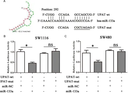
tor (Genewiz, USA). The result-ing vector was called the re- porter vector pmiRGLO-UPAT-wild type (pmiRGLO-UPAT-wt). The corresponding mutants were created by mutating the hsa-miR-133a seed region bin- ding site, which was called the reporter vector pmiRGLO-UPAT- mutated-type (pmiRGLO-UPAT-mut). miR-133a mimic or con-trol was co-transfected with the reporter vectors containing either the targeting sequenc- es or the corresponding mu- tants using Transfection rea- gent (Invitrogen, USA) accord-ing to the protocol. Luciferase activity was measured using the Dual-Luciferase Reporter Assay System (Promega, USA). Tumor formation in nude mice
[image:3.612.92.369.264.586.2]^ 2/2. The animals were sacrificed 4 weeks after injection.
Statistical analysis
Data were expressed as the mean ± SD of at least three independent experiments. Stati- stical analysis was carried out using GraphPad Prism 5 software (GraphPad Software, USA). Student’s t test was performed to analyze the data. Values of P < 0.05 were considered significant.
Results
Inverse expression of UPAT and miR-133a in clinical GC tissues and cells
Real-time PCR revealed that compared with adjacent tissues, tumors exhibited a signifi-cantly higher UPAT expression (Figure 1A) and a significantly lower miR-133a expression (Fi- gure 1B). Pearson expression analysis sugg-
ested an inverse relationship between UPAT and miR-133a expression in tumors (Figure 1C, r = -0.0707, P < 0.001). In vitro, UPAT was sig-nificantly higher expressed in SW1116 and SW480 compared with CW-2 cells (Figure 2A), while miR-133a was expressed significantly lower (Figure 2B).
Knockdown of UPAT inhibits cell proliferation and cell cycle progression in vitro
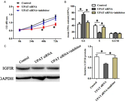
[image:4.612.97.520.75.386.2]ether the effect of knockdown UPAT on prolif-eration of CRC cells reflected cell-cycle arrest, cell-cycle progression was analyzed by flow-cytometry. The results revealed that SW480 cells transfected with UPAT siRNA had an obvi-ous cell-cycle arrest at the G1-G0 phase and had a decreased G2-S phase (Figure 2F). These results indicate that UPAT strongly pro-motes cell proliferation.
LncRNA UPAT acts as a molecular sponge of miR-133a
We performed a search for miRNAs that have complementary base pairing with GAPLINC, using online software program RNAhybrid (https://bibiserv.cebitec.uni-bielefeld.de/rna-hybrid/). The results demonstrated that
miR-133a formed complementary base pairing wi- th UPAT (Figure 3A). To further investigate wh- ether miR-133a is a functional target of UPAT, a dual-luciferase reporter assay was performed in both SW1116 and SW480 cells. We found that co-transfection of pmiRGLO-UPAT-wt and miR-133a mimic strongly decreased the lucifer-ase activity while co-transfection of pmiRGLO-UPAT-mut and miR-133a mimic did not change luciferase activity (Figure 3B and 3C). Thus, these results demonstrate that miR-133a is a UPAT-targeting miRNA.
miR-133a reverses the promoting effects of UPAT in CRC cells
[image:5.612.96.519.73.417.2]miR-133a in UPAT-induced promotion in CRC cells remained unclear. In order to confirm whether UPAT could promote CRC cells through the UPAT-miR-133a-IGF1R axis, we co-trans-fected UPAT siRNA and miR-133a inhibitor or control in SW480 cells. The CCK-8 assays (Figure 4A) and flow cytometry assays (Figure 4B) showed that miR-133a could largely rever- se the promoting effect of UPAT on CRC cell proliferation and cell cycle progression. Wes- tern blot also revealed that the promotion of IGF1R protein expression by UPAT could be largely reversed by miR-133a (Figure 4C). The- se results indicate that miR-133a can reverse the promoting effects of UPAT in CRC cells and UPAT can promote CRC cells through the UPAT-miR-133a-IGF1R axis.
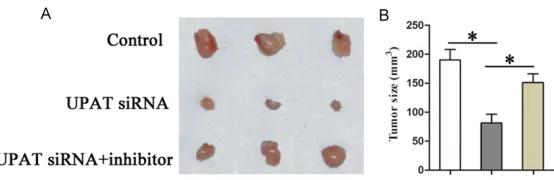
miR-133a reverses the promoting effects of UPAT on tumor growth in xenograft models of CRC
Furthermore, to determine whether UPAT could promote tumor growth in xenograft models of CRC through the UPAT-miR-133a pathway, we assessed tumor growth of xenografts derived from SW480 cells that were co-transfected with UPAT siRNA and miR-133a inhibitor or control prior to subcutaneous injection into nude mice. Our results show that both UPAT knockdown and miR-133a overexpression inhi- bited tumor growth of the SW480 xenograft (Figure 5A and 5B). Collectively, these results support a role for miR-133a in reversal of the promoting effects of UPAT on tumor grow- th in xenograft models of CRC.
diagnostic and therapeutic molecular targets for CRC is particularly crucial.
LncRNAs play critical regulatory roles in diverse cellular processes such as chromatin remodel-ing, transcription, post-transcriptional process-ing, and intracellular trafficking [12]. Recent studies have shown that lncRNAs can act as oncogenes or tumor suppressor genes to affect tumorigenesis [13]. UPAT, whose alias is AOC4P, was originally detected in hepatocellular carci-noma and acted as a tumor suppressor by enhancing Vimentin degradation and suppress-ing the EMT [14]. Additionally, UPAT has been reported to promote colon tumorigenesis by inhibiting degradation of UHRF1 [15]. However, the function and molecular mechanism of UPAT in CRC are still unclear.
In the present study, we found that UPAT expr- ession was inversely correlated to miR-133a expression in colorectal cancer tissues and cells. In addition, knockdown of UPAT inhibited cell proliferation and cell cycle progression of colorectal cancer. Further mechanistic studies revealed that UPAT could sponge endogenous miR-133a and inhibit its activity. These findings suggest that UPAT may promote CRC cell pro- liferation by acting as a molecular sponge of miR-133a.
miR-133a was reported to be downregulated in a variety of cancers, including esophageal squamous cell carcinoma, bladder cancer, pr- ostate cancer, and colorectal cancer [16-19]. Therefore, we examined the role of UPAT siRNA on miR-133a expression level and miR-133a in xenograft models of CRC. A. SW480 cells were transfected with control,
UPAT siRNA, and UPAT siRNA + miR-133a inhibitor. The cells were injected subcutaneously into 9 nude mice per flank. Surgical resections of SW480 xenograft tumors on week 4 for animals were shown. B. Measurements of tumor volumes were shown. Statistically significant differences are indicat-ed: *P < 0.05; Student’s t test.
[image:6.612.94.371.70.160.2]targets. We focused on the target genes IGF1R as it was previously reported to be repressed by 133a [20]. The results showed that miR-133a inhibitor reversed the suppression effects of UPAT siRNA on cell growth and IGF1R expres-sion in CRC cells. We also examined the role of GAPLINC on miR-378 in xenograft models of GC. The results were similar, showing that miR-133a reversed the suppression effects of UPAT siRNA on tumor growth in xenograft models of CRC.
In summary, the present study provides novel evidence of a UPAT-miR-133a-IGF1R regulatory network in CRC and reveals that UPAT is a potential new oncogene for CRC.
Acknowledgements
This work was supported by grants from the Natural Science Foundation of Shandong Pro- vince (ZR2015HL082).
Disclosure of conflict of interest
None.
Address correspondence to: Chenggang Yang, De- partment of Gastrointestinal Surgery, Liaocheng People’s Hospital, 67 Dongchang West Road, Dong- changfu District, Liaocheng 252000, Shandong Province, China. Tel: +86 635-8276110; Fax: +86 635-8966225; E-mail: [email protected]
References
[1] Siegel R, Ma J, Zou Z, Jemal A. Cancer statis-tics, 2014. CA Cancer J Clin 2014; 64: 9-29. [2] Centelles JJ. General aspects of colorectal
can-cer. ISRN Oncol 2012; 2012: 139268. [3] Fatica A, Bozzoni I. Long non-coding RNAs: new
players in cell differentiation and develop-ment. Nat Rev Genet 2014; 15: 7-21.
[4] St Laurent G, Wahlestedt C, Kapranov P. The Landscape of long noncoding RNA classifica-tion. Trends Genet 2015; 31: 239-251. [5] Wang KC, Chang HY. Molecular mechanisms of
long noncoding RNAs. Mol Cell 2011; 43: 904-914.
[6] Bian Z, Jin L, Zhang J, Yin Y, Quan C, Hu Y, Feng Y, Liu H, Fei B, Mao Y, Zhou L, Qi X, Huang S, Hua D, Xing C, Huang Z. LncRNA-UCA1 enhanc-es cell proliferation and 5-fluorouracil renhanc-esis- resis-tance in colorectal cancer by inhibiting miR-204-5p. Sci Rep 2016; 6: 23892.
[7] Yang W, Ning N, Jin X. The lncRNA H19 pro-motes cell proliferation by competitively
bind-ing to miR-200a and derepressbind-ing beta-catenin expression in colorectal cancer. Bio- med Res Int 2017; 2017: 2767484.
[8] Siegel R, Naishadham D, Jemal A. Cancer sta-tistics, 2013. CA Cancer J Clin 2013; 63: 11-30.
[9] Sadanandam A, Lyssiotis CA, Homicsko K, Collisson EA, Gibb WJ, Wullschleger S, Ostos LC, Lannon WA, Grotzinger C, Del Rio M, Lher-mitte B, Olshen AB, Wiedenmann B, Cantley LC, Gray JW, Hanahan D. A colorectal cancer classification system that associates cellular phenotype and responses to therapy. Nat Med 2013; 19: 619-625.
[10] Watanabe T, Itabashi M, Shimada Y, Tanaka S, Ito Y, Ajioka Y, Hamaguchi T, Hyodo I, Igarashi M, Ishida H, Ishiguro M, Kanemitsu Y, Kokudo N, Muro K, Ochiai A, Oguchi M, Ohkura Y, Saito Y, Sakai Y, Ueno H, Yoshino T, Fujimori T, Koinu-ma N, Morita T, Nishimura G, Sakata Y, Taka-hashi K, Takiuchi H, Tsuruta O, Yamaguchi T, Yoshida M, Yamaguchi N, Kotake K, Sugihara K. Japanese society for cancer of the colon and rectum (JSCCR) guidelines 2010 for the treatment of colorectal cancer. Int J Clin Oncol 2012; 17: 1-29.
[11] Uccello M, Malaguarnera G, Basile F, D’Agata V, Malaguarnera M, Bertino G, Vacante M, Dra-go F, Biondi A. Potential role of probiotics on colorectal cancer prevention. BMC Surg 2012; 12 Suppl 1: S35.
[12] Prensner JR, Chinnaiyan AM. The emergence of lncRNAs in cancer biology. Cancer Discov 2011; 1: 391-407.
[13] Wapinski O, Chang HY. Long noncoding RNAs and human disease. Trends Cell Biol 2011; 21: 354-361.
[14] Wang TH, Lin YS, Chen Y, Yeh CT, Huang YL, Hsieh TH, Shieh TM, Hsueh C, Chen TC. Long non-coding RNA AOC4P suppresses hepatocel-lular carcinoma metastasis by enhancing vi-mentin degradation and inhibiting epithelial-mesenchymal transition. Oncotarget 2015; 6: 23342-23357.
[15] Taniue K, Kurimoto A, Sugimasa H, Nasu E, Takeda Y, Iwasaki K, Nagashima T, Okada-Hatakeyama M, Oyama M, Kozuka-Hata H, Hiy-oshi M, Kitayama J, Negishi L, Kawasaki Y, Aki-yama T. Long noncoding RNA UPAT promotes colon tumorigenesis by inhibiting degradation of UHRF1. Proc Natl Acad Sci U S A 2016; 113: 1273-1278.