https://www.scirp.org/journal/wjcd ISSN Online: 2164-5337
ISSN Print: 2164-5329
DOI: 10.4236/wjcd.2019.912080 Dec. 19, 2019 899 World Journal of Cardiovascular Diseases
Evaluation of Left Ventricular Function after
Percutaneous Recanalization of Chronic
Coronary Occlusions: The Role of
Two-Dimensional Speckle Tracking
Echocardiography
Ahmed Emara
1, Shady Zahran
2, Neveen I. Samy
11Faculty of Medicine, Menoufia University,Menoufia, Egypt 2Elagouza Charity Hospital, Giza, Egypt
Abstract
Background: The recanalization of a chronic total coronary occlusion is the possible way to improve left ventricular (LV) function through the recovery of hibernating myocardium. Aim: The aim of this study is to evaluate the role of 2D speckle tracking in evaluation of the left ventricular (LV) systolic func-tion in chronic total occlusion (CTO) patients before and at 1 day as well as 3 months after percutaneous coronary intervention (PCI). Patients and Me-thods: A prospective observational study included 40 patients diagnosed with coronary angiography to have a chronic total occlusion. Percutaneous coro-nary revascularization was performed according to standard practices with the femoral approach. Conventional 2D echocardiography was used to assess LV functions and wall motion abnormalities scoring index (WMAI). Using speckle-tracking echocardiography was to measure global longitudinal strain (GLS) and. Follow-up of patients was done at day 1 and 3 months later after PCI. Results: Forty patients were included in this study, with a mean age of 58.55 ± 7.98 years. GLS and WMAI difference at baseline and follow-up shows a positive correlation with left ventricular ejection fraction (LVEF) changes at baseline and follow-up (p < 0.001). Mean value of baseline GLS (−14.26 ± 0.93) significantly improved at follow-up as GLS was (−15.66 ± 0.92) with p-value < 0.001. Periprocedural complications were contrast in-duced nephropathy (CIN) in 2 (5%), Cardiac tamponade 1 (2.5%) and Emer-gency re-PCI 1 (2.5%). Conclusion: The results of this study provide evi-dence to support the clinical use of 2D-STE to monitor the early changes of LV function. In patients undergoing CTO revascularization, change in GLS How to cite this paper: Emara, A., Zahran,
S. and Samy, N.I. (2019) Evaluation of Left Ventricular Function after Percutaneous Re- canalization of Chronic Coronary Occlusions: The Role of Two-Dimensional Speckle Track- ing Echocardiography. World Journal of Car- diovascular Diseases, 9, 899-914.
https://doi.org/10.4236/wjcd.2019.912080
Received: October 24, 2019 Accepted: December 16, 2019 Published: December 19, 2019
Copyright © 2019 by author(s) and Scientific Research Publishing Inc. This work is licensed under the Creative Commons Attribution International License (CC BY 4.0).
http://creativecommons.org/licenses/by/4.0/
DOI: 10.4236/wjcd.2019.912080 900 World Journal of Cardiovascular Diseases was more sensitive predictors for LV function improvement at 3-month fol-low-up.
Keywords
Chronic Total Occlusions, Left Ventricular Function, Percutaneous Coronary Intervention, Ejection Fraction, Speckle Tracking Echocardiography
1. Introduction
Chronic total occlusion (CTO) is a common condition in patients with coronary artery disease, and represents one of the most challenging targets of lesion reca-nalization for percutaneous coronary interventions (PCIs) [1]. Chronic total oc-clusion (CTO) is defined as an occluded coronary artery presenting as thrombo-lysis in myocardial infarction (TIMI) with grade 0 or 1 flow with an occlusion duration of >3 months [2]. The rationale for the recanalization of a chronic total coronary occlusion is the possible improvement of left ventricular (LV) function through the recovery of hibernating myocardium [3]. Recanalization of CTO le-sions by percutaneous coronary intervention (PCI) reportedly produces benefi-cial effects on symptoms, long-term survival, and incidence of coronary artery bypass grafting (CABG). However, mechanism of the beneficial effects of reca-nalization of CTO still remains unclear [4]. Two-dimensional speckle tracking echocardiography (2D-STE) allows for an angle-independent evaluation of myo-cardial strain, and provides comprehensive information on LV myomyo-cardial con-tractility. Thus, 2D-STE is superior in detecting subtle deteriorations of contrac-tility. These advantages of 2D-STE are useful for the detection of subclinical re-covery of dysfunctional but viable myocardium after CTO-PCI [5]. In this study we aimed to evaluate the role of 2D speckle tracking in evaluation of the LV sys-tolic function in CTO patients at 1 day as well as 3 months after percutaneous coronary intervention.
2. Patients and Methods
DOI: 10.4236/wjcd.2019.912080 901 World Journal of Cardiovascular Diseases in the study were subjected to full medical history evaluation with special em-phasis on history of Diabetes mellitus, Hypertension, dyslipidemia and Cigarette smoking. Clinical examination on admission with special emphasis on Heart rate, rhythm, Systolic and diastolic blood pressures at the time of the study. Twelve-lead resting surface was done to all patients. Laboratory evaluation with special emphasis on lipid profile and Kidney function tests. Percutaneous coro-nary revascularization was performed by operators experienced in the treatment of CTO according to their standard practices with the femoral approach. The operation was considered successful when the residual stenosis was ≤30% of the intraluminal diameter, with TIMI grade III flow and no in hospital complica-tions (death, acute myocardial infarction, or emergency coronary surgery). DAPT was prescribed to all patients for at least 1 year after stent implantations. Revascularization was done through antegrade technique: 1) Antegrade wire es-calation technique (AWE) either done by Sliding technique with Fielder group of wires or penetrating technique with ASAHI conquest and conquest pro wires or drilling technique. Attempts to cross CTO using specific guidewires according to the morphology of the cap. Generally, it starts with a soft and fine-tipped guidewire (1.0 g), coated with polymer. If the crossing is unsuccessful, a slightly heavier wire (4.0 g), also polymer coated, or a sharp, tapered 12-gauge wire was used. If the guidewire enters the true distal lumen (confirmed in two orthogonal projections), the micro catheter is advanced through the occlusion and the gui-dewire is replaced by a traditional one, followed by balloon angioplasty and stent implantation. If the guidewire comes out of the vessel architecture, it must be retracted and redirected. 2) Antegrade dissection reentry was done by using pilot and miracle wires with intentional use of the subintimal space to cross the occlu-sion in leocclu-sions more than 20 mm length.
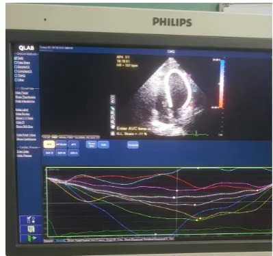
DOI: 10.4236/wjcd.2019.912080 902 World Journal of Cardiovascular Diseases dimensions of an object. When applied to left ventricle, left ventricular deforma-tion is defined by the three normal strains (longitudinal, circumferential, and radial) [8]. Using speckle-tracking echocardiography was to measure global lon-gitudinal strain. The 2D echocardiography images (transmit/receive 1.9/4.0 MHz) were obtained from several views with frame rates of 30 - 90 frames/s. Digital data were stored and analyzed off-line. LV endocardial surface was traced ma-nually, and the speckle tracking width was modified to cover the whole LV wall thickness so as to obtain curves for the peak longitudinal strain of: the inferior septum and lateral wall in the apical four-chamber view (4C-PLS); the inferior and anterior walls in the apical two-chamber view (2C-PLS); and The inferior lateral and anterior septum in the apical three-chamber view (3C-PLS). LV glob-al longitudinglob-al systolic strain (GLS) was cglob-alculated by averaging the peak systolic values of the six LV walls (Figure 1 and Figure 2). All the echocardiographic stu-dies were performed by one echocardiographer. Follow-up of patients was done at day 1 and 3 months later after percutaneous coronary intervention was done by clinical examination and echocardiography as described.
Figure 1. Left ventricular global longitudinal strain pre PCI.
[image:4.595.274.471.544.701.2]DOI: 10.4236/wjcd.2019.912080 903 World Journal of Cardiovascular Diseases
3. Statistical Methods
Data were statistically described in terms of range, mean standard deviation (SD), median, frequencies (number of cases) and percentages when appropriate. Com-parison of quantitative variables between the study groups was done using Mann Whitney U test for independent samples. For comparing categorical data, Chi square (χ2) test was performed. Exact test was used instead when the expected
frequency is less than 5. ROC curves were plotted to determine cutoff values. Correlation between various variables was done using Spearman rank correla-tion equacorrela-tion for non-normal variables. A probability value (p-value) less than 0.05 was considered statistically significant. All statistical calculations were done using computer programs Microsoft Excel 2010 (Microsoft Corporation, NY, USA) and SPSS (Statistical Package for the Social Science, SPSS Inc., Chicago, IL, USA) version 15 for Microsoft Windows.
4. Results
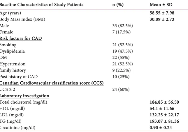
40 patients were included in this study, their ages ranged from 47 to 79 years with a mean 58.55 ± 7.98 years. Males were 33 (82.5%) and females were 7 (17.5%). All patients had at least one risk factor and the most frequent was DM 22 (55%) followed by hypertension 21 (52.5%), and smoking 21 (52.5%) beside other risk factors. Patients who had chest pain with Canadian class score (CCS) ≥ 2 were 24 (60%). Lipid profile of the patients shows that mean levels of choles-terol, HDL, LDL and TG were (184.85 ± 56.50, 54.1 ± 11.66, 132.25 ± 22.17 and 193.07 ± 81.36) mg/dl respectively. Mean creatinine level in the studied patients was 0.90 ± 0.24 mg/dl (Table 1). Regarding the angiographic data, two vessel disease lesions were most frequent 42.5%, then multivessels 32.5% and single vessel 25.5%. The CTO was located in the left anterior descending in 15 (37.5%) patients, the right coronary artery in 16 (40%) patients, and the circumflex artery in 9 (22.5%) patients. Mean of J-CTO score was 1.77 ± 1.02 in CTO vessels. Re-vascularization was done by antegrade approach only through antegrade wire escalation (AWE) in 35 (87.5%) and Antegrade Dissection and Reentry (ADR) in 5 (12.5%). The mean of procedural time was 77.25 ± 18.22 minutes (Table 2).
The echocardiographic findings show that mean of pre-procedural LVEDV 104 ± 32.17 and day 1 post-procedural LVEDV 105 ± 38.85 without statistical difference. Mean of pre-procedural LVESV was 61.52 ± 15.14 and day 1 post- procedural LVESV was 58.22 ± 11.65 with statistical difference (p = 0.031).
Mean value of pre-procedural GLS (−14.22 ± 0.91) and day 1 post-procedural GLS was −14.26 ± 0.93) without significant statistical difference. Mean value of pre-procedural WMSI (1.48 ± 0.31) and day 1 post-procedural WMSI was (1.41 ± 0.17) without significant statistical difference. Mean value of pre-procedural EF% for patients was 53.88% ± 14.30% and the mean value of day 1 post-pro- cedural EF was 54.85% ± 7.63% without statistical difference between them (Table 3).
DOI: 10.4236/wjcd.2019.912080 904 World Journal of Cardiovascular Diseases 61.52 ± 15.14 and follow-up LVESV was 50.60 ± 14.07 with statistical differ-ence (p = 0.047). Mean value of pre-procedural GLS (−14.22 ± 0.91) and fol-low-up GLS was (−15.66 ± 0.92) with significant statistical difference (p < 0.001). Mean value of pre-procedural WMSI (1.48 ± 0.31) and follow-up WMSI was (1.20 ± 0.13) with significant statistical difference (p < 0.001). Mean value of pre-procedural EF% for patients was 53.88% ± 14.30% and the mean value of follow-up EF was 55.54% ± 8.19% without statistical difference between them (Table 4).
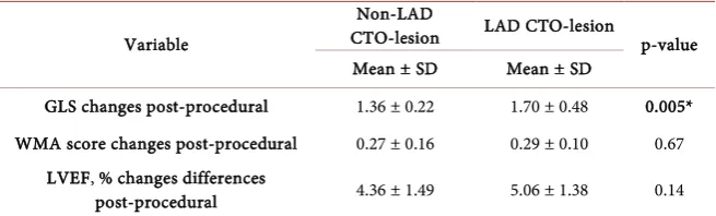
[image:6.595.211.534.303.531.2]Comparison between post-procedural changes in GLS and WMA in LAD and non-LAD CTO-lesions shows that there was significant statistical difference be-tween Non-LAD CTO-lesion and LAD CTO-lesion as regard post-procedural changes in GLS but non-significant in post-procedural WMA score changes and LVEF, % changes (Table 5).
Table 1. Baseline characteristics of study patients.
Baseline Characteristics of Study Patients n (%) Mean ± SD
Age (years)
Body Mass Index (BMI) Male
Female
Risk factors for CAD Smoking
Dyslipidemia DM Hypertension family history Past history of CAD
Canadian Cardiovascular classification score (CCS) CCS ≥ 2
33 (82.5%) 7 (17.5%) 21 (52.5%) 19 (47.5%) 22 (55%) 21 (52.5%) 9 (22.5%) 10 (25%) 24 (60%)
58.55 ± 7.98 30.09 ± 2.73
Laboratory investigation Total cholesterol (mg/dl) HDL (mg/dl)
LDL (mg/dl) TG (mg/dl) Creatinine (mg/dl)
184.85 ± 56.50 54.1 ± 11.66 132.25 ± 22.17 193.07 ± 81.36 0.90 ± 0.24
Table 2. Characteristics of angiographic data.
Characteristics of angiographic data %
Single vessel n (%)
2 vessels n (%)
Multivessels
10 (25%) 17 (42.5%) 13 (32.5%) CTO vessel
LAD n (%)
LCX n (%)
RCA n (%)
15 (37.5%) 9 (22.5%) 16 (40%)
J-CTO score mean ± SD 1.77 ± 1.02
Antegrade approach only n (%)
-Antegrade wire escalation (AWE) n (%)
-Antegrade Dissection and Reentry (ADR) n (%)
40 (100%) 35 (87.5%) 5 (12.5%)
[image:6.595.212.541.562.729.2]DOI: 10.4236/wjcd.2019.912080 905 World Journal of Cardiovascular Diseases
Table 3. Comparison between echocardiographic parameters at pre-procedural and day 1
post-procedural follow-up.
Variables preprocedural Day 1 p-value
Mean ± SD Mean ± SD
LVEF, % LVEDV, ml
LVESV, ml GLS WMSI
53.88 ± 14.30 104 ± 32.17 61.52 ± 15.14 −14.22 ± 0.91 1.48 ± 0.31
54.85 ± 7.63 105 ± 38.85 58.22 ± 11.65 −14.26 ± 0.93 1.41 ± 0.17
0.12 0.46 0.031*
[image:7.595.210.540.270.360.2]0.39 0.11 EDV, end-diastolic volume; e0, early diastolic velocity of mitral annular displacement; GCS, global circum-ferential strain; GLS, global longitudinal strain; LA, left atrium; LVEF, left ventricular ejection fraction; SV, stroke volume; WMSI, wall motion score index.
Table 4. Comparison between echocardiographic parameters pre-procedural and 3 months
follow-up.
Variables preprocedural 3 month follow-up p-value
Mean ± SD Mean ± SD
LVEF, % LVEDV, ml LVESV, ml
GLS WMSI
53.88 ± 14.30 104 ± 32.17 61.52 ± 15.14 −14.22 ± 0.91 1.48 ± 0.31
55.54 ± 8.19 101 ± 45.22 50.60 ± 14.07 −15.66 ± 0.92 1.20 ± 0.13
0.76 0.18 0.047 ˂0.001* ˂0.001* EDV, end-diastolic volume; e0, early diastolic velocity of mitral annular displacement; GCS, global circum-ferential strain; GLS, global longitudinal strain; LA, left atrium; LVEF, left ventricular ejection fraction; SV, stroke volume; WMSI, wall motion score index.
Table 5. Comparison between post-procedural changes in GLS, WMA in LAD, and
non-LAD CTO-lesions.
Variable
Non-LAD
CTO-lesion LAD CTO-lesion p-value
Mean ± SD Mean ± SD
GLS changes post-procedural 1.36 ± 0.22 1.70 ± 0.48 0.005*
WMA score changes post-procedural 0.27 ± 0.16 0.29 ± 0.10 0.67
LVEF, % changes differences
post-procedural 4.36 ± 1.49 5.06 ± 1.38 0.14
Lad: Left Anterior Descending; CTO: chronic total occlusion; GLS: global longitudinal strain; WMA: wall motion abnormality; LVEF: left ventricular ejection fraction.
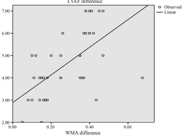
GLS and WMAI difference at baseline and 3 months follow-up shows a posi-tive correlation with LVEF changes at baseline and follow-up with significant statistical difference (p < 0.001) (Table 6, Figure 3 and Figure 4).
[image:7.595.211.539.436.535.2]confi-DOI: 10.4236/wjcd.2019.912080 906 World Journal of Cardiovascular Diseases dence interval CI (0.638 - 0.941).
Periprocedural complications were contrast induced nephropathy in 2 (5%), Cardiac tamponade 1 (2.5%) and Emergency re-PCI 1 (2.5%) (Table 8).
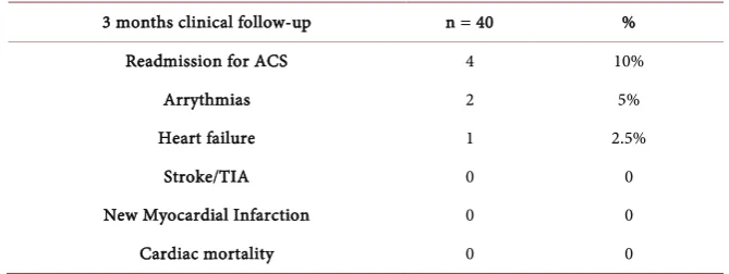
[image:8.595.224.522.209.422.2]Clinical follow-up of patients 3 months post-procedural shows that 2 (10%) patients were readmitted because of chest pain without any new angiographic le-sions and received medical treatment. 2 patients (5%) had paroxysmal AF and one patient developed signs of heart failure. There was no MI, stroke or mortali-ty in the studied patients (Table 9).
Figure 3. Correlations between changes in LVEF by echocardiographic parameters
and changes in GLS.
Figure 4. Correlations between changes in LVEF by echocardiographic parameters
[image:8.595.224.522.472.692.2]DOI: 10.4236/wjcd.2019.912080 907 World Journal of Cardiovascular Diseases
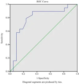
[image:9.595.244.501.320.591.2]Figure 5. Roc curve for GLS difference to predict improved left ventricular EF.
Figure 6. Roc curve for WMSI difference to predict improved left ventricular EF.
Table 6. Correlations between changes in LVEF by echocardiographic parameters and
changes in GLS and WMA.
Variable r p-value
Difference GLS 0.496 0.001*
WMSI difference 0.599 <0.001*
[image:9.595.209.538.654.710.2]DOI: 10.4236/wjcd.2019.912080 908 World Journal of Cardiovascular Diseases
Table 7. Roc curve for GLS and WMSI difference to predict improved left ventricular EF.
Variable AUC CI 95% Cut-off Sensitivity Specificity p-value
GLS 0.82 0.677 - 0.962 1.025 95% 40.7% 0.001*
WMSI 0.79 0.638 - 0.941 0.10 100% 55% 0.002*
[image:10.595.208.539.195.255.2]GLS: Global longitudinal strain; WMSI: Wall motion scoring index; GLS: Global longitudinal strain; WMSI: Wall motion scoring index.
Table 8. Peri-procedural Complications.
Peri-procedural Complications %
-adiac tamponade -mergency re-PCI -Contrast induced nephropathy
n (%) n (%) n (%)
1 (2.5%) 1 (2.5%) 2 (5%)
Table 9. Clinical follow-up after 3-month follow-up across groups.
3 months clinical follow-up n = 40 %
Readmission for ACS 4 10%
Arrythmias 2 5%
Heart failure 1 2.5%
Stroke/TIA 0 0
New Myocardial Infarction 0 0
Cardiac mortality 0 0
5. Discussion
[image:10.595.206.541.286.412.2]revas-DOI: 10.4236/wjcd.2019.912080 909 World Journal of Cardiovascular Diseases cularization, LV function was most commonly assessed by the LV ejection frac-tion using ventriculography [12] or 2D echocardiography [13]. Recently, the use of more accurate non-invasive methods for quantifying LV function, such as cardiac MRI [14] and 2D STE [15], also confirmed the improvement in LV func-tion after CTO PCI. In this study, we aimed to evaluate the role of 2D speckle tracking in evaluation of the LV systolic function in CTO patients before and at 1 day as well as 3 months after percutaneous coronary intervention. To achieve our aim, this prospective observational study was conducted on 40 patients who have ischemic heart disease and diagnosed with coronary angiography to have a chronic total occlusion. Patients were assessed by 2D echocardiography and speckle tracking and were followed-up for recovery of left ventricular function in 3 months after percutaneous coronary revascularization for chronic total occlu-sion.In this study, comparison of mean value of baseline GLS (−14.26 ± 0.93) and 3 moths follow-up GLS were (−15.66 ± 0.92) with significant statistical dif-ference (p < 0.001). Comparison of mean value of baseline WMSI (1.41 ± 0.17) and 3 months follow-up WMSI were (1.20 ± 0.13) with significant statistical dif-ference (p < 0.001). The more significant improvement of GLS and WMSI reflect improvement in the myocardial contractile function based on strain analysis. In accordance with several studies as follows; Choi etal., 2009 reported that longi-tudinal strain analysis by 2D-STE is a quite sensitive and suitable method to evaluate this component of cardiac motion. Multiple retrospective studies have shown the potential benefit of PCI in patients with CTO [11]. Successful treat-ment improves anginal symptoms, exercise tolerance, and LV function. The prospective Total Occlusion Angioplasty Study (TOAST-GISE) showed that re-vascularization of a CTO is associated with relieved angina and reduces the 12- month incidence of cardiac death or MI and the need for CABG [9]. This study found that the LVEF tended to improve at 3 months after percutaneous revascu-larization in patients with CTO. These findings were in agreement with Wang et al. in 2019 that using 2D-STE, GLS was observed to be restored as early as 1 day after CTO-PCI and improvement of LVEF was observed for up to 3 and 6 months. Their results demonstrate that 2D-STE is a reliable way to monitor ear-ly subclinical LV changes [16]. Also, Erdogan etal. in 2013 showed that restor-ing the coronary blood flow in chronic total occlusion patients reduces the left ventricular volumes and improves the left ventricular ejection fraction and the global longitudinal strain of hibernating myocardium. These findings were as-sessed by two-dimensional speckle tracking echocardiography immediately afer and one month after the procedure [15]. Nakachi etal. in 2017 assessed func-tional recovery after PCI to CTO vessels. They reported that the change in lon-gitudinal strain (LS) was more sensitive for removal of ischemia by CTO PCI, indicating the utility of LS to monitor the therapeutic effects of CTO recanaliza-tion [17].
DOI: 10.4236/wjcd.2019.912080 910 World Journal of Cardiovascular Diseases their study showed that the recovery of left ventricular function could be de-tected early post-revascularization of coronary artery disease by either ejection fraction or global longitudinal strain measurements; however the latter is more accurate. Improvement of GLPS is correlated moderately with target vessel re-vascularization involving non-left anterior descending artery. González etal., in 2016 concluded that Successful revascularization of CTO improves regional sys-tolic function determined by WMSI and decreases angina. LS of treated seg-ments tend to decrease after the procedure, with no change in global measures of cardiac function. In the current study, GLS and WMAI difference at baseline and follow-up shows a positive correlation with LVEF changes at baseline and follow-up with significant statistical difference (p < 0.001). This was in line with Staron etal. in 2013 who reported that GLS correlates well with EF measured by echocardiography, and GLS is a superior predictor of outcome compared with LVEF [19]. This could be explained by probable initial appearance of systolic dysfunction in the longitudinal direction, as the longitudinally oriented suben-docardial fibers are more vulnerable to myocardial ischemia and fibrosis. GLS improvement can predict improved left ventricular EF and could be used as a prognstic factor with high sensitivity 95% (p = 0.002). Biopsies of hibernating myocardium always show defects in nearly all cells [20]. The pathological changes include loss of sarcomeres and myofibrils in the center of the cells, absence of contractile material in the perinuclear areas, and presence of cellular debris in the enlarged extracellular space [21]. The dysfunctional heart tissue in CTO pa-tients can be improved after revascularization [22]. Steg and colleagues found that the recovery time of dysfunctional myocardium is dependent on the extent of damage at the cellular level, which is affected by different factors such as the duration and severity of ischemia [23]. The current results were not in agree-ment with several investigators; Henriques etal. in 2016 reported that they did not find an overall benefit for CTO-PCI in terms of LVEF or LVEDV [24]. Ma-shayekhi etal. in 2018 reported that no benefit was seen for CTO-PCI in terms of the segmental wall thickening, LVEF and LVEDV [25]. Also, in the study by Chimura et al. in 2019, LVEF and LVEDV also showed no significant changes both successful and failed PCI groups during the 9 month follow-up, respective-ly [26]. However, since there was report that reduced major adverse coronary event rates at 12 months in CTO-PCI compared with optimal medical therapy. These differences can be explained by small sample size or difference in some end points of the studies. However, longer duration of follow-up in these studies differ from follow-up period in the current study.
tampo-DOI: 10.4236/wjcd.2019.912080 911 World Journal of Cardiovascular Diseases nade is low (approximately 0.5%) [28]. The risk factors predictive of perforation include the use of oversized compliant balloons (balloon-to-artery ratio > 1.2) coupled with relatively high inflation pressure and hydrophilic and stiffer wires, particularly in calcified and tortuous arteries [29].
6. Limitations
The limitations in the current study were small sample size and short duration follow-up and observational not comparative. Also, we didn’t correlate LV func-tions improvement to related CTO vessel, as longitudinal shortening mechanics were correlated with the target vessel revascularization. Since the vascular cov-erage of LAD in myocardium is wider than that of right coronary artery as well as left circumflex artery, the remodeling area of LAD covered myocardial cells is wider when LAD gets ischemia or infarct, this must be considered.
7. Conclusion
CTO-PCI can effectively improve LV function. The results of this study provide evidence to support the clinical use of 2D-STE to monitor the early changes of LV function. In patients undergoing CTO revascularization, change in LS and GLS was more sensitive predictors for LV function improvement at 3-month follow-up.
Disclosure Statement
Data are available at Faculty of Medicine, Menoufia University and Al Agouza Charity Hospital through the authors only as at those places there is no record-ing system for the data of all cases. Only researcher keeps his data.
Conflicts of Interest
The authors declare no conflicts of interest regarding the publication of this pa-per.
References
[1] Stone, G.W., Reifart, N.J., Moussa, I., Hoye, A., Cox, D.A., Colombo, A., Baim, D.S., Teirstein, P.S., Strauss, B.H., Selmon, M., et al. (2005) Percutaneous Recanalization of Chronically Occluded Coronary Arteries: A Consensus Document. Part II. Cir-culation, 112, 2530-2537.https://doi.org/10.1161/CIRCULATIONAHA.105.583716
[2] Sun, D., Wang, J., Tian, Y., Narsinh, K., Wang, H., Li, C., et al. (2012) Multimodali-ty Imaging Evaluation of Functional and Clinical Benefits of Percutaneous Coro-nary Intervention in Patients with Chronic Total Occlusion Lesion. Theranostics, 2, 788-800.https://doi.org/10.7150/thno.4717
[3] Shivalkar, B., Maes, A., Borgers, M., Ausma, J., Scheys, I., Nuyts, J., et al. (1996) Only Hibernating Myocardium Invariably Shows Early Recovery after Coronary Revascularization. Circulation, 94, 308-315.https://doi.org/10.1161/01.CIR.94.3.308
DOI: 10.4236/wjcd.2019.912080 912 World Journal of Cardiovascular Diseases A., Di Mario, C. and Hildick-Smith, D. (2014) Long-Term Follow-up of Elective Chronic Total Coronary Occlusion Angioplasty: Analysis from the U.K. Central Car-diac Audit Database. Journal of the American College of Cardiology, 64, 235-243.
https://doi.org/10.1016/j.jacc.2014.04.040
[5] Geyer, H., Caracciolo, G., Abe, H., et al. (2010) Assessment of Myocardial Mechan-ics Using Speckle Tracking Echocardiography: Fundamentals and Clinical Applica-tions. Journal of the American Society of Echocardiography, 23, 351-369.
https://doi.org/10.1016/j.echo.2010.02.015
[6] Lang, R.M., Badano, L.P., Mor-Avi, V., et al. (2015) Recommendations for Cardiac Chamber Quantification by Echocardiography in Adults: An Update from the Amer-ican Society of Echocardiography and the European Association of Cardiovascular Imaging. Journal of the American Society of Echocardiography, 28, 1-39.e14.
https://doi.org/10.1016/j.echo.2014.10.003
[7] Moller, J.E., Hillis, G.S., Oh, J.K., et al. (2006) Wall Motion Score Index and Ejec-tion FracEjec-tion for Risk StratificaEjec-tion after Acute Myocardial InfarcEjec-tion. American Heart Journal, 151, 419-425.https://doi.org/10.1016/j.ahj.2005.03.042
[8] D’hooge, J., Heimdal, A., Jamal, F., Kukulski, T., Bijnens, B., Rademakers, F., et al. (2000) Regional Strain and Strain Rate Measurements by Cardiac Ultrasound: Prin-ciples, Implementation and Limitations. European Journal of Echocardiography, 1, 154-170. https://doi.org/10.1053/euje.2000.0031
[9] Olivari, Z., Rubartelli, P., Pisicone, F., Ettori, F., Fontanelli, A., Salemme, L., et al. (2003) On Behalf of TOAST-GISE Investigators. Immediate Results and One-Year Clinical Outcome after Percutaneous Coronary Interventions in Chronic Total Occlu-sions (TOAST-GISSE). Journal of the American College of Cardiology, 41, 1672-1678.
https://doi.org/10.1016/S0735-1097(03)00312-7
[10] Braunwald, E. and Bonow, R.O. (2012) Braunwald’s Heart Disease: A Textbook of Cardiovascular Medicine. Saunders, Philadelphia, PA.
[11] Choi, J.O., Cho, S.W., Song, Y.B., Cho, S.J., Song, B.G., Lee, S.C. and Park, S.W. (2009) Longitudinal 2D Strain at Rest Predicts the Presence of Left Main and Three Vessel Coronary Artery Disease in Patients without Regional Wall Motion Abnor-mality. European Journal of Echocardiography, 10, 695-701.
https://doi.org/10.1093/ejechocard/jep041
[12] Werner, G. (2017) A Randomized Multicentre Trial to Evaluate the Utilization of Revascularization or Optimal Medical Therapy for the Treatment of Chronic Total Coronary Occlusions (EuroCTO). EuroPCR, Paris, France.
[13] Piscione, F., Galasso, G., De Luca, G., Marrazzo, G., Sarno, G., Viola, O., Accardo, D. and Chiariello, M. (2005) Late Reopening of an Occluded Infarct Related Artery Improves Left Ventricular Function and Long Term Clinical Outcome. Heart, 91, 646-651.https://doi.org/10.1136/hrt.2004.041152
[14] Kirschbaum, S.W., Baks, T. and van den Ent, M. (2008) Evaluation of Left Ventri-cular Function Three Years after Percutaneous Recanalization of Chronic Total Co-ronary Occlusions. American Journal of Cardiology, 101, 179-185.
https://doi.org/10.1016/j.amjcard.2007.07.060
[15] Erdogan, E., Akkaya, M., Bacaksiz, A., Tasal, A., Sonmez, O., Elbey, M.A., Kul, S., Vatankulu, M.A., Turfan, M. and Goktekin, O. (2013) Early Assessment of Percu-taneous Coronary Interventions for Chronic Total Occlusions Analyzed by Novel Echo-Cardiographic Techniques. Clinics, 68, 1333-1337.
https://doi.org/10.6061/clinics/2013(10)07
DOI: 10.4236/wjcd.2019.912080 913 World Journal of Cardiovascular Diseases Dimensional Speckle Tracking Echocardiography. Herz, 44, 170-174.
[17] Nakachi, T., Kato, S., Kirigaya, H., Iinuma, N., Fukui, K., Saito, N., et al. (2017) Pre-diction of Functional Recovery after Percutaneous Coronary Revascularization for Chronic Total Occlusion Using Late Gadolinium Enhanced Magnetic Resonance Imaging. Journal of Cardiology, 69, 836-842.
https://doi.org/10.1016/j.jjcc.2017.01.002
[18] Rifqi, S., Sungkar, S., Sobirin, M.A., Uddin, I., Furuse, Y., Nugroho, M.A., et al. (2017) Early Recovery of Left Ventricular Function after Revascularization of Co-ronary Artery Disease Detected by Myocardial Strain. Biomedical Research, 28, 1487-1492.
[19] Staron, A., Bansal, M., Kalakoti, P., Nakabo, A., Gasior, Z., Pysz, P., et al. (2013) Speckle Tracking Echocardiography Derived 2-Dimensional Myocardial Strain Pre-dicts Left Ventricular Function and Mass Regression in Aortic Stenosis Patients Undergoing Aortic Valve Replacement. The International Journal of Cardiovascular Imaging, 29, 797-808.
[20] White, H.D. and Braunwald, E. (1998) Applying the Open Artery Theory: Use of Predictive Survival Markers. European Heart Journal, 19, 1132-1139.
https://doi.org/10.1053/euhj.1998.1017
[21] Monteiro, P., Antunes, A., Goncalves, L.M. and Providencia, L.A. (2003) Long- Term Clinical Impact of Coronary Collateral Vessels after Acute Myocardial Infarc-tion. Revista Portuguesa de Cardiologia, 22, 1051-1061.
[22] Joyal, D., Afilalo, J. and Rinfret, S. (2010) Effectiveness of Recanalization of Chronic Total Occlusions: A Systematic Review and Meta-Analysis. American Heart Journal, 160, 179-187. https://doi.org/10.1016/j.ahj.2010.04.015
[23] Steg, P.G., Thuaire, C., Himbert, D., Carrie, D., Champagne, S., Coisne, D., et al. (2004) DECOPI (Des Obstruction-COronaireen Post-Infarctus): A Randomized Multicenter Trial of Occluded Artery Angioplasty after Acute Myocardial Infarc-tion. European Heart Journal, 25, 2187-2194.
https://doi.org/10.1016/j.ehj.2004.10.019
[24] Henriques, J.P., Hoebers, L.P., Ramunddal, T., Laanmets, P., Eriksen, E., Bax, M., et al. (2016) EXPLORE Trial Investigators. Percutaneous Intervention for Concurrent Chronic Total Occlusions in Patients with STEMI: The EXPLORE Trial. Journal of the American College of Cardiology, 68, 1622-1632.
https://doi.org/10.1016/j.jacc.2016.07.744
[25] Mashayekhi, K., Nührenberg, T.G., Toma, A., Gick, M., Ferenc, M., Hochholzer, W., et al. (2018) A Randomized Trial to Assess Regional Left Ventricular Function after Stent Implantation in Chronic Total Occlusion: The REVASC Trial. JACC: Cardiovascular Interventions, 19, 1982-1991.
https://doi.org/10.1016/j.jcin.2018.05.041
[26] Chimura, M., Yamada, S., Yasaka, Y. and Kawai, H. (2019) Improvement of Left Ventricular Function Assessment by Global Longitudinal Strain after Successful Percutaneous Coronary Intervention for Chronic Total Occlusion. PLoS ONE, 14, e0217092. https://doi.org/10.1371/journal.pone.0217092
[27] Patel, V.G., Brayton, K.M. and Tamayo, A. (2013) Angiographic Success and Pro-cedural Complications in Patients Undergoing Percutaneous Coronary Chronic Total Occlusion Interventions: A Weighted Meta-Analysis of 18,061 Patients from 65 Studies. JACC: Cardiovascular Interventions, 6, 128-136.
https://doi.org/10.1016/j.jcin.2012.10.011
DOI: 10.4236/wjcd.2019.912080 914 World Journal of Cardiovascular Diseases Procedural Complications in Patients Undergoing Retrograde Percutaneous Coro-nary Chronic Total Occlusion Interventions: A Weighted Meta-Analysis of 3,482 Patients from 26 Studies. International Journal of Cardiology, 174, 243-248.
https://doi.org/10.1016/j.ijcard.2014.04.004
[29] Gruberg, L., Pinnow, E., Flood, R., et al. (2006) Incidence, Management, and Out-come of Coronary Artery Perforation during Percutaneous Coronary Intervention American Journal of Cardiology, 86, 680-682.