organic papers
o2880
Piet al. C6H8ClN3O2 doi:10.1107/S1600536805025201 Acta Cryst.(2005). E61, o2880–o2881
Acta Crystallographica Section E Structure Reports Online
ISSN 1600-5368
Chlorometronidazole
Wen-Xia Pi, Yu-Min Yang, Huan-Qiu Li and Hai-Liang Zhu*
Institute of Functional Biomolecules, State Key Laboratory of Pharmaceutical Biotechnology, Nanjing University, Nanjing 210093, People’s Republic of China
Correspondence e-mail: [email protected]
Key indicators
Single-crystal X-ray study
T= 298 K
Mean(C–C) = 0.004 A˚
Rfactor = 0.045
wRfactor = 0.128
Data-to-parameter ratio = 16.0
For details of how these key indicators were automatically derived from the article, see http://journals.iucr.org/e.
#2005 International Union of Crystallography
Printed in Great Britain – all rights reserved
In the title compound, 1-(2-chloroethyl)-2-methyl-5-nitro-1H-imidazole, C6H8ClN3O2, the dihedral angle between the imidazole ring and the attached nitro group is 6.5 (1).
Comment
Metronidazole is a widely used antibacterial medicine (Creditoet al., 2000; Mendz & Me´graud, 2002). As a result of its toxicity, its structural modification has recently been an active research topic (Alcaldeet al., 1984). In this paper, we report the structure of chlorometronidazole, (I), in which the hydroxyl group of the metronidazole has been replaced by a chloride group.
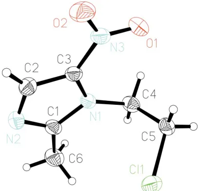
The molecular structure of (I) is shown in Fig. 1. In each molecule, the imidazole ring is a well defined plane with an average deviation of 0.002 (1) A˚ . The nitro N atom lies 0.060 (1) A˚ above the plane; the two other groups attached to the ring are located on the opposite side of the plane, with C4
[image:1.610.228.432.516.712.2]Received 20 July 2005 Accepted 8 August 2005 Online 12 August 2005
Figure 1
0.144 (1) and C6 0.004 (1) A˚ from the ring. The dihedral angle between the imidazole ring and the nitro plane is 6.5 (1).
Experimental
To a pyridine solution (5 ml) of metronidazole (171 mg, 1 mmol) was added SO2Cl2 (149 mg, 1.1 mmol) and Na2CO3. This mixture was
stirred for 3 h at 353 K. The solvents and excess SO2Cl2were then
removed. The residue was recrystallized from chloroform, large crystals suitable for X-ray crystal structure determination being formed in 12 h. These were collected by filtration, washed with chloroform and diethyl ether, and dried in a vacuum desiccator using CaCl2(yield: 98%). Elemental analysis found: C 37.9, H 4.3, N 22.2%.
Calculated for C6H8ClN3O2: C 38.0, H 4.2, N 22.2%. 1
H NMR (500 MHz, DMSO-d6):7.95 (1H,s, broad, —CH—), 4.55 (2H,t, J=
7.2 Hz, —CH2—), 3.35 (2H, t, J = 7.2 Hz, —CH2—), 2.26 (3H, s,
—CH3—).
Crystal data
C6H8ClN3O2
Mr= 189.60
Monoclinic,P21=c a= 12.098 (14) A˚
b= 11.007 (13) A˚
c= 6.295 (7) A˚
= 97.886 (18)
V= 830.3 (16) A˚3
Z= 4
Dx= 1.517 Mg m
3
MoKradiation Cell parameters from 1562
reflections
= 6.3–27.4
= 0.42 mm1
T= 298 (2) K Prism, colorless 0.380.350.30 mm
Data collection
Siemens SMART CCD area-detector diffractometer
’and!scans
Absorption correction: multi-scan (SADABS; Sheldrick, 1996)
Tmin= 0.850,Tmax= 0.890
4602 measured reflections
1778 independent reflections 1293 reflections withI> 2(I)
Rint= 0.032 max= 27.0
h=15!14
k=14!14
l=6!7
Refinement
Refinement onF2 R[F2> 2(F2)] = 0.045
wR(F2) = 0.128
S= 1.02 1778 reflections 111 parameters
H-atom parameters constrained
w= 1/[2
(Fo2) + (0.0716P)2]
whereP= (Fo2+ 2Fc2)/3
(/)max= 0.001
max= 0.26 e A˚
3
min=0.25 e A˚
3
Extinction correction:SHELXL97
Extinction coefficient: 0.039 (6)
All H atoms were positioned geometrically and constrained to ride on their parent atoms, with C—H = 0.96 A˚ . They were treated as riding atoms, withUiso(H) = 1.2Ueq(C).
Data collection:SMART(Siemens, 1996); cell refinement:SAINT; data reduction: SAINT(Siemens, 1996); program(s) used to solve structure:SHELXS97(Sheldrick, 1997a); program(s) used to refine structure: SHELXL97 (Sheldrick, 1997a); molecular graphics:
SHELXTL(Sheldrick, 1997b); software used to prepare material for publication:SHELXTL.
This project was sponsored by the Scientific Research Foundation for the Returned Overseas Chinese Scholars, State Education Ministry.
References
Alcalde, E., Manos, L., Valls, N. & Elguero, J. (1984).J. Heterocycl. Chem.21, 1647–51.
Credito, K. L., Jacobs, M. R. & Appelbaum, P. C. (2000).Diagn. Microbiol. Infec. Dis.38, 181–183.
Mendz, G. L. & Me´graud, F., (2002).Trends Microbiol.10, 370–375. Sheldrick, G. M. (1996).SADABS. University of Go¨ttingen, Germany. Sheldrick, G. M. (1997a). SHELXL97 and SHELXS97. University of
Go¨ttingen, Germany.
Sheldrick, G. M. (1997b).SHELXTL. Version 5.1. Bruker AXS Inc., Madison, Wisconsin, USA.
supporting information
sup-1 Acta Cryst. (2005). E61, o2880–o2881
supporting information
Acta Cryst. (2005). E61, o2880–o2881 [https://doi.org/10.1107/S1600536805025201]
Chlorometronidazole
Wen-Xia Pi, Yu-Min Yang, Huan-Qiu Li and Hai-Liang Zhu
1-(2-chloroethyl)-2-methyl-5-nitro-1H-imidazole
Crystal data
C6H8ClN3O2
Mr = 189.60 Monoclinic, P21/c
Hall symbol: -P 2ybc
a = 12.098 (14) Å
b = 11.007 (13) Å
c = 6.295 (7) Å
β = 97.886 (18)°
V = 830.3 (16) Å3
Z = 4
F(000) = 392
Dx = 1.517 Mg m−3
Mo Kα radiation, λ = 0.71073 Å Cell parameters from 1562 reflections
θ = 6.3–27.4°
µ = 0.42 mm−1
T = 298 K Prism, colorless 0.38 × 0.35 × 0.30 mm
Data collection
Siemens SMART CCD area-detector diffractometer
Radiation source: fine-focus sealed tube Graphite monochromator
φ and ω scans
Absorption correction: multi-scan (SADABS; Sheldrick, 1996)
Tmin = 0.850, Tmax = 0.890
4602 measured reflections 1778 independent reflections 1293 reflections with I > 2σ(I)
Rint = 0.032
θmax = 27.0°, θmin = 1.7°
h = −15→14
k = −14→14
l = −6→7
Refinement
Refinement on F2
Least-squares matrix: full
R[F2 > 2σ(F2)] = 0.045
wR(F2) = 0.128
S = 1.02 1778 reflections 111 parameters 0 restraints
Primary atom site location: structure-invariant direct methods
Secondary atom site location: difference Fourier map
Hydrogen site location: inferred from neighbouring sites
H-atom parameters constrained
w = 1/[σ2(F
o2) + (0.0716P)2]
where P = (Fo2 + 2Fc2)/3
(Δ/σ)max = 0.001
Δρmax = 0.26 e Å−3
Δρmin = −0.25 e Å−3
Extinction correction: SHELXL97, Fc*=kFc[1+0.001xFc2λ3/sin(2θ)]-1/4
Special details
Geometry. All e.s.d.'s (except the e.s.d. in the dihedral angle between two l.s. planes) are estimated using the full covariance matrix. The cell e.s.d.'s are taken into account individually in the estimation of e.s.d.'s in distances, angles and torsion angles; correlations between e.s.d.'s in cell parameters are only used when they are defined by crystal symmetry. An approximate (isotropic) treatment of cell e.s.d.'s is used for estimating e.s.d.'s involving l.s. planes.
Refinement. Refinement of F2 against ALL reflections. The weighted R-factor wR and goodness of fit S are based on F2,
conventional R-factors R are based on F, with F set to zero for negative F2. The threshold expression of F2 > σ(F2) is used
only for calculating R-factors(gt) etc. and is not relevant to the choice of reflections for refinement. R-factors based on F2
are statistically about twice as large as those based on F, and R- factors based on ALL data will be even larger.
Fractional atomic coordinates and isotropic or equivalent isotropic displacement parameters (Å2)
x y z Uiso*/Ueq
Cl1 0.26941 (5) 1.08697 (6) 1.18846 (9) 0.0648 (3) N1 0.25179 (12) 0.97894 (14) 0.7240 (2) 0.0383 (4) N3 0.11217 (14) 0.81284 (17) 0.6867 (3) 0.0481 (5) C3 0.22186 (15) 0.85786 (18) 0.7036 (3) 0.0400 (5) C4 0.18295 (17) 1.08359 (17) 0.7650 (4) 0.0476 (5) H4A 0.1161 1.0839 0.6605 0.057* H4B 0.2240 1.1576 0.7457 0.057* O1 0.03299 (12) 0.88355 (15) 0.6762 (3) 0.0626 (5) O2 0.10093 (14) 0.70113 (14) 0.6819 (3) 0.0704 (5) C5 0.14968 (17) 1.08328 (19) 0.9867 (4) 0.0505 (5) H5A 0.1063 1.0109 1.0052 0.061* H5B 0.1031 1.1534 1.0032 0.061* C2 0.31708 (18) 0.7931 (2) 0.6932 (3) 0.0498 (5) H2A 0.3205 0.7091 0.6798 0.060* N2 0.40514 (14) 0.86834 (17) 0.7051 (3) 0.0534 (5) C1 0.36460 (15) 0.9803 (2) 0.7245 (3) 0.0459 (5) C6 0.43437 (19) 1.0915 (2) 0.7436 (5) 0.0654 (7) H6A 0.4388 1.1231 0.8866 0.098* H6B 0.4016 1.1513 0.6431 0.098* H6C 0.5080 1.0720 0.7133 0.098*
Atomic displacement parameters (Å2)
U11 U22 U33 U12 U13 U23
supporting information
sup-3 Acta Cryst. (2005). E61, o2880–o2881
Geometric parameters (Å, º)
Cl1—C5 1.791 (3) C4—H4B 0.9700
N1—C1 1.364 (3) C5—H5A 0.9700
N1—C3 1.382 (3) C5—H5B 0.9700
N1—C4 1.465 (3) C2—N2 1.344 (3)
N3—O1 1.229 (2) C2—H2A 0.9300
N3—O2 1.237 (3) N2—C1 1.338 (3)
N3—C3 1.407 (3) C1—C6 1.482 (3)
C3—C2 1.364 (3) C6—H6A 0.9600
C4—C5 1.504 (4) C6—H6B 0.9600
C4—H4A 0.9700 C6—H6C 0.9600
C1—N1—C3 105.06 (16) Cl1—C5—H5A 109.4 C1—N1—C4 125.71 (17) C4—C5—H5B 109.4 C3—N1—C4 128.78 (17) Cl1—C5—H5B 109.4 O1—N3—O2 123.09 (19) H5A—C5—H5B 108.0 O1—N3—C3 120.07 (19) N2—C2—C3 110.1 (2) O2—N3—C3 116.84 (19) N2—C2—H2A 125.0 C2—C3—N1 107.24 (18) C3—C2—H2A 125.0 C2—C3—N3 127.3 (2) C1—N2—C2 105.97 (18) N1—C3—N3 125.38 (17) N2—C1—N1 111.66 (18) N1—C4—C5 113.12 (16) N2—C1—C6 123.78 (19) N1—C4—H4A 109.0 N1—C1—C6 124.57 (19)
C5—C4—H4A 109.0 C1—C6—H6A 109.5
N1—C4—H4B 109.0 C1—C6—H6B 109.5
C5—C4—H4B 109.0 H6A—C6—H6B 109.5
H4A—C4—H4B 107.8 C1—C6—H6C 109.5
C4—C5—Cl1 111.37 (16) H6A—C6—H6C 109.5
C4—C5—H5A 109.4 H6B—C6—H6C 109.5