Introduction: Most neurophysiology departments around the world establish their own normative data. However, ethnic differences are not taken into account. Our aim was to establish normal nerve conduction studies (NCS) data for routinely tested nerves in individuals of Pakistani (South Asian) origin and to compare with Western published data. Materials and Methods: One hundred healthy adults’ nerves were assessed, using standardized techniques. Individuals were grouped into age groups. Gender differences were assessed. Results: Of the 100 volunteers, 49 were female and 51 were male. Their mean age was 39.8 years. Findings showed statistically significant prolongation of median distal motor latency (DML) and F‑wave latency with age and reduction of median, ulnar, and sural sensory amplitudes as age increased. Gender differences showed consistent difference in the normal values for median, ulnar, and peroneal DMLs and respective F‑wave latencies, which were significantly shorter in females. Sensory amplitudes of tested upper extremity nerves were significantly lower in males. Comparing with available data, our findings are similar to the Saudi population but significantly different from the American and multiethnic Malaysian populations. Pakistani individuals generally have significantly higher amplitudes and faster conduction velocities with similarities to South Asian studies. Conclusions: We recommend normative NCS parameters for commonly tested nerves for the Pakistani population, using standardized techniques to ensure highest quality testing and outcomes.
Keywords: Nerve conduction studies, normal values, normative data, practices standards, reference values
Developing Normative Reference Values for Nerve Conduction Studies
of Commonly Tested Nerves among a Sample Pakistani Population
Zaitoon Shivji, Anita Jabeen, Safia Awan1, Sara KhanAccess this article online Quick Response Code:
Website:
www.ruralneuropractice.com
DOI:
10.4103/jnrp.jnrp_370_18
Address for correspondence: Dr. Sara Khan, Department of Neurophysiology (Neurology), Aga Khan University Hospital, Karachi, Pakistan. E‑mail: [email protected]
own reference values[3,5] or use established Western
values. The neurophysiology department at the Aga Khan University Hospital (AKUH), Karachi, was established over 20 years ago. The aim of our study is to help establish NCS normative data that could be applicable in the Pakistani population and can be used in neurophysiology departments around the country. We also aim to compare our findings with the existing published data from different populations.
Original Article
Introduction
N
erve conduction studies (NCS) are an important diagnostic tool in the assessment of peripheral nervous system disorders and helps in diagnosis, prognostication, and longitudinal monitoring of a disease process.[1,2] In order to identify abnormalvalues, a set of “normative” or “reference” values need to be determined.[3,4] The latter are derived
from a healthy subject population that approximates the demographics of the patients. Age, gender, temperature, techniques, and possibly ethnicity may affect the results.
With the dearth of published data on reference values that can be widely used, most neurophysiology departments around the world tend to identify their
Departments of Neurophysiology and
1Medicine, Aga Khan
University Hospital, Karachi,
Pakistan
Abs
tract
This is an open access journal, and articles are distributed under the terms of the Creative Commons Attribution-NonCommercial-ShareAlike 4.0 License, which allows others to remix, tweak, and build upon the work non-commercially, as long as appropriate credit is given and the new creations are licensed under the identical terms.
For reprints contact: [email protected]
Materials and Methods
A prospective, descriptive study of willing healthy controls was undertaken over a 3‑year period (2014–2017). Each individual was administered a standard screening questionnaire and only those individuals meeting the inclusion criteria were included in the study. Written consent was obtained. This study was approved by the Ethical Review Committee of the AKUH.
One hundred asymptomatic adult volunteers, over the age of 18 years, willing to consent, and without known sensory symptoms, neurologic disorders, or other medical issues that could affect peripheral nerves such as diabetes mellitus, thyroid disorders, Vitamin B12 deficiency, cancer with or without chemotherapy, or other such medications were enrolled. Participants underwent neurological examination, height and weight determinations, and NCS using standard methodology for sensory (median, ulnar, radial, median palmar, ulnar palmar, and sural) and motor (median, ulnar, peroneal, and tibial) nerves.
For NCS and electromyography, a Nicolet Viking machine was used. Low‑ and high‑pass filters were set at 2 Hz and 10 kHz and 20 Hz and 3 kHz for motor studies and sensory studies, respectively, with a sweep speed of 2 ms/division.
Nerve conduction responses were recorded using standardized techniques. All responses were obtained using supramaximal stimulation and proper placement of electrodes. Data were collected for the following parameters: onset and peak latencies, amplitude, amplitude drop, conduction velocity (CV), and F‑waves. All sensory nerves were examined antidromically. Amplitudes for the sensory nerve action potential (SNAP) were measured from the peak of the negative potential to the peak of the positive potential. SNAP peak and onset latencies were noted. Sensory median and ulnar nerves were stimulated at the wrist and recorded from the 2nd and 5th digits, respectively,
at a distance of 14 cm. Distance for stimulation for palmar responses was 8 cm. The radial nerve was stimulated 10 cm proximal to the active electrode on the dorsolateral aspect of the distal forearm.
Median and ulnar motor nerves were tested with the active electrode over the motor point of the abductor pollicis brevis and the abductor digiti minimi muscles, respectively. In the lower limbs, for the tibial and peroneal nerves, the active electrodes were over the adductor hallucis and extensor digitorum brevis, respectively. The reference electrodes were placed on the tendon of the muscle in question. Distal stimulations
were 8 cm proximal to the active electrode for motor median, ulnar, and peroneal nerves and 9 cm for the tibial nerve. The acceptable limb temperature for performing NCS was ≥32°C. In the event of cool limb temperatures, the participant was warmed up using hot water bags or a heating pad to maintain the temperature, as needed.
Statistical analysis
Statistical analysis was performed using IBM SPSS Statistics for Windows, Version 19.0. (Armonk, NY: IBM Corp). Continuous variables were reported as mean and standard deviation. Independent sample t‑test was used to assess differences between sexes and age groups. Reference value was set to outside the mean ±2 standard deviations. P < 0.05 was taken as cutoff level for statistical significance.
Results
Of the 100 healthy volunteers, 49 (49%) were female and 51 (51%) were male. Their mean age was 39.8 ± 12.3 years (range 18–67). Participants were grouped in the age groups of <30 (n = 24), 30–39 (n = 28), 40–49 (n = 20), and >50 (n = 28) years. Male and female differences were assessed. Normal values for motor nerve and sensory nerve conductions are summarized in Tables 1 and 2, respectively.
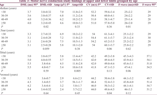
Tables 3 and 4 show the variation in normal values of motor and sensory findings among various age groups.
Variations with age
There is a significant increase in the median distal motor latency and F‑wave latency with age (>40 years) [Table 3]. Median SNAP from digit II and ulnar SNAP from digit V showed significantly higher amplitudes in the younger age groups (<40 years). Sural sensory amplitudes also decreased with increasing age (P < 0.003) [Table 4]. Tibial motor amplitudes decreased with age (P 0.005). Although peroneal motor CVs statistically increased with age, the increase through age groups is not consistent and therefore not clinically significant.
Variations with gender
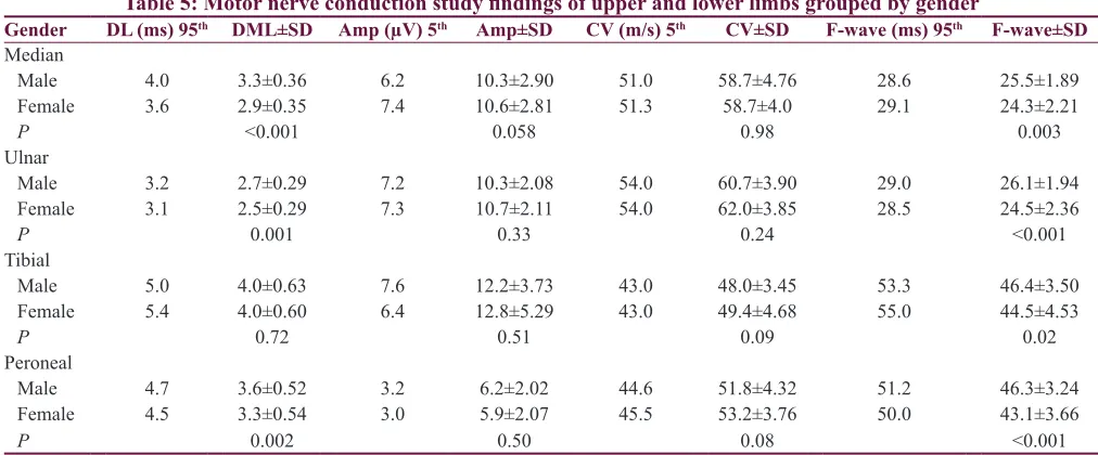
Gender differences (adjusted for height) showed consistent difference in the normal values for median, ulnar, and peroneal distal motor latencies and respective F‑wave latencies, which were significantly shorter in females for median, ulnar, and peroneal nerves [Table 5]. No significant gender differences were noted for motor conduction velocities.
amplitude and longer latency (P = 0.03, amplitude: male = 40.2 ± 19.6 and female = 48.4 ± 18.8, peak latency: male = 1.8 ± 0.15 and female = 1.7 ± 0.14).
Discussion
NCS are an important tool for the evaluation of peripheral nervous system disorders. The main aim of our study
was to provide reference NCS values for commonly tested nerves in normal Pakistani adult volunteers and to compare with published normative values, using standardized techniques. Falck and Stalberg[6] determined
that “Reference values in one laboratory can be used in others when techniques are standardized.” Normative data for NCS to our knowledge have not been formally Table 1: Motor nerve conduction study findings of upper and lower limbs
Distance DML±SD (ms) DML 95th Amp±SD (µV) Amp 5th CV±SD (m/s) CV 5th F‑wave±SD (ms) F‑wave 95th
Median 8.0 3.1±0.40 3.8 10.5±2.8 6.2 58.7±4.3 51 24.9±2.1 29
Ulnar 8.0 2.5±0.30 3.1 10.4±2.0 7.4 61.3±3.9 54 25.3±2.2 29
Tibial 9.0 3.9±0.6 5.0 12.5±4.5 5.7 48.6±4.1 42 45.4±4.1 54
Peroneal 8.0 3.4±0.5 4.5 6.1±2.0 3.2 50.7±3.9 45 44.7±3.7 50
DML: Distal motor latency, Amp: Amplitude, CV: Conduction velocity, SD: Standard deviation, 95th: 95th percentile, 5th: 5th percentile
Table 2: Sensory nerve conduction study findings of upper and lower limbs
Onset DL (95th) Mean±SD Peak DL (95th) Mean±SD Amp (5th) Mean±SD CV (5th) Mean±SD Median
Palmar 1.6 1.3±0.14 2.1 1.9±0.15 40.1 98±39.3 51.6 63±7.3
Digit II 2.7 2.3±0.18 3.5 3.0±0.27 18.0 41±15.1 52.0 60.4±4.5
Ulnar
Palmar 1.5 1.2±0.13 2.0 1.7±0.15 1 8.1 44.1±19.5 53.3 66.1±6.6
Digit V 2.7 2.3±0.24 3.4 3.0±0.30 15.0 35.0±13.0 60.0±5.6
Radial ‑ forearm 2.0 1.6±0.18 2.5 2.1±0.19 25.0 45.5±14.2 60.0 71.7±6.0
Sural ‑ calf 3.0 2.4±0.3 3.8 3.2±0.32 12.0 22.5±8.8 44.0 57±6.3
DL: Distal latency, Amp: Amplitude, CV: Conduction velocity, SD: Standard deviation, 95th: 95th percentile, 5th: 5th percentile
Table 3: Motor nerve conduction study findings of upper and lower limbs grouped by age
Age DML (ms) 95th DML±SD Amp (µV) 5th Amp±SD CV (m/s) 5th CV±SD F‑wave (ms)±SD F‑wave 95th Median (years)
<30 3.7 3.0±0.32 7.0 11.0±2.5 52.2 59.6±3.4 25±2.2 29
30‑39 3.6 3.0±0.37 6.8 11.2±2.6 50.4 60.0±4.1 24±2.2 29
40‑49 4.0 3.2±0.36 6.2 10.2±2.5 51.0 58.1±4.7 25±1.4 28
≥50 4.0 3.2±0.43 4.6 10.0±3.3 51.0 57.8±5.0 26±2.0 29
P 0.02 0.33 0.36 0.01
Ulnar (years)
<30 3.3 2.7±0.33 6.9 10.2±2.2 54 61.3±4.1 25.1±2.2 29
30‑39 3.1 2.4±0.28 7.2 11.0±2.3 54.4 61.1±3.7 25.1±2.4 30
40‑49 3.1 2.6±0.28 7.5 10.5±1.5 54.2 63.2±3.5 25.2±2.2 28
≥50 3.1 2.5±0.28 5.8 10.1±2.0 54 60.1±3.7 25.8±2.2 29
P 0.059 0.38 0.06 0.62
Tibial (years)
<30 5.0 3.8±0.57 5.8 13.4±4.7 42.2 49.2±3.8 45.3±4.3 57.1
30‑39 5.0 4.0±0.55 5.7 14.5±5.1 42.0 49.4±4.5 43.9±4.1 54.1
40‑49 5.5 3.8±0.6 4.5 11.4±2.8 42.0 49.0±4.6 45.6±3.1 51.0
≥50 5.5 4.0±0.7 5.4 10.6±3.8 41.8 47.1±3.3 47±4.1 54.5
P 0.59 0.005 0.13 0.06
Peroneal (years)
<30 5.2 3.6±0.7 2.9 6.6±2.3 44.2 50.4±3.8 44.1±3.2 49.7
30‑39 4.3 3.4±0.5 3.7 56.1±1.7 46.4 52.4±3.3 43.8±4.0 51.4
40‑49 4.2 3.4±0.4 3.2 5.8±1.7 46.0 50.3±3.2 44.1±4.1 54.7
≥50 4.4 3.4±0.52 2.4 5.7±2.2 44.0 49.6±4.5 46±3.3 51.2
P 0.64 0.45 0.04 0.12
performed previously in our population. The reference values currently used in Pakistan are either established by individual institutions based on a very small sample size or based on Western data. We determined that there are some differences between the two groups.
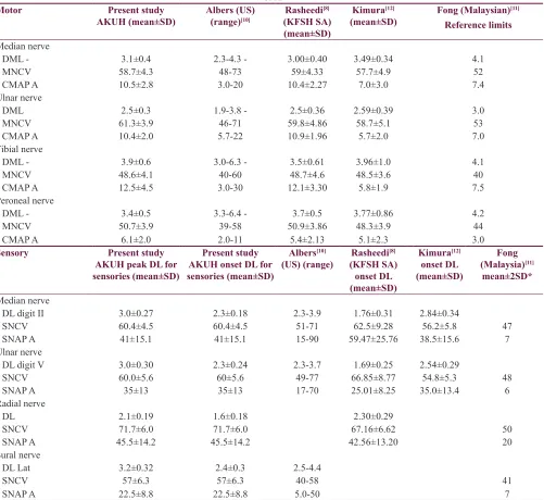
Our reference values were compared to normative nerve conduction values from other population‑based studies
that investigated asymptomatic healthy controls, using standardized techniques. The results of our study show similar findings to that described (taking variation in measurements techniques into consideration) in the Saudi and Indian population and somewhat to the Kuwaiti population[7‑9] but significantly different for sensory
studies in the American and multi‑ethnic Malaysian Table 5: Motor nerve conduction study findings of upper and lower limbs grouped by gender
Gender DL (ms) 95th DML±SD Amp (µV) 5th Amp±SD CV (m/s) 5th CV±SD F‑wave (ms) 95th F‑wave±SD Median
Male 4.0 3.3±0.36 6.2 10.3±2.90 51.0 58.7±4.76 28.6 25.5±1.89
Female 3.6 2.9±0.35 7.4 10.6±2.81 51.3 58.7±4.0 29.1 24.3±2.21
P <0.001 0.058 0.98 0.003
Ulnar
Male 3.2 2.7±0.29 7.2 10.3±2.08 54.0 60.7±3.90 29.0 26.1±1.94
Female 3.1 2.5±0.29 7.3 10.7±2.11 54.0 62.0±3.85 28.5 24.5±2.36
P 0.001 0.33 0.24 <0.001
Tibial
Male 5.0 4.0±0.63 7.6 12.2±3.73 43.0 48.0±3.45 53.3 46.4±3.50
Female 5.4 4.0±0.60 6.4 12.8±5.29 43.0 49.4±4.68 55.0 44.5±4.53
P 0.72 0.51 0.09 0.02
Peroneal
Male 4.7 3.6±0.52 3.2 6.2±2.02 44.6 51.8±4.32 51.2 46.3±3.24
Female 4.5 3.3±0.54 3.0 5.9±2.07 45.5 53.2±3.76 50.0 43.1±3.66
P 0.002 0.50 0.08 <0.001
DML: Distal motor latency, Amp: Amplitude, CV: Conduction velocity, SD: Standard deviation, 95th: 95th percentile, 5th: 5th percentile
Table 4: Sensory nerve conduction study findings of upper and lower limbs grouped by age
Age DL (ms) 95th Peak DL±SD Amp (µV) 5th Amp±SD CV (m/s) 5th CV±SD
Median digit II (years)
<30 3.5 3.0±0.37 18.6 43.6±13.2 52 59.2±4.8
30‑39 3.4 3.0±0.24 34.4 51.0±16.2 53.4 61.7±4.0
40‑49 3.4 3.1±0.19 16.3 35.4±10 53 60.2±3.8
≥50 3.5 3.1±0.21 15.4 32.5±12.0 53 60.2±12
P 0.06 <0.0001 0.27
Ulnar digit V (years)
<30 3.4 3.0±0.38 13.2 37.8±14.5 51 61.6±5.7
30‑39 3.4 3.0±0.21 22.9 39.5±11.6 53 61.2±5.3
40‑49 3.4 3.1±0.31 20.0 34.8±10.1 50 57.4±4.1
≥50 3.5 3.1±0.24 14.0 27.7±11.8 50.4 58.7±6.1
P 0.052 0.003 0.03
Radial (years)
<30 2.5 2.1±0.18 24 47.0±16.7 60 71.4±6.1
30‑39 2.4 2.0±0.18 28.1 49.5±14.5 63 72.0±4.5
40‑49 2.8 2.1±0.24 30 43.5±11.6 57.1 71.5±7.0
≥50 2.6 2.1±0.19 30 41.6±12.5 55.3 72.0±6.5
P 0.38 0.18 0.97
Sural (years)
<30 3.0 2.4±0.20 10.5 24.5±10.8 49 57.7±4.2
30‑39 3.6 3.1±0.27 15.4 26.2±8.2 45.8 57.7±4.2
40‑49 4.0 3.1±0.31 10.1 21.0±7.6 56 57.5±7.1
≥50 4.1 3.2±0.4 9.3 18.2±6.0 51 56.2±8.1
P 0.94 0.003 0.62
population[10‑12] when comparing with published data
[Table 7]. CVs in our participants were comparable to the CVs reported in a study from India;[13] however, we
were not able to compare amplitudes as their measuring technique was baseline to peak. Pakistani individuals generally have significantly increased sensory amplitudes and faster conduction velocities. Sensory distal latencies were slightly increased which we believe could be related to the variations in distal distances used or whether onset or peak latency was used. For this reason, we opted to make note of both latencies.
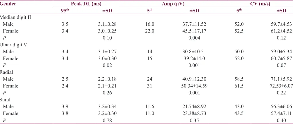
Amplitude drop was also noted as the age increased [Table 4]. This could be explained secondary to the natural loss of axons with aging. Similar to previous studies,[14] SNAP amplitudes decreased with age, significantly for median, ulnar, and sural SNAPs. Females had consistently higher SNAP amplitudes compared to males, most significantly in upper‑limb nerves. This is similar to findings described in other populations.[15,16] Median and ulnar SNAP (digit II and V)
showed statistically significant amplitude decrease in males [Table 6]. Bolton and Carter[17] suggested this to
be possibly because of varying finger circumference. Variation in SNAP amplitude from other studies could also be because of different methods of measurement.[7]
We used peak‑to‑peak amplitude measurement, the peak latency for distal latency, and onset latency for CV of sensory nerves studies. Males had higher normal values for median, ulnar, and peroneal F‑wave latency, which has also been described in a study from India.[9]
Some of these differences may be due to body habitus, genetic factors, lifestyle, and dietary
differences.[8] Furthermore, factors such as the filter
setting of the high frequencies and low frequencies make significant difference in the NCS values; therefore, filter settings should be specified.[18] Albers[10] used filter settings
of 2 Hz and 10 kHz and 20 Hz and 2 kHz for motor and sensory studies, respectively, for their laboratory.
Another important factor that can make a difference is the need for standardized techniques. They include accurate and standardized distal distances, supramaximal stimulation but preventing excessive stimulation or overstimulation. Temperature is an important factor. Electrode application and impedance is another technical factor to consider. Standardization of techniques ensures quality of nerve conduction studies. It helps in comparing studies over periods of time with easier comparison of outcome measures and effectiveness of treatment.[18]
There is considerable mention in literature about the effect of age, gender, height, and other individual related variables, but little information on how ethnicity affects the NCS. Fong et al.[11] have demonstrated the
ethnic/racial differences in their article. Yet, others have failed to demonstrate that ethnicity[19,20] makes a
difference. Therefore, as suggested by Lahoria et al. “highly developed reference values with correction for applicable variables may be used for cohorts of mixed ethnicity.”[20]
Normative reference values of commonly tested peripheral nerves were established for a sample healthy adult population in Karachi, Pakistan. Values for sensory NCS for most nerves tested varied from published data.[10‑12] This study showed that amplitudes were higher
and CVs were faster. Our data have similarities to the Table 6: Sensory nerve conduction study findings of upper and lower limbs grouped by gender
Gender Peak DL (ms) Amp (µV) CV (m/s)
95th ±SD 5th ±SD 5th ±SD
Median digit II
Male 3.5 3.1±0.28 16.0 37.7±11.52 52.0 59.7±4.53
Female 3.4 3.0±0.25 22.0 45.5±17.17 52.5 61.2±4.52
P 0.10 0.004 0.12
Ulnar digit V
Male 3.4 3.1±0.27 14 30.8±10.51 50.0 59.0±5.34
Female 3.4 3.0±0.30 15 39.2±14.0 52.0 60.7±5.87
P 0.02 0.001 0.07
Radial
Male 2.5 2.2±0.18 24 40.9±12.30 58.5 71.1±5.92
Female 2.4 2.1±0.21 31 50.34±14.59 61.5 72.53±6.07
P 0.26 0.001 0.22
Sural
Male 3.9 3.2±0.34 11.6 21.74±8.92 43.0 56.3±6.06
Female 3.8 3.2±0.30 11.0 23.38±8.73 43.5 57.4±7.11
P 0.78 0.35 0.40
South Asian studies we reviewed. The motor studies, though, were comparable to reported normative nerve conduction values, with variations according to sex and age. Amplitude variations could be explained by the different methods of amplitude measurement, distal latency variations by differences in distal distances used, techniques and equipment settings, genetic factors, lifestyle, dietary differences, and possibly ethnicity. It is, therefore, highly recommended and encouraged to obtain our own reference values for NCS. It would be interesting to do a normal NCS for the South Asian population.
Financial support and sponsorship
Nil.
Conflicts of interest
There are no conflicts of interest.
References
1. Benatar M, Wuu J, Peng L. Reference data for commonly used sensory and motor nerve conduction studies. Muscle Nerve
2009;40:772‑94.
2. Mallik A, Weir AI. Nerve conduction studies: Essentials and pitfalls in practice. J Neurol Neurosurg Psychiatry 2005;76 Suppl 2:ii23‑31.
Table 7: Comparison of current study data of motor and sensory nerves with published data from other regions of the world
Motor Present study
AKUH (mean±SD) Albers (US) (range)[10] Rasheedi [8] (KFSH SA) (mean±SD)
Kimura[12]
(mean±SD) Fong (Malaysian) [11]
Reference limits
Median nerve
DML ‑ 3.1±0.4 2.3‑4.3 ‑ 3.00±0.40 3.49±0.34 4.1
MNCV 58.7±4.3 48‑73 59±4.33 57.7±4.9 52
CMAP A 10.5±2.8 3.0‑20 10.4±2.27 7.0±3.0 7.4
Ulnar nerve
DML 2.5±0.3 1.9‑3.8 ‑ 2.5±0.36 2.59±0.39 3.0
MNCV 61.3±3.9 46‑71 59.8±4.86 58.7±5.1 53
CMAP A 10.4±2.0 5.7‑22 10.9±1.96 5.7±2.0 7.0
Tibial nerve
DML ‑ 3.9±0.6 3.0‑6.3 ‑ 3.5±0.61 3.96±1.0 4.1
MNCV 48.6±4.1 40‑60 48.7±4.6 48.5±3.6 40
CMAP A 12.5±4.5 3.0‑30 12.1±3.30 5.8±1.9 7.5
Peroneal nerve
DML ‑ 3.4±0.5 3.3‑6.4 ‑ 3.7±0.5 3.77±0.86 4.2
MNCV 50.7±3.9 39‑58 50.9±3.86 48.3±3.9 44
CMAP A 6.1±2.0 2.0‑11 5.4±2.13 5.1±2.3 3.0
Sensory Present study
AKUH peak DL for sensories (mean±SD)
Present study AKUH onset DL for sensories (mean±SD)
Albers[10]
(US) (range) Rasheedi [8] (KFSH SA)
onset DL (mean±SD)
Kimura[12] onset DL (mean±SD)
Fong (Malaysia)[11]
mean±2SD*
Median nerve
DL digit II 3.0±0.27 2.3±0.18 2.3‑3.9 1.76±0.31 2.84±0.34
SNCV 60.4±4.5 60.4±4.5 51‑71 62.5±9.28 56.2±5.8 47
SNAP A 41±15.1 41±15.1 15‑90 59.47±25.76 38.5±15.6 7
Ulnar nerve
DL digit V 3.0±0.30 2.3±0.24 2.3‑3.7 1.69±0.25 2.54±0.29
SNCV 60.0±5.6 60±5.6 49‑77 66.85±8.77 54.8±5.3 48
SNAP A 35±13 35±13 17‑70 25.01±8.25 35.0±13.4 6
Radial nerve
DL 2.1±0.19 1.6±0.18 2.30±0.29
SNCV 71.7±6.0 71.7±6.0 67.16±6.62 50
SNAP A 45.5±14.2 45.5±14.2 42.56±13.20 20
Sural nerve
DL Lat 3.2±0.32 2.4±0.3 2.5‑4.4
SNCV 57±6.3 57±6.3 40‑58 41
SNAP A 22.5±8.8 22.5±8.8 5.0‑50 7
3. Dorfman LJ, Robinson LR. AAEM minimonograph #47:
Normative data in electrodiagnostic medicine. Ff. Muscle Nerve
1997;20:4‑14.
4. Peng L, Wuu J, Benatar M. Developing reference data for nerve conduction studies: An application of quantile regression. Muscle Nerve 2009;40:763‑71.
5. Chen S, Andary M, Buschbacher R, Del Toro D, Smith B, So Y,
et al. Electrodiagnostic reference values for upper and lower limb nerve conduction studies in adult populations. Muscle
Nerve 2016;54:371‑7.
6. Falck B, Stålberg E. Motor nerve conduction studies: Measurement principles and interpretation of findings. J Clin Neurophysiol 1995;12:254‑79.
7. Shehab DK. Normative data of nerve conduction studies in the
upper limb in Kuwait: Are they different from the western data? Med Princ Pract 1998;7:203‑8.
8. Rasheedi J, Ali Q, Shivji Z, Shah M, Al‑Said Y, Al‑Falah A,
et al. Normative nerve conduction study (NCS) Data in the Saudi population. P3.176, 2015 AAN annual meeting supplement.
Neurology 2015;84:82.
9. Pawar SM, Taksande AB, Singh R. Normative data of upper limb nerve conduction in Central India. Indian J Physiol Pharmacol
2011;55:241‑5.
10. Albers JW. Nerve Conduction Manual, University of Michigan.
Available from: https://wiki.med.umich.edu/download/
attachments/66716003/SECTION04.pdf. [Last accessed on 2019
Feb 06].
11. Fong SY, Goh KJ, Shahrizaila N, Wong KT, Tan CT. Effects
of demographic and physical factors on nerve conduction study values of healthy subjects in a multi‑ethnic asian population.
Muscle Nerve 2016;54:244‑8.
12. Kimura J. Electrodiagnosis in Diseases of Nerve and Muscle: Principles and Practice. Philadelphia: FA Davis; 2001. p. 3.
13. McKnight J, Nicholls PG, Loretta D, et al. Reference values for nerve function assessments among a study population in northern
India – III: Sensory and motor nerve conduction. Neurol Asia 2010;15:39‑54.
14. Tong HC, Werner RA, Franzblau A. Effect of aging on sensory nerve conduction study parameters. Muscle Nerve 2004;29:716‑20.
15. Alemdar M. Effects of gender and age on median and ulnar
nerve sensory responses over ring finger. J Electromyogr Kinesiol 2014;24:52‑7.
16. Fujimaki Y, Kuwabara S, Sato Y, Isose S, Shibuya K, Sekiguchi Y, et al. The effects of age, gender, and body
mass index on amplitude of sensory nerve action potentials: Multivariate analyses. Clin Neurophysiol 2009;120:1683‑6.
17. Bolton CF, Carter KM. Human sensory nerve compound action
potential amplitude: Variation with sex and finger circumference. J Neurol Neurosurg Psychiatry 1980;43:925‑8.
18. Dillingham T, Chen S, Andary M, Buschbacher R, Del Toro D, Smith B, et al. Establishing high‑quality reference
values for nerve conduction studies: A report from the
normative data task force of the American Association of Neuromuscular and Electrodiagnostic Medicine. Muscle
Nerve 2016;54:366‑70.
19. Buschbacher RM, Koch J. Race effect on nerve conduction
studies: A comparison between 50 blacks and 50 whites. Arch Phys Med Rehabil 1999;80:536‑9.
20. Lahoria R, Litchy W, Dyck PJ, et al. Reference Attributes
of Nerve Conduction and Ethnicity: Comparison in Northern
Plain Indians, Mexican Americans and Caucasians (P6.097).