Ganesh et al. World Journal of Pharmaceutical and Life Sciences
FORMULATION AND EVALUATION OF LYCOPENE NIOSOMES WITH SPECIAL
EMPHASIS ON STABILITY
Ganesh N.S*, K. Bhagya Lakshmi and Vineeth Chandy
Department of Pharmaceutics, T. John College of Pharmacy, Gottigere, Bengaluru-560083.
Article Received on 15/03/2017 Article Revised on 04/04/2017 Article Accepted on 25/04/2017
INTODUCTION
Nutritional therapy and phyto-therapy have emerged as new concepts of health aid in recent years. The term nutraceuticals was coined from “nutrition” and “pharmaceutical” by Stephen Defelice MD, founder and chairman of the foundation for innovation in medicine, Cranford.[1]
Consumption of nutraceuticals from plant origin has become most popular to improve health, and to prevent and treat diseases. Nutraceuticals are naturally derived bioactive compounds that are found in foods, dietary and herbal products and have health promoting, disease preventing and medicinal properties.[2,3]
Some popular phyto nutraceuticals include lycopene from tomato, glucosamine from ginseng, curcumin from turmeric, etc.[4]
The main disadvantage of nutraceuticals is its stability towards environmental factors such as heat, moisture, light etc. Nowadays to overcome the stability issues of nutraceuticals many innovative methods have been developed like nanotechnology.[5]
Lycopene is a member of caroteniod family and it is naturally occurring compound having a strong antioxidant property which neutralizes the free radicals generated from oxygen. It is hydrophobic compound,
having poor absorption and stability. It consists of two isomeric forms that are cis-lycopene and trans-lycopene.[6,7] It is susceptible to chemical changes such as oxidation followed by degradation or isomerization when exposed to light, heat and oxygen, and hence has a short storage life if not stored properly.[8]
In processing steps of lycopene such as homogenization and extraction, lycopene appears to be stable except for the initial loss in the process. Stability of lycopene on storage for longer period shows that at room temperature and 4oC no change in the content, but shows loss of content at higher temperature (more than 100oC).[9]
Lycopene is a primary carotenoid in human plasma which does not show any vitamin A activity but has strong anti oxidant activity.[10] This perhaps is an indication of its biological importance in the human defense system. It also act as anti mutagenic against prostate cancer, anti aging and also prevent cardiovascular diseases, diabetes.[11]
Major beneficial actions attributed to lycopene are that it quenches singlet oxygen, traps peroxyl radicals, inhibits peroxidation, inhibits oxidative DNA damage, and stimulates gap junction communication.[12]
World Journal of Pharmaceutical and Life Sciences
WJPLS
www.wjpls.org SJIF Impact Factor: 4.223
*Corresponding Author: Ganesh N.S.
Department of Pharmaceutics, T. John College of Pharmacy, Gottigere, Bengaluru-560083.
ABSTRACT
The objective of the work was to formulate and evaluate lycopene niosomes with special emphasis on stability. Niosomes was prepared by thin layer hydration and ether injection method using cholesterol, span 20, span 60. The drug-polymer incompatibility was carried out by FTIR studies. The formulated niosomes were then incorporated into 1% Carbopol gel for topical administration. Evaluation studies like drug content, entrapment efficiency, spredability, and in-vitro permeation release studies were performed. From the FTIR studies, the drug-polymers compatibility was confirmed, that the polymer did not interfere with the drug used. Entrapment efficiency varied from 58.56 to 98.92 %. The most ideal formulation was F3 since it showed good In vitro permeation release of 94.65 % at the end of 12 hours. From this study it could be concluded that the formulated Lycopene niosomal gel showed good and effective release.
Thus to prevent the degradative reactions and enhance the stability, lycopene are formulated into microcapsules, microemulsions, microspheres, nanoparticles etc.[1,13]
Niosomes are lamellar structures that are microscopic in size. They constitute of non ionic surfactant of alkyl or dialkyl polyglycerol ether class and cholesterol with subsequent hydration in aqueous media.[14] The surfactant molecules tend to orient themselves in such a way that the hydrophilic ends of the non-ionic surfactant point outwards, while the hydrophobic ends face each other to form the bilayer.[15] Since the structure of the niosome accommodate hydrophilic, lipophilic as well as ampiphilic drug moieties, they can be used as delivery device for various drugs which are lipophilic and having very short storage life. Addition of cholesterol results in an ordered liquid phase formation which gives the rigidity to the bilayer, and results in less leaky niosomes. Dicetyl phosphate is known to increase the size of vesicles, provide charge to the vesicles, and thus shows increase entrapment efficiency. Other charge inducers are stearylamine and diacylglycerol, which also help in electrostatic stabilization of the vesicles. Hence lycopene is formulated into niosomes.
MATERIALS AND METHOD
Lycopene (Rank Chem Pvt Lmt), cholesterol, non ionic surfactants such as span 20, span 60, diethyl ether, chloroform, Pot. Dihydrogen orthophosphate, sodium hydroxide, methanol.
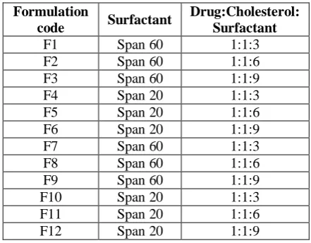
Table 1: Formulation table for Lycopene niosomes.
Formulation
code Surfactant
Drug:Cholesterol: Surfactant
F1 Span 60 1:1:3
F2 Span 60 1:1:6
F3 Span 60 1:1:9
F4 Span 20 1:1:3
F5 Span 20 1:1:6
F6 Span 20 1:1:9
F7 Span 60 1:1:3
F8 Span 60 1:1:6
F9 Span 60 1:1:9
F10 Span 20 1:1:3
F11 Span 20 1:1:6
F12 Span 20 1:1:9
Method of preparation of Niosomes of Lycopene Thin film hydration technique
1. A thin film was prepared from the mixture of vesicles forming ingredients that is Cholesterol and nonionic surfactants such as span 60, span 20 by dissolving in volatile organic solvent (chloroform: methanol 3:2) in a round bottom flask. The organic solvent is removed at room temperature using rotary evaporator leaving a thin layer of solid mixture which is deposited on the walls of flask.[13]
2. The dried surfactant film was hydrated with aqueous phase (Phosphate buffer pH 7.4) at 600C with agitation at 100rpm for 1 hr. The resulted vesicles suspension were kept for swelling at room temperature.[13]
Ether Injection method
Cholesterol, lycopene and non-ionic surfactants i.e. Span 60 and Span 20 were taken in prescribed ratio in a 50 ml beaker. The mixture was dissolved in diethyl ether and the solution was slowly injected into beaker containing phosphate buffer pH 7.4. The temperature maintained during this process was 550-600. The differences in temperature between phases cause rapid vaporization of ether resulting in spontaneous vesiculation.[16]
Formulation of niosomal gel
Te required quantity of carbopol 934 was weighed and dispersed in a small amount of distilled water to prepare an aqueous dispersion. The aqueous dispersion was allowed to hydrate for 4-5hours. Required quantity of Niosomal suspension was added and properly dispersed. The pH was adjusted by addition of 1% (w/v) triethanolamine solution. The final weight of the gel was adjusted with distilled water.[16]
Evaluation of Lycopene niosomes gel Preparation of standard graph of Lycopene
1st Stock: 100 mg of Lycopene was accurately weighed
into 100 ml volumetric flask and dissolved in small quantity of Chloroform, finally the volume was made up to 100 ml with Chloroform(1000 μg/ml).
2nd Stock: 1 ml of the above solution was pipette out into another 100 ml volumetric flask and the volume was made up to 100 ml with Chloroform (10 μg/ml).
From standard solution of 2nd stock i.e., 2ml, 4ml, 6ml, 8ml and 10ml were pipette into 10 ml volumetric flasks. The volume was made up with Chloroform. The spectrum of this solution was run in 200-800 nm range in UV-Visible spectrophotometer. The λ max of Lycopene was found to be 486 nm. The absorbance of each concentration was measured at 486 nm using Chloroform as blank. This was performed in triplicates and average value was reported.
Drug content determination
The amount of drug contained in the Niosomes was determined by dissolving 1ml of the formulation in 9 ml of chloroform and the volume was made up to 100 ml with chloroform. The mixture was analysed by a UV-Visible spectrophotometer at 486 nm against chloroform as a blank.[17]
Entrapment Efficiency (%EE)
the resulting solution was assayed by a UV spectrophotometer (Shimadzu- 1800, Japan) at 486 nm.
The percentage of drug encapsulation was calculated by the following equation:
Entrapment efficiency (%) = [(Ct − Cf) / Ct] × 100
Where Ct is the concentration of total drug and Cf is the concentration of unentrapped drug.[17]
Photo microscopy
All batches of niosomes prepared were observed under light microscope.[17]
Spreading efficiency
To determine spredabilty value spredability apparatus was used. Spredability apparatus contains wooden block having two glass plates. Initially gel sample was placed between the two glass plates. Weight near about 500 g was putted on top plate which expelled the air and to form uniform gel layer. Time required to move upper plate by 10 cm distance was noted, lesser the time required for dragging the upper plate better is the spredability value. Speradability value was determined with the help of formula-
S = M × L / T Where,
S is the Spreadability value, L is the Length of the glass slide,
M is the Weight tied to the upper plate, T is the Time taken to separate the glass slides.[18]
In-Vitro Permeation Study
The in vitro skin permeation study was carried out using a Franz diffusion cell with egg membrane. The membrane with effective diffusion area of 2.0 cm2 was mounted between the donor and receptor compartments of the diffusion cell. The volume of receptor medium was 110ml of PBS (pH 7.4) thermo stated at 37 ± 1 °C, which was continuously stirred at 100 rpm throughout the experiment. 1gm of Lycopene niosomal gel was applied on membrane and placed between the compartments. At 0.5, 1, 1.5, 2, 3, 4, 5, 6, 7, 8, 9, 10, 11 and 12 hr intervals 5ml of the solution in receptor compartment was removed and replaced immediately with equal volume of fresh buffer. Samples were analyzed using UV spectrophotometer and the data was recorded.[18]
RESULTS AND DISCUSSION
Determination of Solubility
Lycopene was freely soluble in chloroform, n-hexane, diethyl ether, insoluble in water ethanol, methanol.
Determination of Melting point
Melting point of Lycopene was determined by using Thale’s tube method by taking a small amount of Lycopene in a capillary tube closed at one end and placed in Thale’s tube containing liquid petroleum and temperature at which drug melts was found to be 1700C.
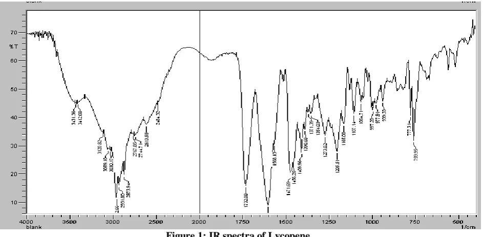
IR Spectroscopy
The IR spectra of pure drug was carried out and the graph shows characteristic absorption peaks of functional peaks of Lycopene.
Figure 1: IR spectra of Lycopene.
Calibration curve in chloroform
UV spectrum of Lycopene in chloroform shows absorbance maxima (λ max) at wavelength 486nm. The standard graph of drug were prepared in chloroform,
Figure 2: Standard graph of Lycopene.
Drug content determination
The drug content for the formulations was carried out as per procedure. The amount of drug contained in the Niosomes was determined by dissolving 1ml of the formulation in 9 ml of chloroform and the volume was made up to 100 ml with chloroform. The mixture was analysed by a UV-Visible spectrophotometer at 486 nm against chloroform as a blank. Drug content of all the formulations were found to be in the range of 46.52 – 92.23% (Table 4.1). This indicates a good vehicle for the release of the drug. Also no degradation of the drug was seen.
Photomicroscopy
Microscopic image of formulated niosomes were viewed by light microscopy under 45X (Figure 4.3). The pictures showed the formation of small and spherical shape vesicles with drug entrapped inside them.
Figure 3: Microscopic image of niosomal formulaion.
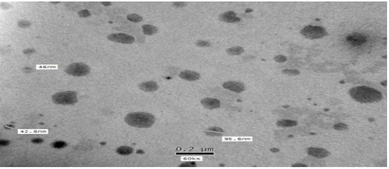
SEM of Niosomes
Scanning electron microscopy of Lycopene loaded niosomal formulations was carried out, the shape of vesicles was found to be spherical. This suggests that the formulation has spherical vesicles and also stable. (Figure 4.4).
Figure 4: SEM of Lycopene Niosomes.
Entrapment efficiency (EC)
The amount of drug entrapped into the niosomal formulation was determined. Percentage entrapment efficiency was conducted by the centrifuge method. The niosome dispersion obtained was centrifuged (REMI LJ 01, Mumbai, India) at 7000 rpm for 45 min. The clear
drug was entrapped in the niosome formulations with higher concentration of Span 60, due to the presence of long chain in the span 60. However formulation F3 has highest drug entrapment of 98.92 %.
Spredability
The spredability of formulated niosomal gels were determined by spredability apparatus. Spredability apparatus contains wooden block having two glass plates. Initially gel sample was placed between the two glass plates. Weight near about 500 g was kept on top plate which expelled the air and to form uniform gel layer. Time required to move upper plate by 10 cm distance was noted, lesser the time required for dragging the upper plate better is the spredability value. Speradability value was determined with the spredability formula. The spredability was in the range of 5.45 to 15.98 gm.cm/sec (Table 4.1). Results showed that good spredability of gel with higher concentration of span 60.
In vitro permeation study
The in vitro skin permeation study was carried out using a Franz diffusion cell with egg membrane. The membrane with effective diffusion area of 2.0 cm2 was mounted between the donor and receptor compartments of the diffusion cell. The volume of receptor medium
was 110ml of PBS (pH 7.4) thermo stated at 37 ± 1 °C, which was continuously stirred at 100 rpm throughout the experiment. 1gm of Lycopene niosomal gel was applied on membrane and placed between the compartments. At 0.5, 1, 1.5, 2, 3, 4, 5, 6, 7, 8, 9, 10, 11 and 12 hr intervals 5ml of the solution in receptor compartment was removed and replaced immediately with equal volume of fresh buffer. Samples were analyzed using UV spectrophotometer and the data was recorded. The In-vitro permeation studies results of all the formulations were calculated. However formulation F3 had highest drug release at the end of 12th hour (Figure 5.15, 5.16).
Stability data of Optimized Lycopene niosomal gel
Optimized batch was kept for accelerated stability at 400C 75% RH. The samples were withdrawn with predetermined days i.e. 30, 60, 90 days. The amount of drug contained in the gel was determined by dissolving 1gm of the formulation in 9 ml of chloroform and the volume was made up to 100 ml with chloroform. The
mixture was analysed by a UV-Visible
spectrophotometer at 486 nm against chloroform as a blank. The drug content was found after 30 days – 90.97%, 60 days - 89.14%, 90 days - 87.43 % (Table. 5.10). There was no change in appearance of niosomal gel. The lycopene niosomal gel formulation was found to be stable.
Table 2: Evaluation table for the Lycopene Niosomal gel.
Formulation Code
%Entrapment Efficiency
% Drug Content
Spreadability
(gm⋅cm/sec)
F1 87.87±0.16 82.51±0.01 11.65
F2 96.45±0.21 86.92±0.07 12.98
F3 98.92±0.012 92.23±0.10 15.2
F4 61.91±0.06 46.52±0.1 9.56
F5 72.87±0.03 72.16±2.1 6.87
F6 74.56±0.27 68.60±0.02 8.94
F7 90.93±0.34 76.71±3.0 9.98
F8 97.54±0.16 86.21±0.81 11.91
F9 98.48±0.03 90.74±0.04 12.56
F10 58.56±0.09 50.21±0.1 5.45
F11 69.98±0.04 61.92±.0.01 7.56
F12 77.43±0.02 81.23±0.06 9.87
Table 3: Stability data for the Lycopene niosomal gel.
Sampling day
Appearance Drug Content
30 Red Colour 90.97±0.290
60 Red Colour 89.14±0.12
90 Red Colour 87.43±0.47
CONCLUSION
Niosome formulation could be conveniently prepared by thin layer hydration method and ether injection method using span 60, span 20, and cholesterol at different concentrations. The drug content of all niosome gel formulations were found to be uniform with low SD values and the results were reproducible. The entrapment efficiency of niosome gel formulation increased with increase in span 60 concentration. The In-vitro diffusion rate was studied by Franz diffusion cell. The drug release from niosomal gel was dependent on concentration of cholesterol and surfactant. Lycopene niosomal gel was also found to be quite stable at 40±5oC over a period of 90 days. Formulation F3 shows maximum encapsulation efficiency of 98.92 % and drug release of 94.65 % after 12 hr have been attained. Niosomal gel formulation with higher concentration of span 60 had more encapsulation efficiency compared with higher concentration of span 20 because of the presence of long chain in structure in span 60. This work has established the foundation for future study on the possibility and effective formulation of Lycopene niosomal gel for topical administration.
REFERENCES
1. Thakur Varun.Niosomes and Liposomes-Vesicuular approach towards transdermal drug delivery. International journal of pharmaceutical and chemical science, 2012; 1(3): 981-93.
2. Shashi Pandey Rai. Medicinal plants derived nutraceuticals. International Journal of Pharma and Bio science, 2011; 2(4): 419-41.
3. Pandey M, Verma RK, Saraf SA. Nutraceuticals: New era of medicine and health. Asian Journal of Pharmaceutical and Clinical Research, 2010; 3: 11– 5.
4. Manisha Pandey. Nutraceuticals: new era of medicine and health. Asian Journal of Pharmaceutical and Clinical Research, 2010; 3(1): 11-15.
5. Whitman M. Understanding the perceived need for complementary and alternative nutraceuticals lifestyle issues, 2001; 5: 190-94.
6. Tamler R, Mechanick JI. Dietary Supplements and Nutraceuticals in the Management of Andrologic Disorders. Endocrinology and Metabolism Clinics of North America, 2007; 36: 533–52.
7. Shahzad T, Ahmad I, Choudhry S, Saeed MK, Khan MN. Dpph free radical scavenging activity of tomato, cherry tomato and watermelon: Lycopene extraction, purification and quantification. Int J Pharm Pharm Sci, 2014; b 6(2): 223–8.
8. LU Zhufen, CHEN Yanzhong. Lycopene
nanoparticles coated with microemulsion to improve stability. Advanced Materials Research, 2015; 1120-1121: 897-902.
9. Rao A V., Ray MR, Rao LG. Lycopene. Advances in Food and Nutrition Research, 2006; 51: 99–164.
10. Maryam Mohammadi. Formulation of
nanaliposomal vitamin D3 for potential application. Advanced pharmaceutical bulletin, 2014; 4(2): 569-75.
11. Wang XD. Lycopene metabolism and its biological significance. Vol. 96, American Journal of Clinical Nutrition, 2012.
12. Uchegbu IF, Vyas SP. Non-ionic surfactant based vesicles (niosomes) in drug delivery. International Journal of Pharmaceutics, 1998; 172: 33–70. 13. Lohumi Ashutosh, Rawat Suman, Sarkar Sidhyartha,
Sipai Altaf bhai. YMV. A Novel Drug Delivery System: Niosomes Review. J Drug Deliv Ther, 2012; 2(5): 129–35.
14. Rajera R, Nagpal K, Singh SK, Mishra DN. Niosomes: a controlled and novel drug delivery system. Biol {and} Pharm Bull, 2011; 34(7): 945– 53.
15. Moghassemi S, Hadjizadeh A. Nano-niosomes as nanoscale drug delivery systems: An illustrated review. Journal of Controlled Release, 2014; 185: 22–36.
16. Yadav JD, Kulkarni PR, Vaidya K a, Shelke GT. Niosomes: a review. J Pharm Res, 2011; 2(3): 632– 6.
17. Shirsand SB, Para MS, Nagendrakumar D, Kanani KM, Keerthy D. Formulation and evaluation of Ketoconazole niosomal gel drug delivery system. Int J Pharm Investig, 2012; 2(4): 201–7.